Journal of Virology logoLink to Journal of Virology
letter
. 2006 Feb;80(3):1610–1612. doi: 10.1128/JVI.80.3.1610-1612.2006

Egress of Alphaherpesviruses

Thomas C Mettenleiter 1,1,*, Tony Minson 1,1
PMCID: PMC1346970  PMID: 16415038

In their recent papers on the assembly of alphaherpesviruses, Wild et al. (13) and Leuzinger et al. (3) propose that the egress of herpesvirus capsids from the nucleus and formation of the enveloped herpesvirus virion occur by two alternative routes.

The first route is similar to that described in earlier proposals (7) and involves primary envelopment of intranuclear capsids at the inner nuclear membrane followed by transport of these primary virions through the endoplasmic reticulum (ER) and secretory Golgi pathway to the cell surface. This mode of herpesvirus release has been challenged in the last few years, since it does not account for a wealth of biochemical, genetic, and morphological data (reviewed in reference 5) which clearly demonstrate a lack of identity between primary and mature virus particles. The former contain proteins homologous to the herpes simplex virus UL31 and UL34 products which are not present in any of the hitherto-analyzed mature herpesvirus virions, whereas important constituents of mature virions, among them major tegument proteins, are absent from primary virions (reviewed in reference 4). Moreover, the lipid composition of the mature virus envelope differs strikingly from that of the nuclear membrane (10), and viral envelope proteins engineered to be retained in the ER do not become part of the mature virion (12). These well-documented differences between primary enveloped and mature virions are best explained by an envelopment-deenvelopment-reenvelopment pathway (8) which entails budding of intranuclear DNA-filled capsids at the inner nuclear membrane followed by fusion of the primary envelope with the outer nuclear membrane, thus transferring capsids through the nuclear membrane into the cytoplasm where they acquire their final tegument and envelope by budding into a presumably trans-Golgi compartment (2, 9, 11, 14). While not every aspect of this model has been proven, it explains the relevant observations outlined above, which any alternative model has to take into account as well.

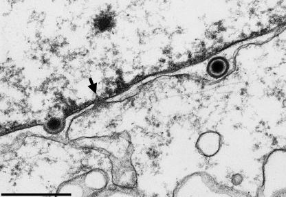
The second route of egress proposed by Wild et al. (13) and Leuzinger et al. (3) involves the dilation of nuclear pores resulting in direct access of capsids to the cytoplasm and envelopment in the Golgi compartment. Indeed, the authors consider that most of the enveloped virions observed in the perinuclear space arise by budding from the cytoplasm into the outer nuclear membrane/ER and hence that naked nucleocapsids are apparently capable of budding into any membrane. These proposals also raise interesting issues. A number of genetic defects (e.g., deletions in UL31/UL34) result in failure of nuclear egress (1, 6), implying that these gene functions may be responsible for nuclear pore dilation. It would be interesting to see whether the authors observe pore dilation in cells infected with mutant viruses lacking these proteins. In extensive studies at relevant time points after infection, we did not observe a similar impairment of nuclear pores (Fig. 1). Furthermore, many mutations that result in the accumulation of capsids in the cytoplasm by a failure of cytoplasmic envelopment (e.g., defects in UL36, UL37, or UL11 and their homologues [reviewed in reference 5)] have been described, yet cells infected with these mutants contain enveloped virions in the perinuclear space. Thus, different functions are clearly required for budding into these different compartments, and this is entirely consistent with the observation that enveloped virions in different compartments have quite different compositions and morphologies.

FIG. 1.

FIG. 1.

Electron micrograph of BHK21 cells 12 h after infection with HSV-1. The arrow denotes an intact nuclear pore next to a capsid undergoing primary envelopment (left) and a primary enveloped virion in the perinuclear space (right). The nuclear side is on the top, and the cytoplasmic side is on the bottom. Bar, 500 nm. (Electron micrograph courtesy of Harald Granzow, Friedrich-Loeffler-Institut, Insel Riems, Germany).

The electron microscopic observations reported in these two papers challenge current views of the egress and maturation of alphaherpesvirus particles and emphasize the need for further study. However, the models proposed take little account of the weight of biochemical, genetic, and morphological evidence in the literature and cannot be readily reconciled with this evidence.

REFERENCES

  • 1.Fuchs, W., B. G. Klupp, H. Granzow, N. Osterrieder, and T. C. Mettenleiter. 2002. The interacting UL31 and Ul34 gene products of pseudorabies virus are involved in egress from the host cell nucleus and represent components of primary enveloped but not of mature virions. J. Virol. 76:364-378. [DOI] [PMC free article] [PubMed] [Google Scholar]
  • 2.Granzow, H., B. G. Klupp, W. Fuchs, J. Veits, N. Osterrieder, and T. C. Mettenleiter. 2001. Egress of alphaherpesviruses: comparative ultrastructural study. J. Virol. 75:3675-3684. [DOI] [PMC free article] [PubMed] [Google Scholar]
  • 3.Leuzinger, H., U. Ziegler, E. M. Schraner, C. Fraefel, D. L. Glauser, I. Heid, M. Ackermann, M. Mueller, and P. Wild. 2005. Herpes simplex virus type 1 envelopment follows two diverse pathways. J. Virol. 79:13047-13059. [DOI] [PMC free article] [PubMed] [Google Scholar]
  • 4.Mettenleiter, T. C. 2002. Herpesvirus assembly and egress. J. Virol. 76:1537-1547. [DOI] [PMC free article] [PubMed] [Google Scholar]
  • 5.Mettenleiter, T. C. 2004. Budding events in herpesvirus morphogenesis. Virus Res. 106:167-180. [DOI] [PubMed] [Google Scholar]
  • 6.Reynolds, A., B. Ryckman, J. Baines, Y. Zhou, L. Liang, and R. Roller. 2001. UL31 and UL34 proteins of herpes simplex virus type 1 form a complex that accumulates at the nuclear rim and is required for envelopment of nucleocapsids. J. Virol. 75:8803-8817. [DOI] [PMC free article] [PubMed] [Google Scholar]
  • 7.Roizman, B., and D. Knipe. 2001. Herpes simplex viruses and their replication, p. 2399-2460. In D. Knipe and P. M. Howley (ed.), Fields virology, 4th ed. Lippincott, Williams and Wilkins, Philadelphia, Pa.
  • 8.Skepper, J., A. Whiteley, H. Browne, and A. Minson. 2001. Herpes simplex virus nucleocapsids mature to progeny virions by an envelopment-deenvelopment-reenvelopment pathway. J. Virol. 75:5697-5702. [DOI] [PMC free article] [PubMed] [Google Scholar]
  • 9.Turcotte, S., J. Letellier, and R. Lippé. 2005. Herpes simplex virus type 1 capsids transit by the trans-Golgi network, where viral glycoproteins accumulate independently of capsid egress. J. Virol. 79:8847-8860. [DOI] [PMC free article] [PubMed] [Google Scholar]
  • 10.Van Genderen, I. L., R. Brandimarti, M. Torrisi, G. Campadelli-Fiume, and G. van Meer. 1994. The phospholipid composition of extracellular herpes simplex virions differs from that of the host cell nuclei. Virology 200:831-836. [DOI] [PubMed] [Google Scholar]
  • 11.Whealy, E. M., J. P. Card, R. P. Meade, A. K. Robbins, and L. W. Enquist. 1991. Effect of brefeldin A on alphaherpesvirus membrane protein glycosylation and virus egress. J. Virol. 65:1066-1081. [DOI] [PMC free article] [PubMed] [Google Scholar]
  • 12.Whiteley, A., B. Bruun, T. Minson, and H. Browne. 1999. Effects of targeting herpes simplex virus type 1 gD to the endoplasmic reticulum and trans-Golgi network. J. Virol. 73:9515-9520. [DOI] [PMC free article] [PubMed] [Google Scholar]
  • 13.Wild, P., M. Engels, C. Senn, K. Tobler, U. Ziegler, E. Schranker, E. Loepfe, M. Ackermann, M. Mueller, and P. Walther. 2005. Impairment of nuclear pores in bovine herpesvirus 1-infected MDBK cells. J. Virol. 79:1071-1083. [DOI] [PMC free article] [PubMed] [Google Scholar]
  • 14.Zhu, Z., M. D. Gershon, Y. Hao, R. T. Ambron, C. A. Gabel, and A. A. Gershon. 1995. Envelopment of varicella-zoster virus: targeting of viral glycoproteins to the trans-Golgi network. J. Virol. 69:7951-7959. [DOI] [PMC free article] [PubMed] [Google Scholar]
J Virol. 2006 Feb;80(3):1610–1612.

Author's Reply

Peter Wild 2

In a series of recent publications (8, 17, 18), we have challenged the current model of herpesvirus envelopment, which predicts an unlikely series of energy-consuming steps, i.e., envelopment of capsids at the inner nuclear membrane (INM), deenvelopment by fusion with the outer nuclear membrane (ONM), and reenvelopment at the trans-Golgi network (9).

The Golgi complex plays a crucial role in secretion of proteins and herpesvirus egress. One of its functions is packaging of secretory products into membrane-bound entities that deliver the product to the cell surface for releasing via exocytosis. There is striking evidence that herpesviruses are packaged into such entities (1, 4, 7, 8, 13, 18), which are referred to as transport vacuoles (8, 17, 18). There is also clear evidence that capsids bud at any site of the Golgi complex, including trans-Golgi vacuoles (7, 8, 17, 18). These two entirely different processes demand different routes of transportation of capsids to the outside of Golgi membranes and of virions into cisternae, respectively. The route of virions into Golgi cisternae is considered likely to involve intraluminal transportation (8, 18) through a continuum (18) from the perinuclear space (PNS), via the rough ER to Golgi cisternae. The route of nuclear capsids into the cytoplasm is considered to be via impaired nuclear pores (8, 17).

The deenvelopment theory is based (i) on phenotypes at the ONM that have characteristics of budding rather than of fusion and (ii) on proteins detected only in virions within the PNS. Yet virions within the PNS are inside-out particles. The luminal layer of the envelope faces the luminal layer of the nuclear membranes, the site at which the fission machinery, which is antagonistic to the fusion machinery, is active during budding. At this stage, it is unlikely that the viral envelope can fuse with the ONM. If it did, why would it not fuse with the INM? The INM and ONM are similar and would probably be identical after viral egress has proceeded, because the constituents of the INM are inserted into the ONM as a result of fusion. ONM constituents would need to recycle to the INM to maintain equilibrium between the two membranes. The surface of virus particles within the PNS and rough ER is covered by a dense substance (1, 4, 5, 8, 11, 13, 16-18). UL31 and UL34 have been shown to be incorporated into both nuclear membranes and, less convincingly, into virions within the PNS (11). They might play a role in intraluminal transportation per se or in combination with other proteins that are removed within Golgi cisternae so that additional sugars can be inserted (10).

To support the deenvelopment theory, Mettenleiter and Minson argue that the lipid composition of mature virions differs from that of host cell nuclear membranes (15). This study, however, failed to demonstrate identity of lipid compositions between viral envelopes and Golgi membranes. In fact, the divergence in lipid composition between viral envelope, nuclear membrane, and Golgi membranes is best explained by the dual site of envelope origin.

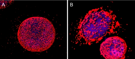
Mettenleiter and Minson question impairment of nuclear pores to be the gateway for capsids to exit the nucleus because they did not find evidence for it. Impairment of the nuclear envelope has been shown earlier (2, 6). Alterations in nuclear pores can be demonstrated by electron microscopy (8, 17) and, very easily, by immunolabeling and confocal microscopy (Fig. 1). Studies on lamin, UL31, and UL34 also support the idea of a dual-exit pathway of capsids (12).

FIG. 1.

FIG. 1.

HeLa cells grown on coverslips were infected with herpes simplex virus type 1 (multiplicity of infection of 2), incubated for 10 h, fixed with paraformaldehyde, and immunostained using monoclonal antibodies against nuclear pore complex proteins. Samples were analyzed using a confocal microscope, and images were deconvolved. (A) A mock-infected cell shows a rather regular distribution of nuclear pore complex proteins. (B) Nuclear pore complex proteins are accumulated at the nuclear surface and within the cytoplasm in a herpes simplex virus type 1-infected cell, clearly indicating dramatic changes of nuclear pores.

Many reports show data that support the dual-pathway theory and/or question the theory of envelopment at the trans-Golgi network (see, e.g., references 3, 5, 13, and 16). Moreover, some data can be explained only by the dual-pathway theory, e.g., glycoproteins that accumulate at the trans-Golgi network independently of capsid egress (14).

REFERENCES

  • 1.Avitabile, E., S. Di Gaeta, M. R. Torrisi, P. L. Ward, B. Roizman, and G. Campadelli-Fiume. 1995. Redistribution of microtubules and Golgi apparatus in herpes simplex virus-infected cells and their role in viral exocytosis. J. Virol. 69:7472-7482. [DOI] [PMC free article] [PubMed] [Google Scholar]
  • 2.Borchers, K., and M. Oezel. 1993. Simian agent 8 (SA8): morphogenesis and ultrastructure. Zentbl. Bakteriol. 279:526-536. [DOI] [PubMed] [Google Scholar]
  • 3.Card, J. P., L. Rinaman, R. B. Lynn, B. H. Lee, R. P. Meade, R. R. Miselis, and L. W. Enquist. 1993. Pseudorabies virus infection of the rat central nervous system: ultrastructural characterization of viral replication, transport, and pathogenesis. J. Neurosci. 13:2515-2539. [DOI] [PMC free article] [PubMed] [Google Scholar]
  • 4.Di Lazzaro, C., G. Campadelli-Fiume, and M. R. Torrisi. 1995. Intermediate forms of glycoconjugates are present in the envelope of herpes simplex virions during their transport along the exocytic pathway. Virology 214:619-623. [DOI] [PubMed] [Google Scholar]
  • 5.Granzow, H., B. G. Klupp, W. Fuchs, J. Veits, N. Osterrieder, and T. C. Mettenleiter. 2001. Egress of alphaherpesviruses: comparative ultrastructural study. J. Virol. 75:3675-3684. [DOI] [PMC free article] [PubMed] [Google Scholar]
  • 6.Haines, H., and R. J. Baerwald. 1976. Nuclear membrane changes in herpes simplex virus-infected BHK-21 cells as seen by freeze-fracture. J. Virol. 17:1038-1042. [DOI] [PMC free article] [PubMed] [Google Scholar]
  • 7.Homman-Loudiyi, M., K. Hultenby, W. Britt, and C. Soderberg-Naucler. 2003. Envelopment of human cytomegalovirus occurs by budding into Golgi-derived vacuole compartments positive for gB, Rab 3, trans-Golgi network 46, and mannosidase II. J. Virol. 77:3191-3203. (Erratum, 77:8179.) [DOI] [PMC free article] [PubMed] [Google Scholar]
  • 8.Leuzinger, H., U. Ziegler, C. Fraefel, E. M. Schraner, D. Glauser, I. Held, M. Ackermann, M. Müller, and P. Wild. 2005. Herpes simplex virus 1 envelopment follows two diverse pathways. J. Virol. 79:13047-13059. [DOI] [PMC free article] [PubMed] [Google Scholar]
  • 9.Mettenleiter, T. C. 2004. Budding events in herpesvirus morphogenesis. Virus Res. 106:167-180. [DOI] [PubMed] [Google Scholar]
  • 10.Poliquin, L., G. Levine, and G. C. Shore. 1985. Involvement of Golgi apparatus and a restructured nuclear envelope during biogenesis and transport of herpes simplex virus glycoproteins. J. Histochem. Cytochem. 33:875-883. [DOI] [PubMed] [Google Scholar]
  • 11.Reynolds, A. E., E. G. Wills, R. J. Roller, B. J. Ryckman, and J. D. Baines. 2002. Ultrastructural localization of the herpes simplex virus type 1 UL31, UL34, and US3 proteins suggests specific roles in primary envelopment and egress of nucleocapsids. J. Virol. 76:8939-8952. [DOI] [PMC free article] [PubMed] [Google Scholar]
  • 12.Simpson-Holley, M., R. C. Colgrove, G. Nalepa, J. W. Harper, and D. M. Knipe. 2005. Identification and functional evaluation of cellular and viral factors involved in the alteration of nuclear architecture during herpes virus 1 infection. J. Virol. 79:12840-12851. [DOI] [PMC free article] [PubMed] [Google Scholar]
  • 13.Stannard, L. M., S. Himmelhoch, and S. Wynchank. 1996. Intra-nuclear localization of two envelope proteins, gB and gD, of herpes simplex virus. Arch. Virol. 141:505-524. [DOI] [PubMed] [Google Scholar]
  • 14.Turcotte, S., J. Letellier, and R. Lippe. 2005. Herpes simplex virus type 1 capsids transit by the trans-Golgi network, where viral glycoproteins accumulate independently of capsid egress. J. Virol. 79:8847-8860. [DOI] [PMC free article] [PubMed] [Google Scholar]
  • 15.van Genderen, I. L., R. Brandimarti, M. R. Torrisi, G. Campadelli, and G. van Meer. 1994. The phospholipid composition of extracellular herpes simplex virions differs from that of host cell nuclei. Virology 200:831-836. [DOI] [PubMed] [Google Scholar]
  • 16.Whealy, M. E., J. P. Card, R. P. Meade, A. K. Robbins, and L. W. Enquist. 1991. Effect of brefeldin A on alphaherpesvirus membrane protein glycosylation and virus egress. J. Virol. 65:1066-1081. [DOI] [PMC free article] [PubMed] [Google Scholar]
  • 17.Wild, P., M. Engels, C. Senn, K. Tobler, U. Ziegler, E. M. Schraner, E. Loepfe, M. Ackermann, M. Mueller, and P. Walther. 2005. Impairment of nuclear pores in bovine herpesvirus 1-infected MDBK cells. J. Virol. 79:1071-1083. [DOI] [PMC free article] [PubMed] [Google Scholar]
  • 18.Wild, P., E. M. Schraner, D. Cantieni, E. Loepfe, P. Walther, M. Mueller, and M. Engels. 2002. The significance of the Golgi complex in envelopment of bovine herpesvirus 1 (BHV-1) as revealed by cryobased electron microscopy. Micron 33:327-337. [DOI] [PubMed] [Google Scholar]

Articles from Journal of Virology are provided here courtesy of American Society for Microbiology (ASM)

RESOURCES