Abstract
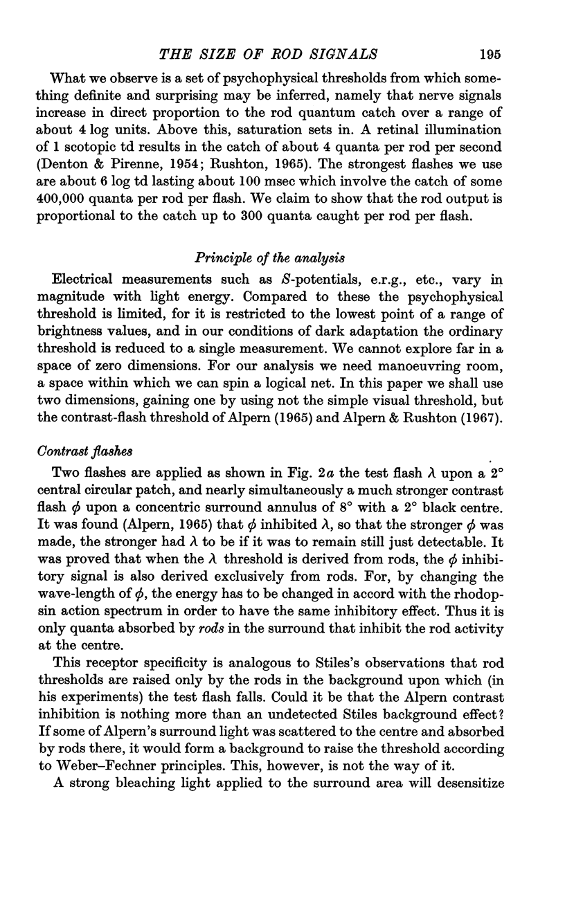
1. This investigation is based upon Alpern's (1965) contrast flash observations. The threshold for the test flash λ (Fig. 2a) is raised if a second flash ϕ falls on the annular surround. Moreover, if λ excites rods at threshold, it is only the rods in the surround that contribute to the threshold rise.
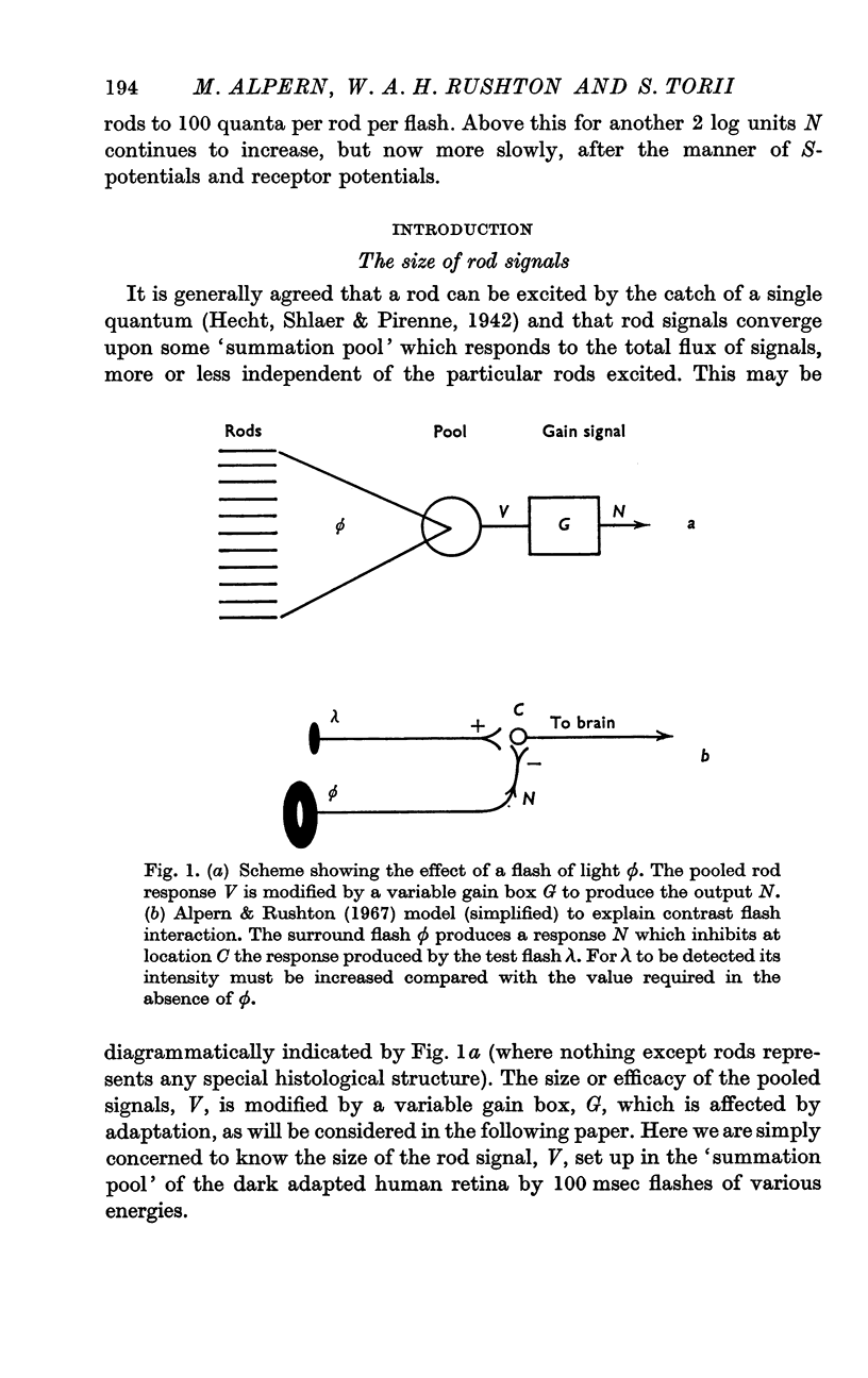
2. The possibility that the rise in λ threshold might be due to light physically scattered from surround to centre we exclude by several different experiments. We conclude (Fig. 1b) that the ϕ flash sets up a nerve signal N which is conducted to some place C where it inhibits the signal from the centre.
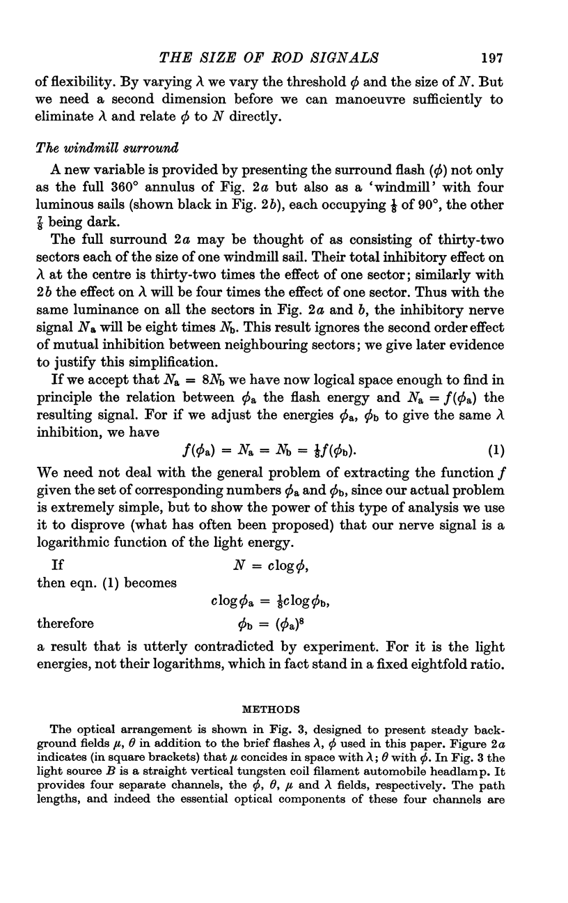
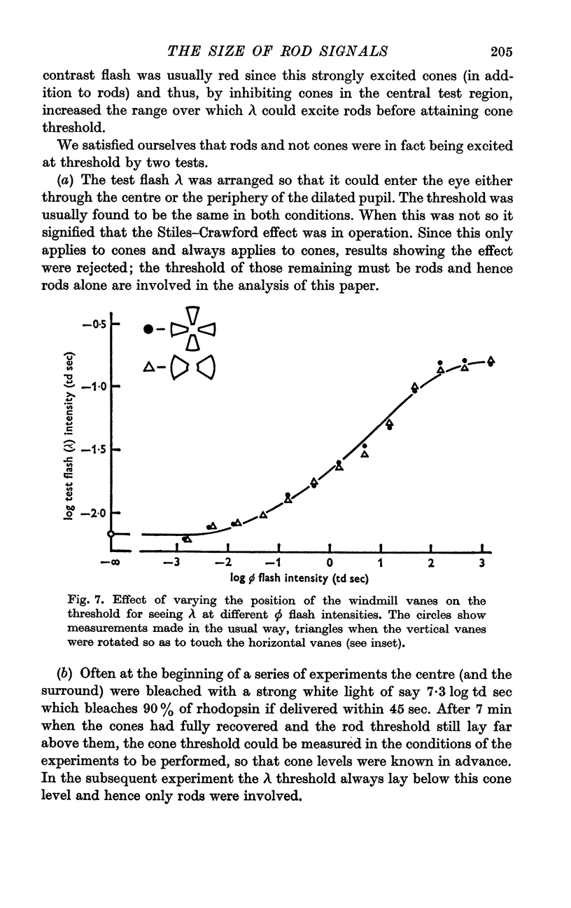
3. If the luminous surround, instead of being a full circle (Fig. 2a) consists only of the sectors shown black in Fig. 2b, that occupy 1/m of the surround area, it is found (in the physiological range) that the light/area on those sectors must be m times as great to produce the same threshold rise at centre, i.e. the total surround illumination must remain the same.
4. This result would obviously follow if N, the inhibitory nerve signal, were proportional to the total surround illumination. We have established the converse; the signal must be proportional to the quantum catch.
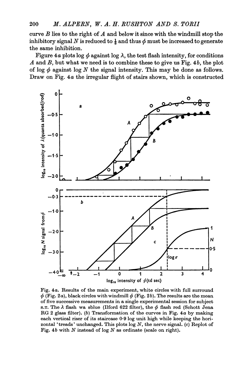
5. Light can be increased indefinitely, nerve signals cannot. When ϕ increases sufficiently, N saturates in the same way that S-potentials and receptor potentials saturate, namely according to N = ϕ/(ϕ + σ) where σ, the semi-saturation constant is about 200 td sec, or 800 quanta absorbed per rod per flash.
6. Thus the nerve signal N is proportional to the quantum catch over 4 log units in the physiological range, namely from 1 quantum per 100 rods to 100 quanta per rod per flash. Above this for another 2 log units N continues to increase, but now more slowly, after the manner of S-potentials and receptor potentials.
Full text
PDF
Selected References
These references are in PubMed. This may not be the complete list of references from this article.
- ALPERN M. ROD-CONE INDEPENDENCE IN THE AFTER-FLASH EFFECT. J Physiol. 1965 Feb;176:462–472. doi: 10.1113/jphysiol.1965.sp007561. [DOI] [PMC free article] [PubMed] [Google Scholar]
- ALPERN M., RUSHTON W. A. THE SPECIFICITY OF THE CONE INTERACTION IN THE AFTER-FLASH EFFECT. J Physiol. 1965 Feb;176:473–482. doi: 10.1113/jphysiol.1965.sp007562. [DOI] [PMC free article] [PubMed] [Google Scholar]
- Alpern M., Rushton W. A. The nature of rise in threshold produced by contrast-flashes. J Physiol. 1967 Apr;189(3):519–534. doi: 10.1113/jphysiol.1967.sp008182. [DOI] [PMC free article] [PubMed] [Google Scholar]
- DENTON E. J., PIRENNE M. H. The absolute sensitivity and functional stability of the human eye. J Physiol. 1954 Mar 29;123(3):417–442. doi: 10.1113/jphysiol.1954.sp005062. [DOI] [PMC free article] [PubMed] [Google Scholar]
- Naka K. I. Computer assisted analysis of S-potentials. Biophys J. 1969 Jun;9(6):845–859. doi: 10.1016/S0006-3495(69)86422-2. [DOI] [PMC free article] [PubMed] [Google Scholar]
- Naka K. I., Rushton W. A. S-potentials from luminosity units in the retina of fish (Cyprinidae). J Physiol. 1966 Aug;185(3):587–599. doi: 10.1113/jphysiol.1966.sp008003. [DOI] [PMC free article] [PubMed] [Google Scholar]
- Naka K. I., Rushton W. A. The generation and spread of S-potentials in fish (Cyprinidae). J Physiol. 1967 Sep;192(2):437–461. doi: 10.1113/jphysiol.1967.sp008308. [DOI] [PMC free article] [PubMed] [Google Scholar]
- RUSHTON W. A. THE SENSITIVITY OF RODS UNDER ILLUMINATION. J Physiol. 1965 May;178:141–160. doi: 10.1113/jphysiol.1965.sp007620. [DOI] [PMC free article] [PubMed] [Google Scholar]
- Rushton W. A., Gubisch R. W. Glare: its measurement by cone thresholds and by the bleaching of cone pigments. J Opt Soc Am. 1966 Jan;56(1):104–110. doi: 10.1364/josa.56.000104. [DOI] [PubMed] [Google Scholar]
- Werblin F. S., Dowling J. E. Organization of the retina of the mudpuppy, Necturus maculosus. II. Intracellular recording. J Neurophysiol. 1969 May;32(3):339–355. doi: 10.1152/jn.1969.32.3.339. [DOI] [PubMed] [Google Scholar]


