Abstract
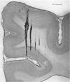
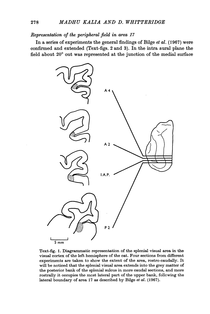
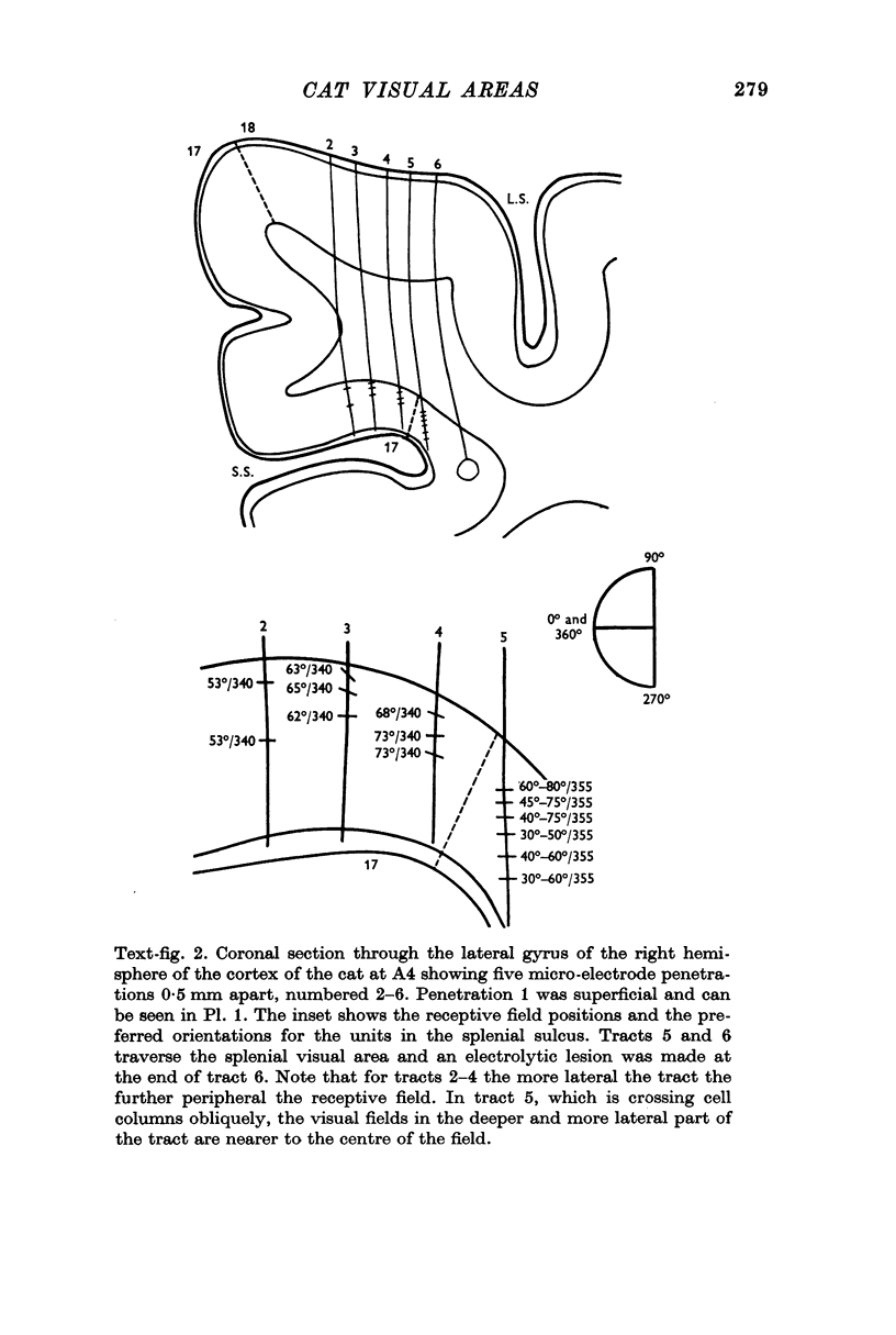
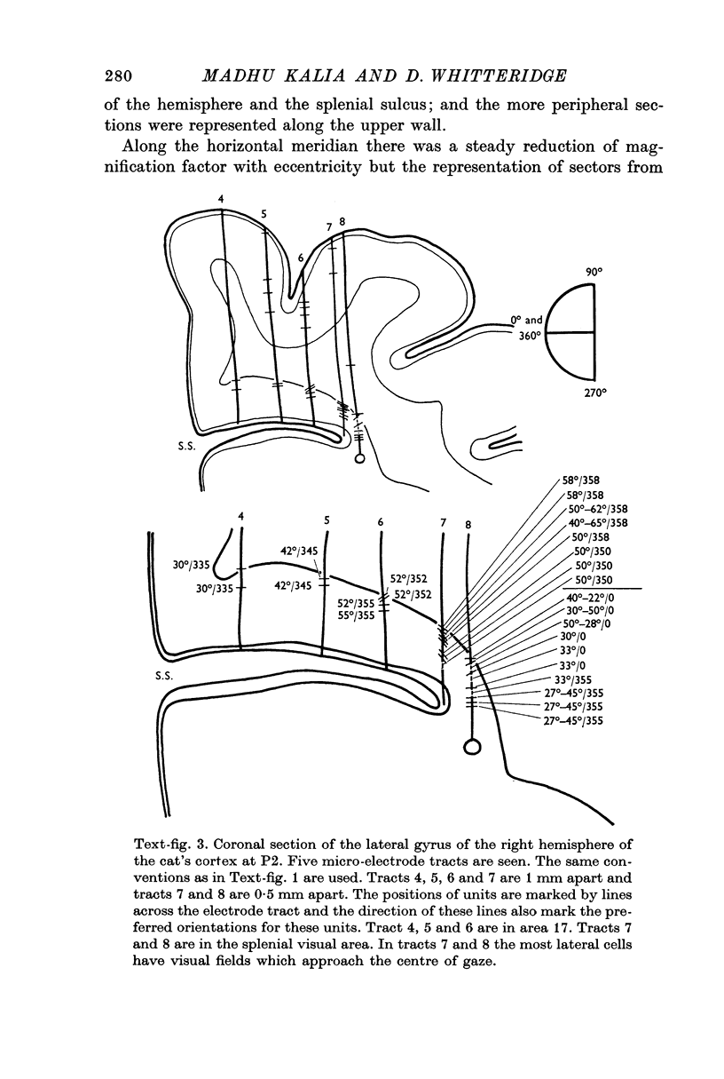
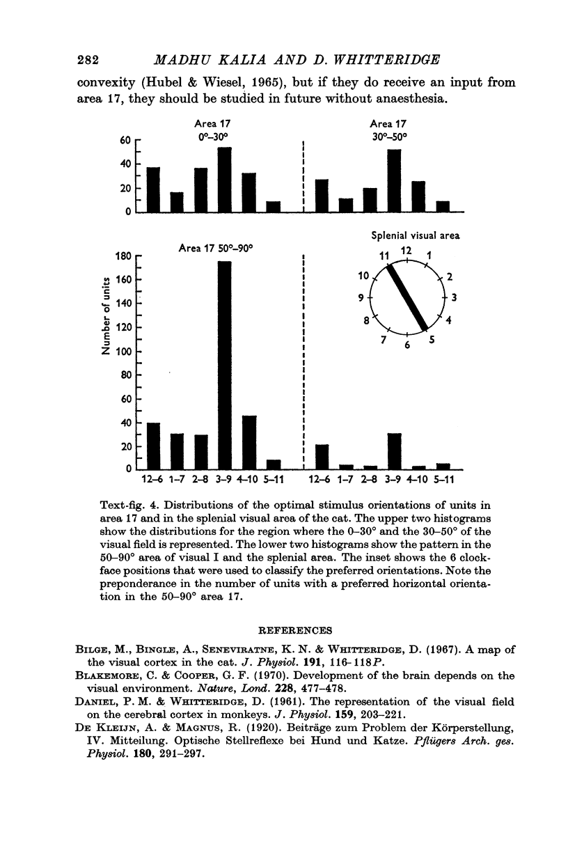
1. The extreme periphery of the visual field is represented in the upper wall of the splenial sulcus where the sulcus is horizontal, and in its anterior wall more posteriorly where the sulcus runs downwards and laterally. About half the cells whose fields lie between 50 and 90° from the area centralis have a sharply horizontal preferred orientation.
2. Beyond the lateral edge of visual I there is a narrow band of visual cortex in which the receptive fields return towards the area centralis as one moves 1-1·5 mm laterally. Their receptive fields are usually about 20-30° degrees across, but all orientations are found. The more central fields may be binocular and those at the area centralis may be as small as 1° in diameter.
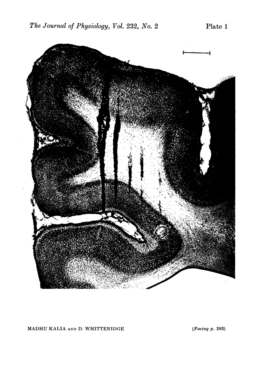
3. This band has been called the splenial visual area. It does not seem to have properties corresponding to those of visual II nor of visual III.
Full text
PDF
Images in this article
Selected References
These references are in PubMed. This may not be the complete list of references from this article.
- Bilge M., Bingle A., Seneviratne K. N., Whitteridge D. A map of the visual cortex in the cat. J Physiol. 1967 Jul;191(2):116P–118P. [PubMed] [Google Scholar]
- Blakemore C., Cooper G. F. Development of the brain depends on the visual environment. Nature. 1970 Oct 31;228(5270):477–478. doi: 10.1038/228477a0. [DOI] [PubMed] [Google Scholar]
- DANIEL P. M., WHITTERIDGE D. The representation of the visual field on the cerebral cortex in monkeys. J Physiol. 1961 Dec;159:203–221. doi: 10.1113/jphysiol.1961.sp006803. [DOI] [PMC free article] [PubMed] [Google Scholar]
- HUBEL D. H., WIESEL T. N. RECEPTIVE FIELDS AND FUNCTIONAL ARCHITECTURE IN TWO NONSTRIATE VISUAL AREAS (18 AND 19) OF THE CAT. J Neurophysiol. 1965 Mar;28:229–289. doi: 10.1152/jn.1965.28.2.229. [DOI] [PubMed] [Google Scholar]
- HUBEL D. H., WIESEL T. N. Receptive fields, binocular interaction and functional architecture in the cat's visual cortex. J Physiol. 1962 Jan;160:106–154. doi: 10.1113/jphysiol.1962.sp006837. [DOI] [PMC free article] [PubMed] [Google Scholar]
- Heath C. J., Jones E. G. The anatomical organization of the suprasylvian gyrus of the cat. Ergeb Anat Entwicklungsgesch. 1971;45(3):3–64. [PubMed] [Google Scholar]
- Nikara T., Bishop P. O., Pettigrew J. D. Analysis of retinal correspondence by studying receptive fields of binocular single units in cat striate cortex. Exp Brain Res. 1968;6(4):353–372. doi: 10.1007/BF00233184. [DOI] [PubMed] [Google Scholar]
- OTSUKA R., HASSLER R. [On the structure and segmentation of the cortical center of vision in the cat]. Arch Psychiatr Nervenkr Z Gesamte Neurol Psychiatr. 1962;203:212–234. doi: 10.1007/BF00352744. [DOI] [PubMed] [Google Scholar]
- Wilson M. E., Cragg B. G. Projections from the lateral geniculate nucleus in the cat and monkey. J Anat. 1967 Sep;101(Pt 4):677–692. [PMC free article] [PubMed] [Google Scholar]
- Woolsey C. N. Comparative studies on cortical representation of vision. Vision Res. 1971;Suppl 3:365–382. doi: 10.1016/0042-6989(71)90051-4. [DOI] [PubMed] [Google Scholar]


