

Abstract
1. Membrane current was derived from simultaneous measurements of interstitial `dark' voltages and conductivities along the radial axis of frog photoreceptor cells. Membrane current was subsequently resolved into its component ionic currents or fluxes by means of ionic substitution and by the use of inhibitors of active transport.
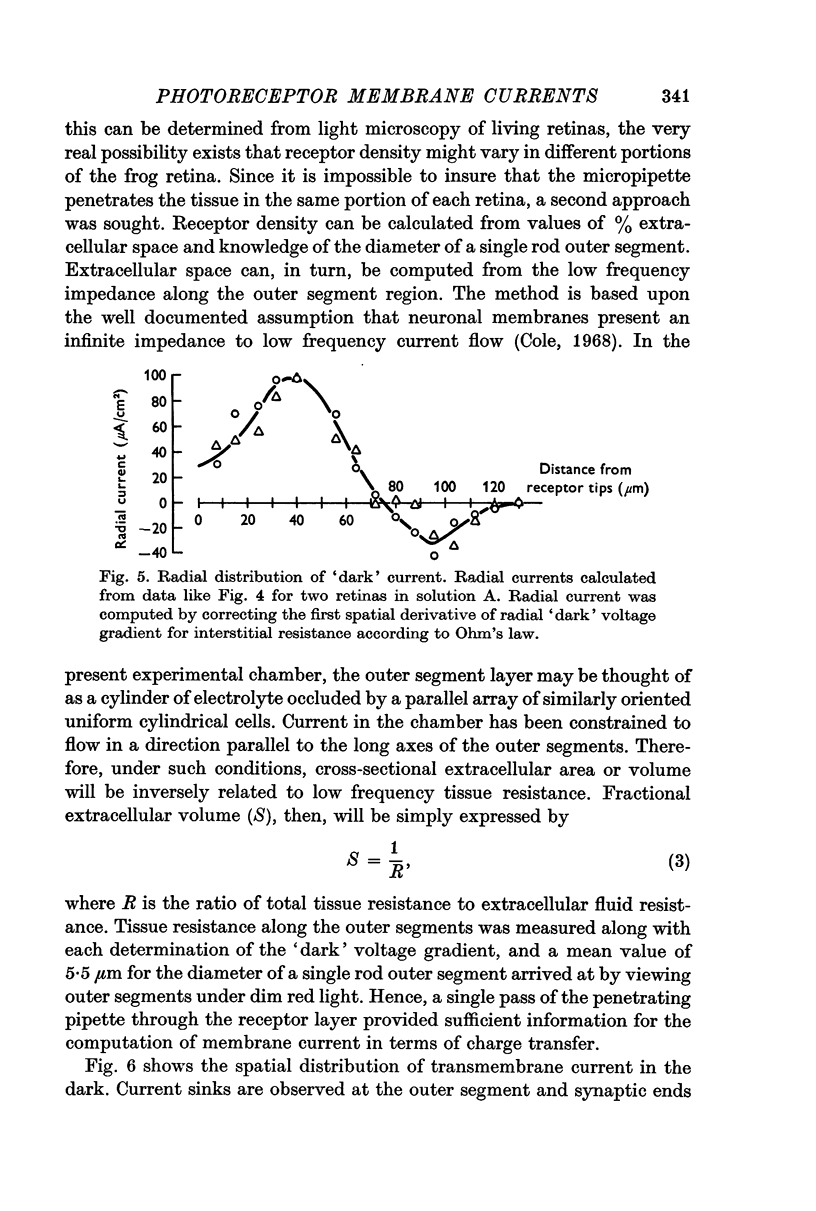
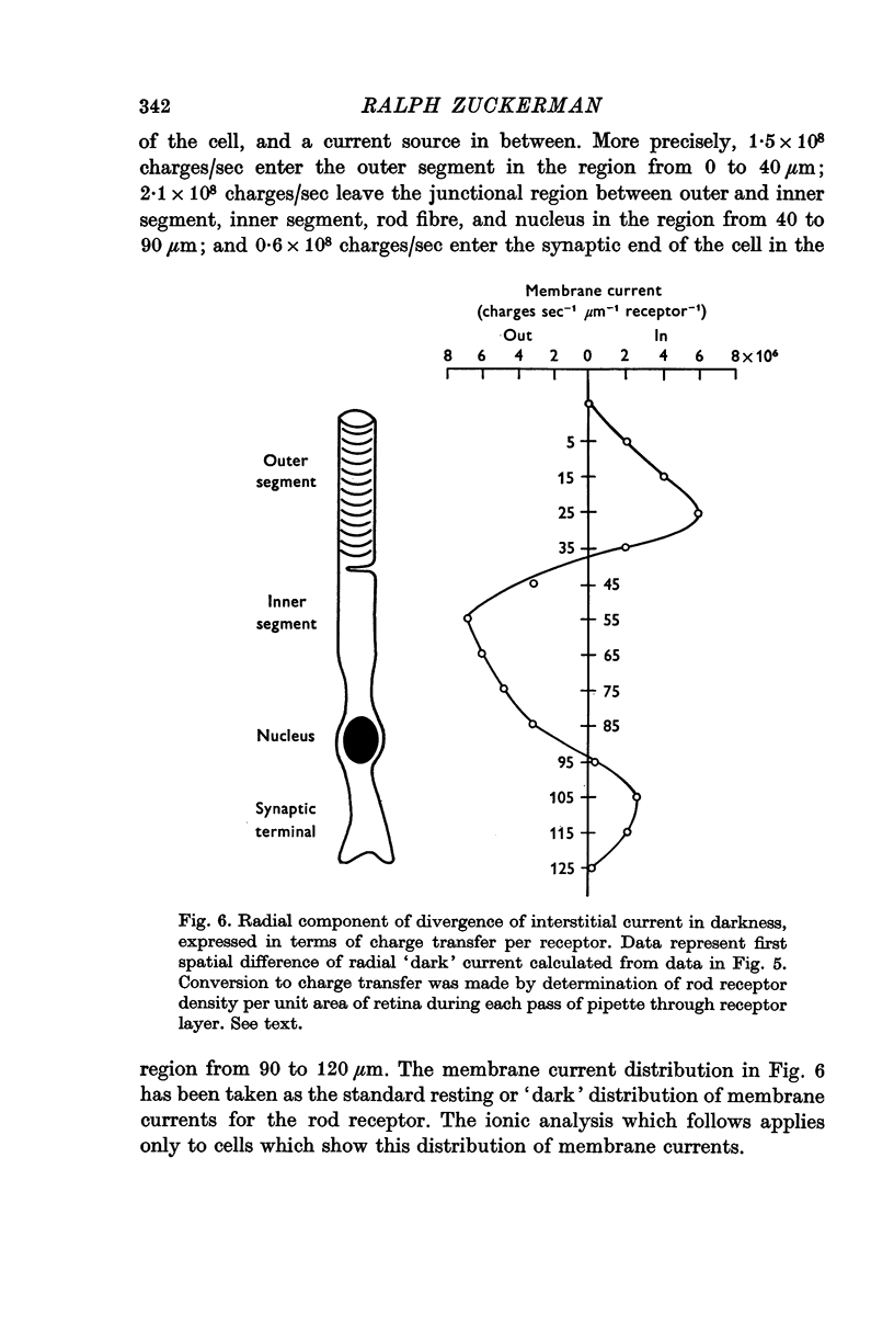
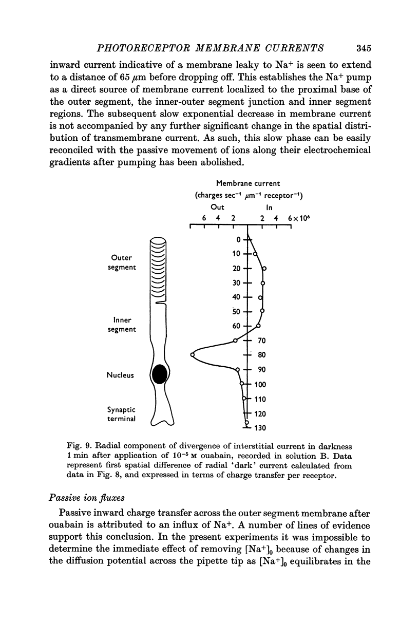
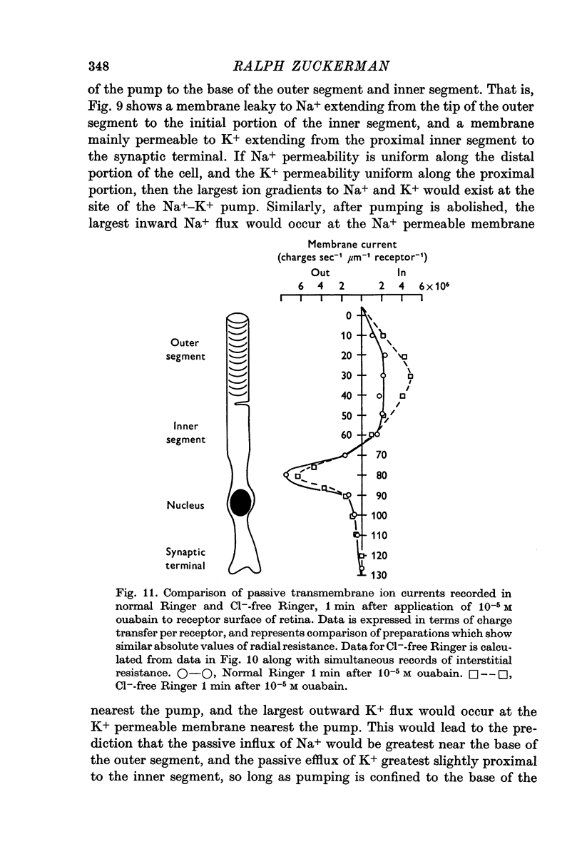
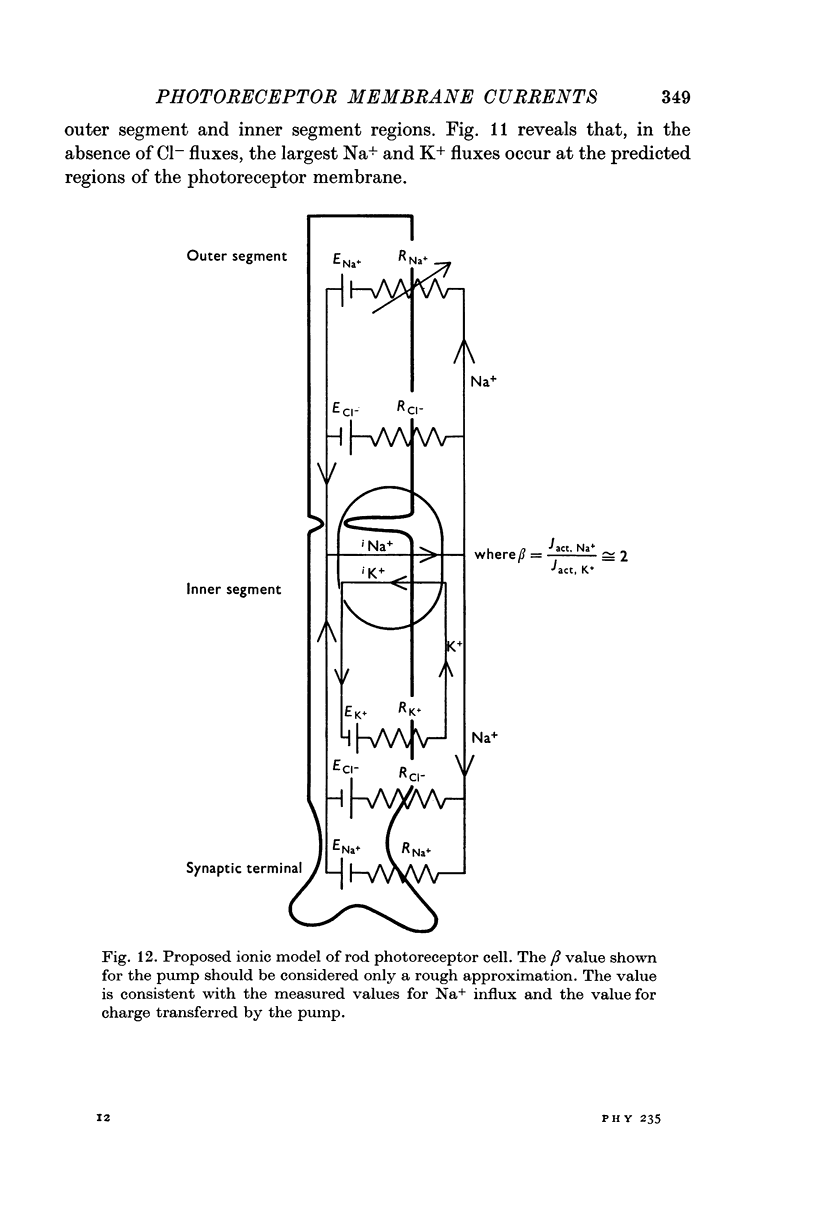
2. The plasma membrane of the frog rod outer segment was found to be permeable to Na+ and Cl-, with a ratio of Na+ to K+ permeabilities higher than that found in most neuronal cells. A net inward flux of 1·5 × 108 Na+/sec.rod flows across the outer segment plasma membrane in the dark.
3. The proximal portion of the rod receptor, extending from the proximal region of the inner segment to the synaptic terminal, is mainly permeable to K+, although some degree of Na+ permeability is also presumed.
4. A hyperpolarizing electrogenic Na pump was localized to the base of the outer segment and inner segment of the cell. The pump transfers at least 108 charges/sec out of the cell at this level, the pump current dividing and re-entering the cell at both the outer segment and proximal portion of the photoreceptor including the synaptic terminal.
5. These findings have been incorporated into an ionic model of the photoreceptor, and its implications for cellular functioning considered.
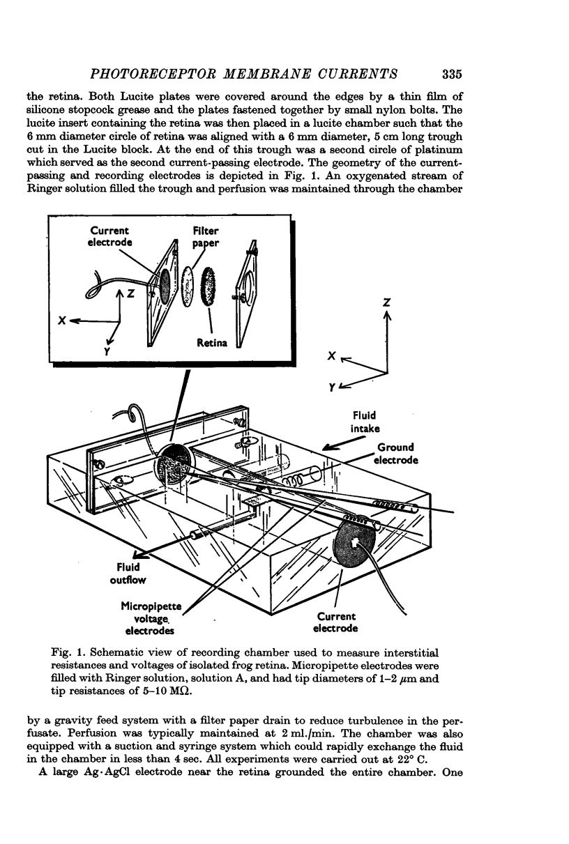
Full text
PDF
Selected References
These references are in PubMed. This may not be the complete list of references from this article.
- Bortoff A. Localization of slow potential responses in the Necturus retina. Vision Res. 1964 Dec;4(11):627–635. doi: 10.1016/0042-6989(64)90048-3. [DOI] [PubMed] [Google Scholar]
- Brierley G. P., Fleischmen D., Hughes S. D., Hunter G. R., McConnell D. G. On the permeability of isolated bovine retinal outer segment fragments. Biochim Biophys Acta. 1968 Aug;163(1):117–120. doi: 10.1016/0005-2736(68)90041-2. [DOI] [PubMed] [Google Scholar]
- Cavaggioni A., Sorbi R. T., Turini S. Efflux of potassium from the isolated frog retina: a study of the photic effect. J Physiol. 1972 Apr;222(2):427–445. doi: 10.1113/jphysiol.1972.sp009807. [DOI] [PMC free article] [PubMed] [Google Scholar]
- Cervetto L., MacNichol E. F., Jr Inactivation of horizontal cells in turtle retina by glutamate and aspartate. Science. 1972 Nov 17;178(4062):767–768. doi: 10.1126/science.178.4062.767. [DOI] [PubMed] [Google Scholar]
- Hagins W. A., Penn R. D., Yoshikami S. Dark current and photocurrent in retinal rods. Biophys J. 1970 May;10(5):380–412. doi: 10.1016/S0006-3495(70)86308-1. [DOI] [PMC free article] [PubMed] [Google Scholar]
- Korenbrot J. I., Cone R. A. Dark ionic flux and the effects of light in isolated rod outer segments. J Gen Physiol. 1972 Jul;60(1):20–45. doi: 10.1085/jgp.60.1.20. [DOI] [PMC free article] [PubMed] [Google Scholar]
- NILSSON S. E. AN ELECTRON MICROSCOPIC CLASSIFICATION OF THE RETINAL RECEPTORS OF THE LEOPARD FROG (RANA PIPIENS). J Ultrastruct Res. 1964 Jun;10:390–416. doi: 10.1016/s0022-5320(64)80018-6. [DOI] [PubMed] [Google Scholar]
- Sillman A. J., Ito H., Tomita T. Studies on the mass receptor potential of the isolated frog retina. II. On the basis of the ionic mechanism. Vision Res. 1969 Dec;9(12):1443–1451. doi: 10.1016/0042-6989(69)90060-1. [DOI] [PubMed] [Google Scholar]
- Tomita T. Electrical activity of vertebrate photoreceptors. Q Rev Biophys. 1970 May;3(2):179–222. doi: 10.1017/s0033583500004571. [DOI] [PubMed] [Google Scholar]
- Toyoda J., Hashimoto H., Anno H., Tomita T. The rod response in the frog and studies by intracellular recording. Vision Res. 1970 Nov;10(11):1093–1100. doi: 10.1016/0042-6989(70)90026-x. [DOI] [PubMed] [Google Scholar]
- Toyoda J., Nosaki H., Tomita T. Light-induced resistance changes in single photoreceptors of Necturus and Gekko. Vision Res. 1969 Apr;9(4):453–463. doi: 10.1016/0042-6989(69)90134-5. [DOI] [PubMed] [Google Scholar]
- Zuckerman R. Mechanisms of photoreceptor current generation in light and darkness. Nat New Biol. 1971 Nov 3;234(44):29–31. doi: 10.1038/newbio234029a0. [DOI] [PubMed] [Google Scholar]