Abstract
1. Anatomical studies have shown the cortex of the posterior bank of the superior temporal sulcus to receive a projection from visual cortical areas, including areas 17, 18 and 19. In this paper the response of single neurones in this area to simple visual stimulation is reported. Ten monkeys were studied.
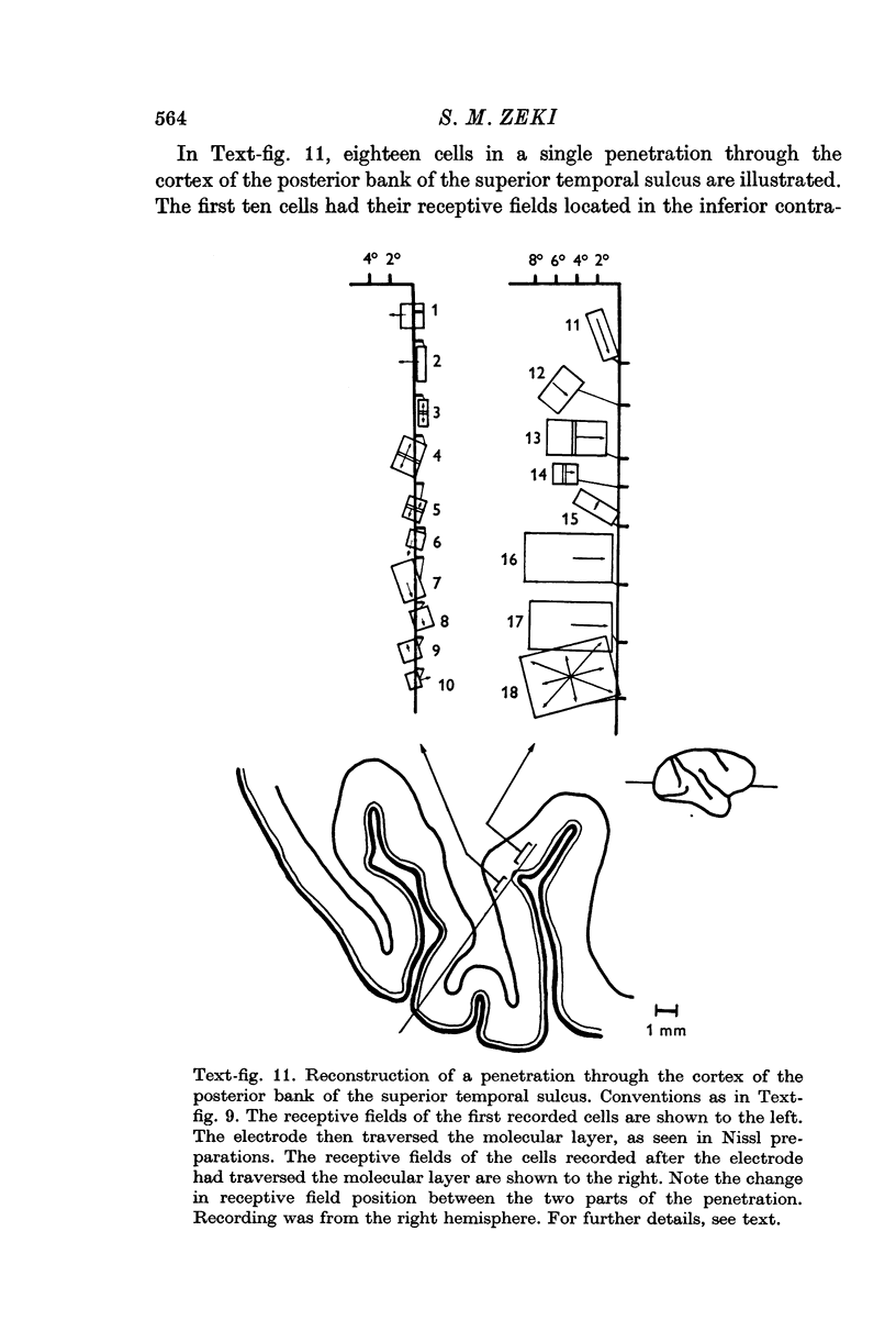
2. A clear but relatively crude topographic representation of the visual field was found. There was a large variation in the size of the receptive fields of individual cells, even in a single penetration. Some cells, with the central parts of their receptive fields located from between 1 and 5° from the centre of gaze had receptive fields averaging about 10° × 10° or even larger. Other cells with central receptive fields had much smaller field sizes.
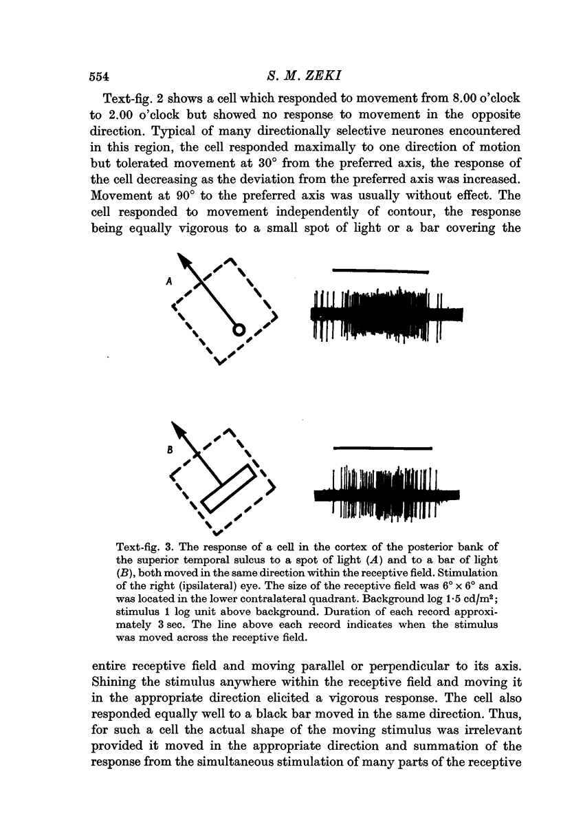
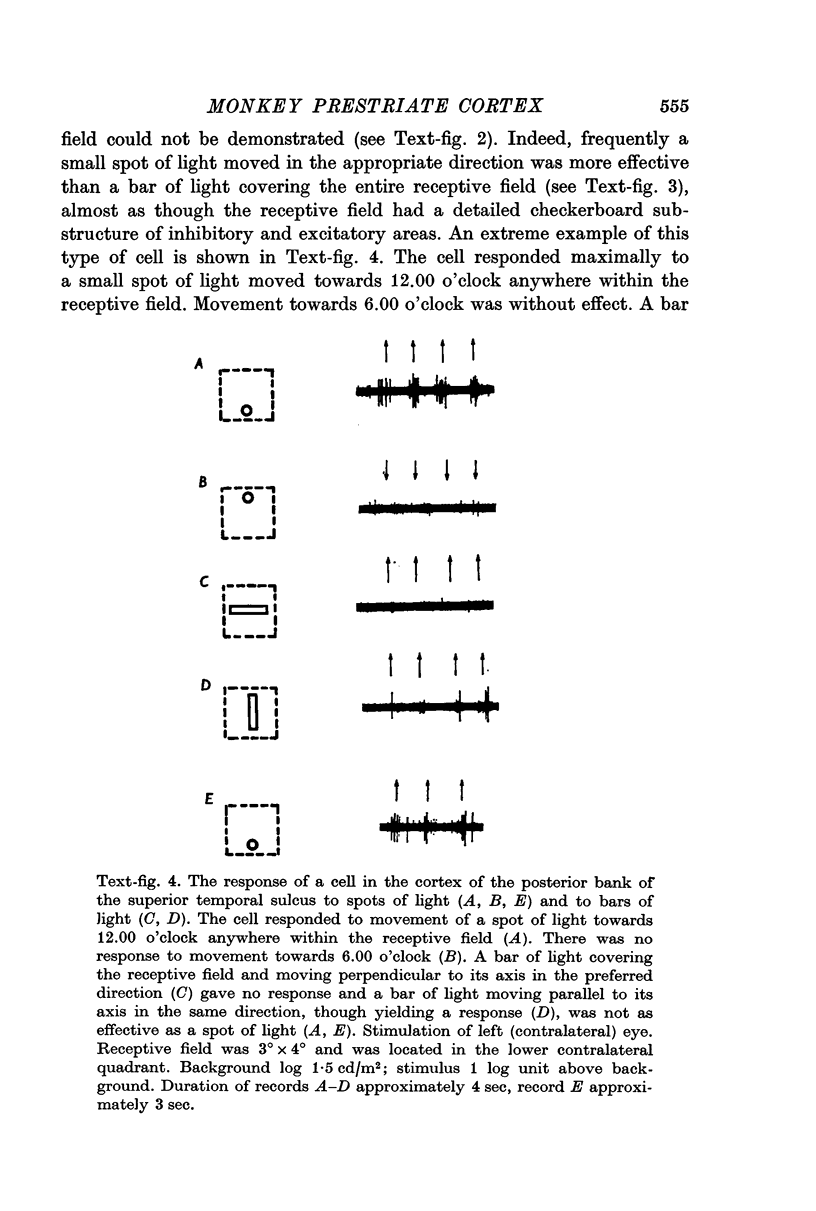
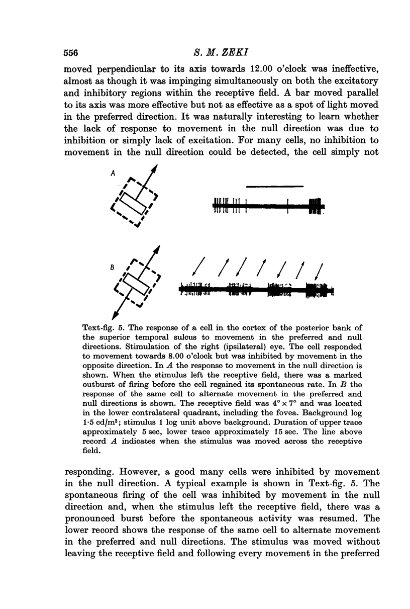
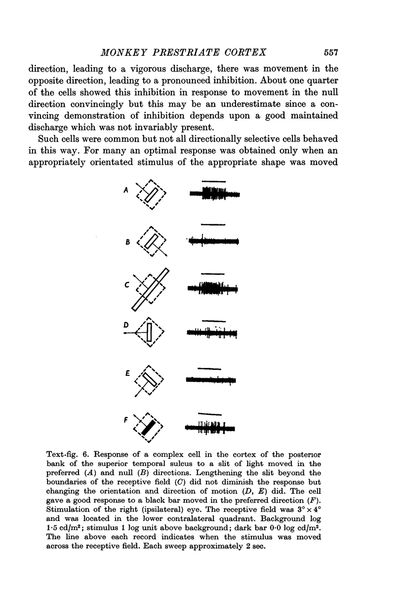
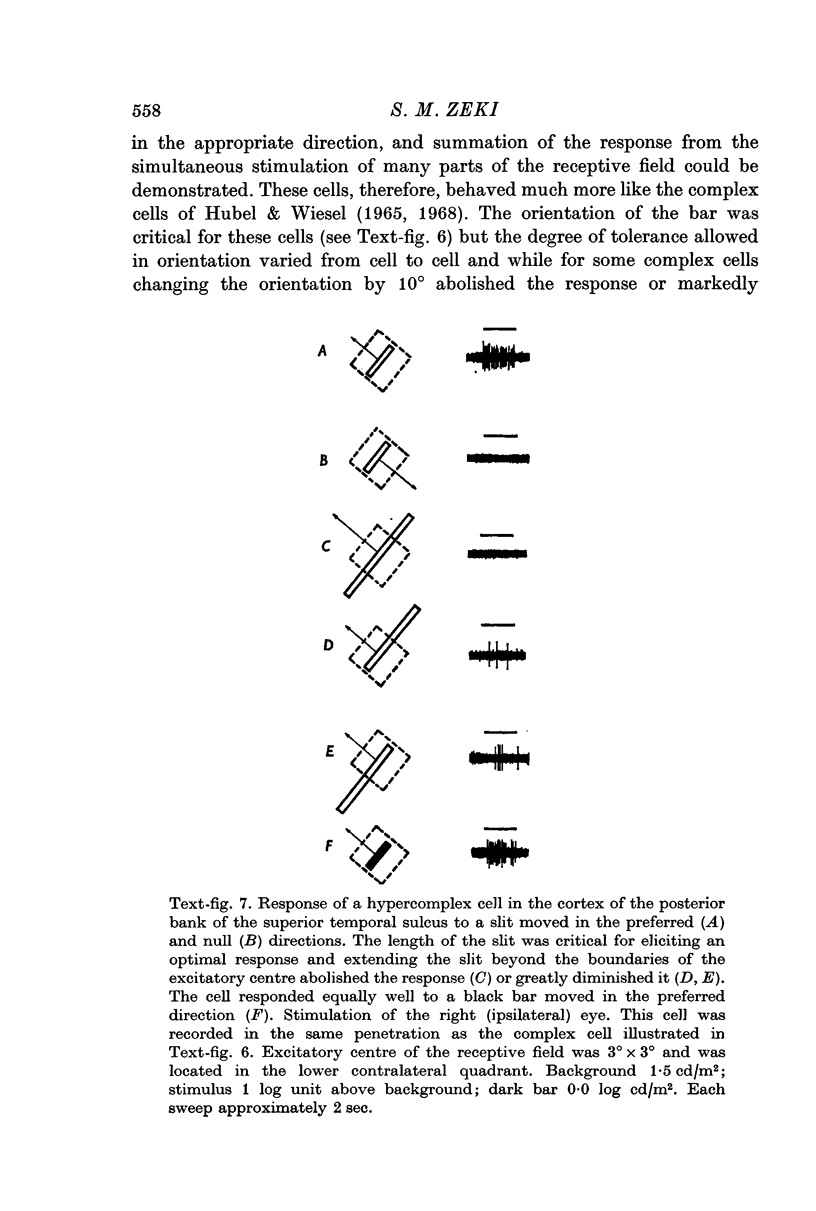
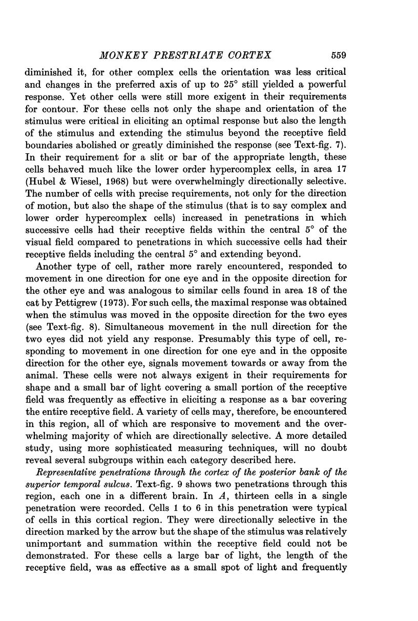
3. Two main types of neurones were encountered, with subdivisions within each type. The first type responded to movement irrespective of form. These could be subdivided into neurones which responded to movement in any direction within the receptive field and neurones which responded to movement in one direction only (directionally selective neurones). Another type of cell was responsive to both contour and movement, much like the complex and lower order hypercomplex cells. Almost all such neurones were directionally selective.
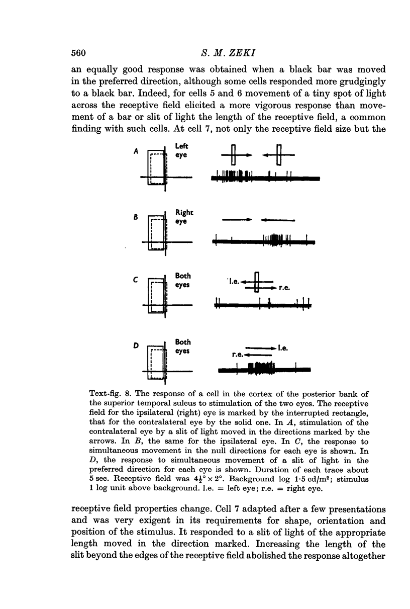
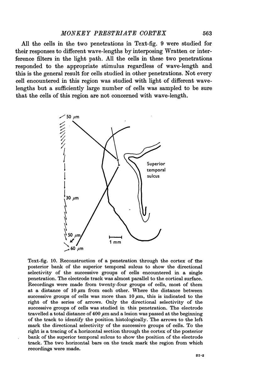
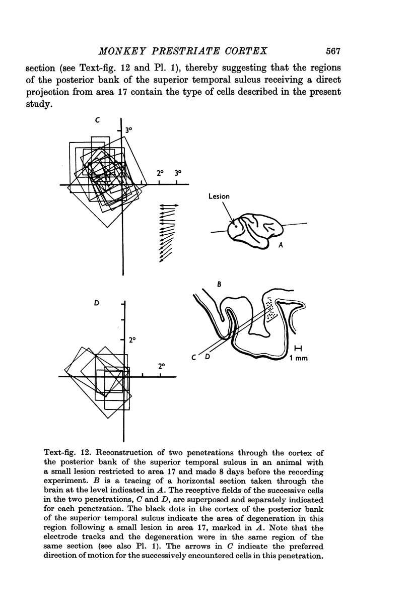
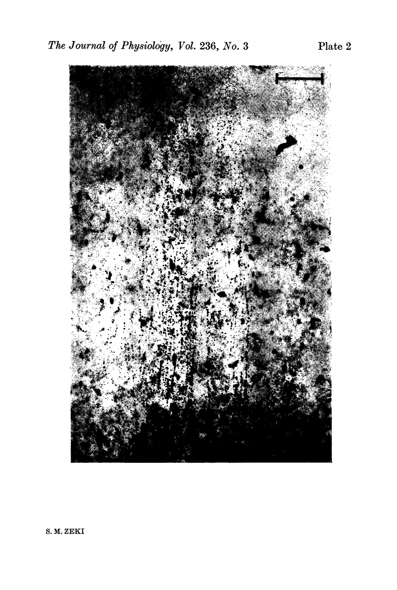
4. In oblique penetrations through this cortical region, there tended frequently to be an orderly shift in preferred directions of motion, thus suggesting the possibility of a columnar organization for movement.
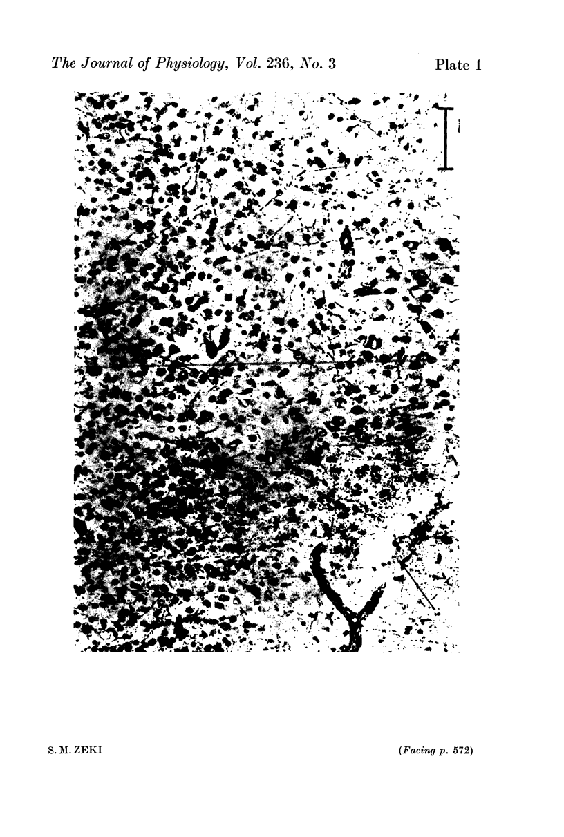
5. Combined anatomical (degeneration) and electrophysiological experiments showed that these types of neurones are found in those regions of the posterior bank of the superior temporal sulcus receiving a direct projection from area 17.
Full text
PDF