Abstract
A variable surface protein, Vmm, of the bovine pathogen Mycoplasma mycoides subsp. mycoides small colony type (M. mycoides SC) has been identified and characterized. Vmm was specific for the SC biotype and was expressed by 68 of 69 analyzed M. mycoides SC strains. The protein was found to undergo reversible phase variation at a frequency of 9 × 10⁻⁴ to 5 × 10⁻⁵ per cell per generation. The vmm gene was present in all of the 69 tested M. mycoides SC strains and encodes a lipoprotein precursor of 59 amino acids (aa), where the mature protein was predicted to be 36 aa and was anchored to the membrane by only the lipid moiety, as no transmembrane region could be identified. DNA sequencing of the vmm gene region from ON and OFF clones showed that the expression of Vmm was regulated at the transcriptional level by dinucleotide insertions or deletions in a repetitive region of the promoter spacer. Vmm-like genes were also found in four closely related mycoplasmas, Mycoplasma capricolum subsp. capricolum, M. capricolum subsp . capripneumoniae, Mycoplasma sp. bovine serogroup 7, and Mycoplasma putrefaciens. However, Vmm could not be detected in whole-cell lysates of these species, suggesting that the proteins encoded by the vmm-like genes lack the binding epitope for the monoclonal antibody used in this study or, alternatively, that the Vmm-like proteins were not expressed.
Mycoplasma mycoides subsp. mycoides small colony type (M. mycoides SC) causes a severe respiratory disease in cattle, contagious bovine pleuropneumonia (CBPP). It is the only bacterial disease included in the A list of communicable animal diseases of the Office International des Epizooties (OIE) and is the most important animal disease in Africa, affecting at least 27 countries (3, 5, 82). CBPP disappeared from Europe at the end of the 19th century but reappeared sporadically and affected several countries in an epizootic in southern Europe between 1983 and 1999.
The CBPP agent was first isolated and described in 1898 (72), and the organism was classified into the genus Mycoplasma (32) nearly 70 years later. Mycoplasmas belong to the class Mollicutes, whose members lack a cell wall and are known as the smallest self-replicating organisms (62, 79). Phylogenetic classification has grouped M. mycoides SC with five closely related and highly pathogenic mycoplasmas into the M. mycoides cluster (26, 74, 94). The M. mycoides cluster comprises Mycoplasma capricolum subsp. capricolum (M. capricolum), Mycoplasma capricolum subsp. capripneumoniae (M. capripneumoniae), M. mycoides subsp. capri (M. capri), M. mycoides subsp. mycoides large colony type (M. mycoides LC), M. mycoides SC, and Mycoplasma sp. bovine serogroup 7. The species Mycoplasma cottewii, Mycoplasma yeatsii, and Mycoplasma putrefaciens are sometimes included in the phylogenetic M. mycoides cluster, which is based on the 16S ribosomal DNA sequences (43). In this article we will only refer to the classical M. mycoides cluster, which excludes the three latter species.
The members of the M. mycoides cluster share many antigenic properties, and serological cross-reactions are often observed. Yet these infectious agents are strictly host specific, and the typical lung lesions of CBPP are solely formed after infection by M. mycoides SC. Consequently, any antigenic epitope that is unique for M. mycoides SC may be of importance for the pathogenicity of this organism and is therefore an interesting subject for research. Hitherto, very little is known about the pathogenicity of M. mycoides SC. The polysaccharide capsule and oxidative damage from hydrogen peroxide production may play important roles in CBPP infection (18, 49, 58, 64, 69, 75). Recently, it was suggested that the lipoprotein encoded by the lppB gene affects the virulence of M. mycoides SC (92).
Although the mortality rate is high among animals affected by CBPP, many become chronic carriers of the disease, and it is clear that M. mycoides SC, like many other pathogenic mycoplasmas, must have sophisticated mechanisms for virulence and persistence in the host.
Variable surface antigens are widely described for mycoplasmas (23, 79) and for other bacteria (44). It is generally believed that variable surface proteins are a means to enhance colonization and to adapt to the host tissue environment at various stages of infection. They have been shown to play a role in adhesion, immunomodulation, and substrate binding (e.g., see references 57, 66, 89, 93, and 97). Variable surface proteins can undergo phase variation, i.e., reversible ON/OFF switch of the expression, or antigenic variation, meaning that alternative phenotypes of the protein are expressed in the offspring of a bacterial cell, thus creating a heterogeneous population. Antigenic variation of proteins often involves DNA rearrangements of repetitive regions, thereby creating new combinations of structural domains and changes in size (12, 13, 24, 60, 61, 93, 95, 98, 99). Epitope masking (83, 88, 96) is another process that contributes to phase variation of proteins on the bacterial surface.
A number of different strategies to regulate phase variation have been reported for mycoplasmas. Some mechanisms are quite complex and involve site-specific DNA inversion, which causes an alternate expression of one gene in a gene family, as described for the vsa genes of M. pulmonis (11, 86). Also, the vsp genes of Mycoplasma bovis seem to be regulated by site-specific DNA inversions (59, 61), and the avg/vpma genes for Mycoplasma agalactiae involve gene rearrangements, although the exact mechanism is still unknown (37, 39). Expression of the vlhA genes of Mycoplasma synoviae is regulated by gene conversion (73). Frameshift mutations in poly(A) or repetitive motifs causing gene truncation were shown to control the ON/OFF switch in the pvpA gene of Mycoplasma gallisepticum (13), the p78 gene of Mycoplasma fermentans (89), and the vaa gene of Mycoplasma hominis (97). The number of repetitive units in putative transcriptional activators caused the phase variation of the pMGA genes of M. gallisepticum (38) and the vlhA genes of Mycoplasma imitans (63). Regulation strategies where the length of the promoter spacer controls the expression have been described for the maa2 gene of Mycoplasma arthritidis (93) and for the vlp genes of Mycoplasma hyorhinis (24, 95).
A phase-variable protein that contributes to a dynamic surface architecture of M. mycoides SC was characterized in this study. We suggest the name Vmm, for variable protein of M. mycoides, and describe the promoter mutation that causes the phase variation. Vmm was studied because it contains an epitope that is specific for M. mycoides SC and it gave the first evidence of intraclonal variability in M. mycoides SC.
MATERIALS AND METHODS
Mycoplasma strains and isolates. (i) Cultivation.
All mycoplasma isolates and reference strains were propagated in liquid medium as described earlier (77), unless otherwise stated. M. mycoides SC strain M223/90 was cultured in F medium (14).
(ii) Strains used to assess specificity of MAb 5G1.
The specificity of the monoclonal antibody (MAb) 5G1 was determined with 28 type and reference strains of mycoplasmas, ureaplasmas, and acholeplasmas that have been isolated in ruminants, as specified in Table 1. In all, 145 other isolates of the M. mycoides cluster were also used to test MAb 5G1, comprising 69 isolates of M. mycoides SC (of which 11 were from France, 5 from Portugal, 37 from Italy, 6 from Spain, and 10 from Africa), 32 isolates of M. mycoides LC, 4 isolates of Mycoplasma sp. bovine serogroup 7, 5 isolates of M. capri, and 35 isolates of M. capricolum. Also, two field isolates of M. yeatsii and three field isolates of M. bovis were used in the tests.
TABLE 1.
Type and reference strains of mycoplasmas isolated in ruminants that were used to assess the specificity of MAb 5G1
| Organism | Strainc | Refer- ence | NCTC no.a | ATCC no.b |
|---|---|---|---|---|
| Acholeplasma laidlawii | PG8T | 31 | 10116 | 23206 |
| Acholeplasma axanthum | S743T | 91 | 10138 | 25176 |
| Acholeplasma modicum | PG49T | 56 | 10134 | 29102 |
| M. agalactiae | PG2T | 32 | 10123 | |
| Mycoplasma alkalescens | PG51T | 56 | 10135 | 29103 |
| Mycoplasma anatis | 1340T | 81 | 10156 | 25524 |
| Mycoplasma arginini | G230T | 8 | 10129 | 23838 |
| M. bovigenitalium | PG11T | 32 | 10122 | 19852 |
| M. bovirhinis | PG43T | 55 | 10118 | 27748 |
| M. bovis | PG45 DonettaT | 41 | 10131 | 25523 |
| Mycoplasma californicum | ST-6T | 52 | 10189 | 33461 |
| Mycoplasma canadense | 275CT | 54 | 10152 | 29418 |
| M. capricolum subsp. capricolum | California KidT | 90 | 10154 | 27343 |
| M. capricolum subsp. capripneumoniae | F38T | 34 | 10192 | |
| Mycoplasma conjunctivae | HRC 581T | 7 | 10147 | 25834 |
| Mycoplasma gallinarum | PG16T | 32 | 10120 | 19708 |
| M. gallisepticum | PG31T | 33 | 10115 | 19610 |
| M. mycoides subsp. capri | PG3T | 30 | 10137 | |
| M. mycoides subsp. mycoides LC | Y goatR | 27 | 11706 | |
| M. mycoides subsp. mycoides SC | PG1T | 32 | 10114 | |
| Mycoplasma ovipneumoniae | Y98T | 19 | 10151 | 29419 |
| M. putrefaciens | KS1T | 90 | 10155 | 15718 |
| Mycoplasma sp. bovine serogroup 11 | 2DR | 55 | ||
| Mycoplasma sp. bovine serogroup 7 | PG50R | 55 | ||
| Mycoplasma verecundum | 107T | 40 | 10145 | 27862 |
| Ureaplasma diversum sero- group A | A417T | 48 | 10182 | 43321 |
| U. diversum serogroup B | D48 | |||
| U. diversum serogroup C | T74 | 47 | 49783 |
NCTC, National Collection of Type Cultures and Pathogenic Fungi (London, United Kingdom).
ATCC, American Type Culture Collection.
T, type strain; R, reference strain.
(iii) Strains used to prepare subclones of phenotypic variants.
M. mycoides SC strain Afadé is a bovine isolate from 1968 that originated in Chad. The strain used in this study is from the CIRAD-EMVT culture collection (CIRAD-EMVT Animal Health Program, TA 30/G Campus International de Baillarguet, Montpellier, France). Strain Afadé was proved to be highly pathogenic by experimental infections (1, 9) and is used to prepare antigen for a complement fixation test, which is the reference test for CBPP that is presently recommended by OIE (4).
M. mycoides SC strain B17 was formerly used to prepare the antigen for the recommended complement fixation test. It was isolated from a zebu in Chad in 1967, and the pathogenicity level is still unknown. Also strain B17 was provided by CIRAD-EMVT.
M. mycoides SC strain T1/44 is widely used as live vaccine in Africa (4). This strain has low pathogenicity and is resistant to streptomycin. T1/44 is a derivative of T1, which was isolated in Tanzania in 1952. The strain used in this study was received from the CIRAD-EMVT collection, batch EMVT 002 PANVAC-MVT, June 1996.
(iv) Preparation of direct lineages of phenotypic variants.
Direct lineages of subcloned variants were prepared as follows: fresh broth cultures of organisms from the strain stocks were passed through a 0.45-μm-pore-size filter and were thereafter serially diluted and plated on agar. After 4 days of incubation, single colonies that were positive for MAb 5G1 were located by aligning them with corresponding colored dots on a nitrocellulose membrane screened by colony immunostaining (see below). The colonies were picked with Pasteur pipettes and propagated at 37°C in 1 ml of broth medium for 4 days. After three successive steps of subcloning, resulting broth cultures were stored at −80°C for further characterization by Western blot analysis, except 100 μl that was replated as above. After 4 days of incubation, the few reverting negative colonies were located by aligning them with corresponding Ponceau-stained dots on colony blots and were subsequently purified by several rounds of subcloning as described above. Negative colonies of the third generation of 5G1 negative subclones, were propagated in 1 ml of broth and were stored at −80°C, except 100 μl that was replated. Reverting 5G1 positive subclones were prepared as above. Oscillating phenotypic switches were quantitatively monitored and were expressed as a fraction of switched phenotype per cell per generation.
(v) Strain used for construction of the phagemid library.
A low-passage isolate of M. mycoides SC referred to as strain M223/90 was used to make a phagemid library. The aim was to obtain a library that could reveal pathogenic features of M. mycoides SC. Strain M223/90 is a bovine isolate from pleural fluid that originated in Tanzania in 1990 (15).
Antibodies and immunobinding assays. (i) MAbs.
MAb 5G1 was prepared from M. mycoides SC type strain PG1 as described in reference 17, in collaboration between AFSSA-Site de Lyon and the Istituto Zooprofilattico della Lombardia e dell'Emilia. Mouse ascites fluid containing MAb 5G1 was sterile filtered through a filter with a pore size of 0.1 μm, and it was subsequently used for the experiments in this study. The specificity of MAb 5G1 was first determined by dot immunobinding assay (77) with the type and reference strains in Table 1 (strain F38 excepted). It was further tested on the 145 isolates of the M. mycoides cluster mentioned above, on the two isolates of M. yeatsii, and on the three M. bovis isolates. The specificity of MAb 5G1 was also investigated by colony immunostaining and Western blotting on reference strains of the M. mycoides cluster. The affinity of MAb 5G1 for strain F38 was determined by Western blotting only. M. mycoides SC strains that were negative for MAb 5G1 by dot immunobinding were also reanalyzed by colony immunostaining.
(ii) Colony immunostaining.
Freshly grown mycoplasma colonies were transferred to nitrocellulose membranes by placing the membranes on the surface of agar plates. The membranes were gently removed and blocked in Tris-buffered saline B (TBS-B) (TBS contains 50 mM and 0.2 M NaCl; TBS-B is TBS supplemented with 10% horse serum), before they were incubated with MAb 5G1 in TBS-B at a concentration of 9 μg/ml for 1.5 h at ambient temperature. Unbound antibody was removed by three washings in TBS-T (TBS and 0.05% Tween 20) and one washing in TBS. Thereafter, the membranes were incubated with peroxidase-conjugated rabbit anti-mouse antibodies (DAKO A/S, Glostrup, Denmark) in TBS-B (2.6 μg/ml) for 1.5 h, followed by three washes in TBS-T and one wash in TBS. Colonies expressing Vmm were specifically identified by an enzymatic color reaction with 4-chloro-1-naphthol that gives dark blue color (10, 83). The membranes were finally stained with Ponceau S solution (Sigma Diagnostics Inc., St. Louis, Mo.), which unspecifically stains proteins red, to reveal the colonies that were negative for MAb 5G1.
(iii) Western blotting, Triton X-114 fractionation, and supernatants.
Western blotting was carried out as previously described (57) after sodium dodecyl sulfate-polyacrylamide gel electrophoresis (SDS-PAGE) in 5 to 15% gradient or in homogenous 18% polyacrylamide gels. Triton X-114 phase fractionation was performed as described earlier (78). Supernatants of mycoplasma cultures were obtained by two successive centrifugation steps at 31,000 × g, followed by one filtration through a 0.20-μm-pore-size filter and two filtrations through a 0.10-μm-pore-size filter and finally a fivefold concentration by dialysis against polyethylene glycol (PEG) 6000 in phosphate-buffered saline (PBS) solution.
Electron microscopy.
Colonies of M. mycoides SC strain Afadé were grown on agar plates, resuspended in PBS, and transferred to a microcentrifuge tube. The cells were concentrated by centrifugation and resuspended in a small volume of PBS. One drop of cell suspension was placed on a 400-mesh nickel grid for 1 min before being washed three times in PBS and blocked with 0.5% bovine serum albumin (BSA) in PBS (PBS-BSA) for 5 min. The grid was transferred to a 50-μl drop of MAb 5G1 in PBS (28 μg/ml) and was left for 1 h at room temperature. After three washes in PBS-BSA, the grid was incubated with the secondary antibody (15-nm colloidal gold-labeled goat-anti-mouse; optical density, 3.5; Amersham Pharmacia Biotech, Uppsala, Sweden) diluted 1:20 in PBS-BSA for 1 h at room temperature. Another three washes in PBS were performed, and the cells were fixed with 2% glutaraldehyde in PBS during 10 min, washed three times in PBS, and once quickly in sterile water, before negative staining with 1% ammonium molybdate. Finally the grids were dried and examined by electron microscopy.
[U-14C]palmitic acid labeling of lipoproteins.
Metabolic labeling of lipoproteins in M. mycoides SC strain PG1, which expresses Vmm, and in strain M223/90, which is Vmm deficient (see “Occurrence of Vmm in field isolates of M. mycoides SC and other mycoplasmas” in Results), was performed by growth in the prescence of [U-14C]palmitic acid, as described by Cheng et al. (20). Whole-cell lysates containing the labeled proteins were analyzed by Western blotting, and the incorporation of [U-14C]palmitic acid was detected with MAb 5G1 and exposure of Kodak Biomax MR-1 film (Amersham Pharmacia Biotech).
Phage display. (i) Phagemid vectors, suppressor strain, and helper phage.
Two gene VIII-based phagemid vectors, pG8PL0 and pG8SPA0 (51), were used to produce a phagemid library. Vector pG8PL0 has the lacZ promoter (β-galactosidase of Escherichia coli) and the pelB signal sequence (pectate lyase of Erwinia chrysanthemi), while pG8SPA0 has a promoter and a signal sequence that originate from the spa gene region (protein A of Staphylococcus aureus). Both vectors contain a tag in fusion to gene VIII, which binds human serum albumin (HSA). The tag is out of frame with gene VIII, which means that some of the randomly fragmented mycoplasma inserts need to restore the open reading frame (ORF) to give an expression of phage protein VIII.
Phages and phagemids were propagated in an E. coli suppressor strain for the TGA codon, strain CDJ64/Δ14, with the genetic markers Δ(lacpro) nalA rif valR thi trpT(Su9)/F′lacpro (35, 84). The E. coli strain was grown in Luria broth (LB) or on Luria agar (LA) (Sigma), prepared according to the manufacturer's description. Selective media or agar plates contained ampicillin at a final concentration of 60 mg/liter unless otherwise stated. Soft agar consisted of LB medium and 0.5% agarose. Helper phage R408 (Promega Corp., Madison, Wis.) was used for the production of phage stocks.
(ii) Construction of the phagemid library.
A whole-genome phage display library of M. mycoides SC strain M223/90 was constructed in a mixture of phagemid vectors pG8PL0 and pG8SPA0 (51). The phagemid vectors were digested with SnaBI and dephosphorylated with calf intestine alkaline phosphatase. Equal amounts of the two vectors were pooled. Genomic DNA from M. mycoides SC strain M223/90 was prepared and purified by proteinase K lysis and phenol and chloroform extractions. The DNA was randomly fragmented by sonication until the majority of the fragments had sizes ranging from 1,000 to 2,000 bp and were thereafter treated with T4 DNA polymerase to give blunt ends and were subsequently phosphorylated with T4 polynucleotide kinase. Approximately 25 μg of the blunt-ended and phosphorylated fragments was ligated with Ready-to-Go ligase to 2.5 μg of the vector mixture. The ligated DNA was purified by phenol and chloroform extractions and precipitated with sodium acetate in ethanol, and the phagemid vectors carrying the mycoplasma inserts were dissolved in 16 μl of sterile H2O. E. coli strain CDJ64/Δ14 was transformed with the phagemid constructs by electroporation. The transformants were immediately transferred to 150 ml of LB medium supplemented with 2% glucose, and the culture was incubated at 37°C. After 1 h of incubation for phenotypic expression, 1 ml was removed to determine the titer by viable count. Ampicillin was added to the remainder to a final concentration of 50 mg/liter. The culture was grown overnight, and 2 ml of the culture was thereafter infected with helper phage R408 at a multiplicity of infection of 200. The infected E. coli was mixed with soft agar and poured on selective LA plates, which were incubated at 37°C overnight. Phagemid particles were eluted from the soft agar by standard procedures, and the phage stock was divided into aliquots and stored at −80°C until use.
(iii) Affinity pannings to identify the 5G1 epitope.
Affinity selection of phage that displayed mycoplasma peptides recognized by MAb 5G1 was made by three subsequent pannings. Two Maxisorp microtiter wells (Nalge Nunc International, Roskilde, Denmark) were coated with 125 μg of protein G in 250 μl of coating buffer (0.05 M Na2CO3 [pH 9.7]) for 1 h at room temperature. The wells were rinsed three times with PBS containing 0.05% Tween 20 before addition of 250 μl of MAb 5G1 (4.5 mg/ml) diluted 1:75 in PBS. Serum from BALB/c mice was used instead of MAb 5G1 as a negative control. The MAbs and BALB/c serum were immobilized for at least 1 h, and the wells were thereafter rinsed six times with PBS-Tween and blocked with PBS-Tween for 10 min. Meanwhile, the HSA binding region of the phagemids was blocked by incubating the phage stock with HSA (100 μg/ml). After 1 h, 200 μl of the phage stock was transferred to each coated well, and the panning proceeded for 4 h at room temperature. To wash away unspecifically bound phage, wells were rinsed 30 times in PBS-Tween before elution of the captured phage with 200 μl of sodium citrate buffer (50 mM Na citrate, 140 mM NaCl, [pH 2]). The eluates were neutralized with 40 μl of 2 M Tris-HCl buffer (pH 8.7), serially diluted in LB medium, and immediately used to infect 150 μl of overnight culture of E. coli CDJ64/Δ14. The infected cultures were incubated for 30 min at room temperature and were thereafter spread on selective LA plates. After incubation of the plates overnight, the colonies were counted and 150 colonies were transferred to a new selective LA plate to perform colony blot screening. New phage stocks were produced by resuspending the rest of the colonies from the 5G1 panning in LB medium, infecting the bacteria with helper phage R408, and processing as described above. A conjugate of HSA and horseradish peroxidase (HRP) was used in the colony blot screening to detect the tag. Replica blots were screened with MAb 5G1 to identify the clones that expressed the 5G1 epitope.
Analysis of the vmm gene. (i) DNA sequencing.
The sequences of the mycoplasma inserts from 27 phagemid clones that were positive for the tag and for MAb 5G1 by colony blot screening were determined. The phagemids were purified by the Wizard Plus SV Miniprep DNA purification system (Promega), and the inserts were first sequenced with the ALBP primer (all primers are listed in Table 2), which is complementary to the HSA binding region of the vector. Depending on in which vector the mycoplasma DNA was inserted, the phagemids were then sequenced with the Sasekv and Nypel primers that are complementary to the spa and pelB signal sequences, respectively. DNA sequencing in this study was performed with the ALFexpress DNA Sequencer and the Thermo Sequenase fluorescent-labeled primer cycle sequencing kit with 7-deaza-dGTP (Amersham Pharmacia Biotech), according to the manufacturer's description. DNA sequences were assembled with ASSEMGEL, software in the PC/Gene package (Intelligenetics Inc., Mountain View, Calif.).
TABLE 2.
Oligonucleotides for DNA sequencing, PCR, Southern blotting, and 5′ RACE
| Primer | Orientation | Sequence | 5′ label |
|---|---|---|---|
| ALBP | Reverse | 5′-GCCATACTGCTTTAGTTCATTGAT-3′ | Cy5a |
| Sasekv | Forward | 5′-TATCTGGTGGCGTAACACCTGCT-3′ | Cy5 |
| Nypel | Forward | 5′-CCTATTGCCTACGGCAGCCGCTGG-3′ | Cy5 |
| 5′f-5G1 | Forward | 5′-AGCAGCTAGAATTTATGCACT-3′ | |
| 3′r-5G1 | Reverse | 5′-ACAAAGATGATATTTTAGATCAG-3′ | |
| 5′-USP-5G1 | Forward | 5′-CGTTGTAAAACGACGGCCAGTTAGTCAGTTGATTAAGTGTAG-3′ | |
| 3′-RSP-5G1 | Reverse | 5′-CACAGGAAACAGCTATGACCCCATATCTAGTACTCTTATTC-3′ | |
| USP | Forward | 5′-CGTTGTAAAACGACGGCCAG-3′ | Cy5 |
| RSP | Reverse | 5′-CACAGGAAACAGCTATGACC-3′ | Cy5 |
| 5G1-insert probe | Forward | 5′-GCGTGTGGTGATAGATCAA-3′ | DIG |
| vmmRT1 | Reverse | 5′-GAATATGACCACTGTCATCATAATCAGC-3′ | |
| vmmRT2 | Reverse | 5′-TGAATCAGCTGGTTTATCAGAAGTTCC-3′ | |
| postlinkRT | Forward | 5′-AAGGACTGCTATCAACGCAGAGTACGCGGG-3′ | |
| postlinkPCR | Forward | 5′-AAGGACTGCTATCAACGCAGAGT-3′ |
Cy5-labeled primers were used for DNA sequencing with the ALFexpress DNA sequencer.
(ii) Analysis of the vmm flanking regions in subcloned phenotypic variants.
A high-quality template for sequencing the vmm gene and its flanking regions of cloned phase variants was produced by nested PCR with the primers 5′f-5G1 and 3′r-5G1 in the first reaction and primers 5′-USP-5G1 and 3′-RSP-5G1 in the second reaction. Amplifications were performed in a reaction mixture consisting of 10 mM Tris-HCl buffer (pH 8.3), 2 mM MgCl2, 50 mM KCl, 0.8 mM deoxynucleoside triphosphate, 10 pmol of each primer, 1 U of AmpliTaq DNA polymerase (Roche Molecular Systems, Inc., Branchburg, N.J.), and 1 μl of highly diluted DNA. The vmm gene region was amplified for 30 cycles, with denaturation for 20 s at 95°C, annealing for 20 s at 58°C for primers 5′f-5G1 and 3′r-5G1 or at 65°C for primers 5′-USP-5G1 and 3′-RSP-5G1, and elongation for 1 min at 72°C. The amplicons were sequenced with the USP and RSP sequencing primers, as described above.
(iii) mRNA analysis.
Total RNA was isolated with Trizol Reagent (Life Technologies, Gaithersburg, Md.) from 200 ml of culture of M. mycoides SC strain PG1. First-strand synthesis and 5′ rapid amplification of cDNA ends (RACE) of the transcript were performed as follows: 5 μg of total RNA was incubated for 10 min at 70°C with 2 pmol of primer vmmRT1 and 4 pmol of primer postlinkRT in a total volume of 11 μl. The first strand was synthesized at 42°C for 90 min after addition of 2 μl of deoxynucleoside triphosphate (10 mM), 2 μl of dithithreitol (100 mM), 1 μl of PowerScript Reverse Transcriptase (Clontech, Palo Alto, Calif.), and 4 μl of the 5× first-strand buffer provided with the enzyme. Subsequently, the transcript was amplified in a seminested PCR, using 5 μl of the cDNA and primers vmmRT1 and postlinkPCR in the first reaction. The PCR mixture contained 10 mM Tris-HCl buffer (pH 8.3), 2.5 mM MgCl2, 50 mM KCl, 0.8 mM deoxynucleoside triphosphate, 10 pmol of each primer, and 1 U of AmpliTaq DNA polymerase. The reactions were amplified with 5 cycles of 94°C for 10 s and 72°C for 3 min, followed by 5 cycles of 94°C for 10 s, 70°C for 15 s, and 72°C for 3 min and finally 25 cycles of 94°C for 10 s, 68°C for 15 s, and 72°C for 3 min. Primers vmmRT2 and postlinkPCR were used in the second PCR, where 0.1 μl of the first amplicon served as template. The reaction mixture was prepared as in the first reaction; however, the primer postlinkPCR was not added until 15 cycles of 94°C for 15 s and 72°C for 1 min 20 s were completed. Another 25 cycles at 94°C for 15 s, 70°C for 15 s, and 72°C for 1 min 20 s were then performed. The final amplicon was DNA sequenced with the vmmRT2 primer and the Thermo Sequenase Cy5 Dye Terminator Kit (Amersham Pharmacia Biotech) as described by the manufacturer. The sequences were analyzed with the ALFexpress DNA sequencer.
(iv) Southern blot hybridizations.
The occurrence of the vmm gene in 18 M. mycoides SC strains that represent the different IS1296 hybridization patterns (21), as well as in three other M. mycoides SC strains, was assessed by Southern blotting. Chromosomal DNA was digested to completion with the restriction enzyme HindIII, and the fragments were separated by electrophoresis on 0.8% agarose gels. Southern blotting was performed as described elsewhere (60, 76). Hybridizations of the digoxigenin (DIG)-labeled probe 5G1-insert probe (Table 2) were carried out at 44°C. Stringent washes were performed at the hybridization temperature in 0.2× SSC (1× SSC buffer is 0.15 M NaCl plus 0.015 M sodium citrate) supplemented with 0.1% (wt/vol) SDS, and the hybridized probe was detected with the DIG nucleic acid detection kit, as described by the manufacturer (Roche Diagnostics GmbH, Mannheim, Germany). The occurrence of the vmm gene in the type strains of the closely related species of the M. mycoides cluster and the species M. putrefaciens, M. cottewii (28), M. bovis, M. agalactiae, Mycoplasma bovirhinis, Mycoplasma bovigenitalium, and Mycoplasma primatum (29) (a primate mycoplasma closely related to M. agalactiae and M. bovis) was analyzed with the 5G1-insert probe as described above and with the 5G1-PCR probe at a hybridization temperature of 68°C. The DIG-labeled 5G1-PCR probe was produced by nested PCR with the primers 5′f-5G1 and 3′r-5G1 in the first reaction and DNA of strain PG1 as the template, followed by amplification with primers 5G1-insert probe and 3′-RSP-5G1 as described above. The reaction mixtures were prepared with the PCR DIG Probe Synthesis Kit (Roche Diagnostics).
Protein analyses of Vmm. (i) Preparation and Western blotting of phage proteins.
To verify that the affinity pannings had selected phage that displayed the true target protein for MAb 5G1 and not a false epitope, the different hybrid proteins of the phage were analyzed by Western blotting. A crude protein preparation of phage was made by precipitating the phages with PEG, as follows: E. coli clones with the recombinant phagemids were inoculated in 15 ml of LB medium supplemented with ampicillin, and the cultures were grown overnight. The cells were pelleted and dissolved in 5 ml of fresh LB medium containing ampicillin, and the culture was then infected with 100 μl of helper phage R408 (1011 PFU/ml). Production of hybrid phage particles was allowed for 4 h at 37°C before the samples were centrifuged at 12,000 × g, and the supernatant was filtered through a 0.45-μm-pore-size sterile filter. Phages were precipitated by adding 500 μl of 20% PEG 8000 in 2.5 M NaCl to 3.5 ml of the phage stock, incubating the mixture at room temperature for 45 min, and centrifuging the samples at 12,000 × g. The pellet was dissolved in 25 μl of PBS before addition of 25 μl of 2× SDS gel loading buffer (85). Western blotting was performed after separation by SDS-PAGE in a 12% gel.
(ii) Protein prediction.
Prediction of the structural organization of Vmm was performed with the SignalP V2.0 World Wide Web Server (70, 71), ProfileScan of PROSITE Patterns at the ISREC-Server of the Swiss Institute for Experimental Cancer Research and the Swiss Institute for Bioinformatics (45), and TMpred at the European Molecular Biology Network (46). Similarity searches were done with BLASTN and BLASTP at the server of the National Center for Biotechnology Information (2).
Nucleotide sequence accession number.
The nucleotide sequence for the vmm gene has been assigned the GenBank accession number AF428142.
RESULTS
Vmm is a phase-variable, surface-located lipoprotein.
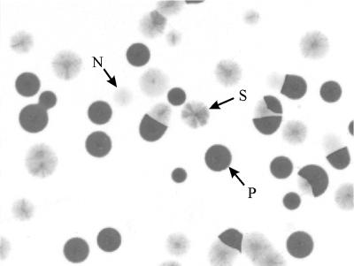
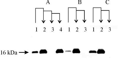
Colony immunostainings of M. mycoides SC with MAb 5G1 showed that this MAb targets a surface-exposed protein, Vmm, that undergoes high-frequency phenotypic variation (Fig. 1). Immunoblotting of whole-cell proteins from ON and OFF variants of direct clonal lineages (Fig. 2) resulted in a single band for stock culture and positive clones, whereas no protein band was detected in negative clones. This proved that the phenotypic variation observed in the colony immunostaining was a result of reversible phase variation of the Vmm expression and not a consequence of epitope masking or size variation. The switching rate of Vmm was similar in the three strains Afadé, B17, and T1/44, and it was calculated to be 7 × 10⁻⁴ to 9 × 10⁻⁴ per cell per generation for ON-to-OFF reversion and 5 × 10⁻⁵ to 9 × 10⁻⁵ per cell per generation for OFF-to-ON reversion.
FIG. 1.
Colony immunostaining with MAb 5G1 of M. mycoides SC strain B17, showing variable expression of the surface protein Vmm among and within colonies. The population consists of colonies that are positive for MAb 5G1 and express the Vmm protein (P), negative colonies (N), and sectored colonies (S) where mutations during growth have induced ON/OFF switching of the Vmm expression. The colonies are derived from a single colony that was cultured in broth, filtered, and plated on agar. The scanned colony blot was processed in CorelDRAW 9.0.
FIG. 2.
Whole-cell proteins of serially subcloned phenotypic variants from three M. mycoides SC strains that were separated in an 18% polyacrylamide gel, immunoblotted, and detected with MAb 5G1. Sequential phenotypic transitions of the Vmm expression are indicated as arrows that represent direct lineages of the subcloned variants. Lanes 1 were stock populations of the strain; lanes 2 were ON-type subclones; lanes 3 were OFF-type revertant subclones; and lane 4 was an ON-type double revertant subclone. Panel A shows samples from strain Afadé, panel B shows strain B17, and panel C shows strain T1/44. The scanned blot was processed with Corel Photo-Paint 9.0 and CorelDRAW 9.0.
Vmm migrated like a 16-kDa protein in SDS-PAGE, and the protein appeared to be identical for the analyzed strains Afadé, B17, and T1/44, as shown in Fig 2. Separation of Vmm on a high-resolution-gradient gel showed that the protein occurred in two sizes, approximately 15 and 17 kDa (Fig. 3). Vmm was present in the Triton X-114 phase and absent in the aqueous phase and the supernatant, suggesting that it is a membrane-associated protein. Metabolic incorporation of [U-14C]palmitic acid in strains PG1 and M223/90 and analysis of whole-cell proteins by Western blotting proved that Vmm is a lipoprotein. Detection of the blot with MAb 5G1 gave a clear band of approximately 16 kDa for strain PG1 but no band for strain M223/90. Similarly, this band was observed among the lipoproteins of PG1 in the autoradiographic film but was absent from the lipoproteins of strain M223/90 (data not shown). The absence of Vmm in strain M223/90 is explained below.
FIG. 3.
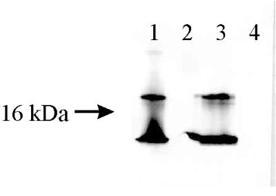
Immunoblots detected with MAb 5G1, showing the occurrence of protein Vmm in whole-cell proteins (lane 1), culture supernatant (lane 2), Triton X-114 phase (lane 3), and aqueous phase (lane 4). The samples were prepared from M. mycoides SC strain T1/44. The scanned blot was processed with Corel Photo-Paint 9.0 and CorelDRAW 9.0.
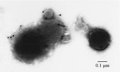
Immunoelectron microscopy of strain Afadé showed that MAb 5G1 targets a protein at the surface of the mycoplasma membrane; hence, Vmm is a surface-located lipoprotein (Fig. 4). The phase variation of Vmm was clearly observed as some gold-labeled bacteria and some negative bacteria, all originating from one clone. It can also be concluded from the electron micrographs that the protein is expressed at low levels in strain Afadé, although it cannot be excluded that the capsular layer limits the accessibility of the membrane proteins, thereby hindering the antibodies from binding Vmm.
FIG. 4.
Electron micrograph after immunostaining of M. mycoides SC strain Afadé with MAb 5G1. The antibody binds surface molecules of cells that express Vmm. The size of the gold particles is 15 nm. The scanned photo was processed with Corel Photo-Paint 9.0 and CorelDRAW 9.0.
Occurrence of Vmm in field isolates of M. mycoides SC and other mycoplasmas.
Analysis of 69 M. mycoides SC isolates by dot immunobinding assay showed that 31 of the isolates were clearly positive for MAb 5G1, including the type strain PG1. Thus, these strains were expressing Vmm. No significant reaction occurred with the other 38 M. mycoides SC strains; however, 37 of these revealed a positive subpopulation by colony immunostaining. A positive subpopulation was defined as expression of Vmm in at least 0.01% of the cells. Only strain M223/90 from Tanzania was negative by both methods, and it was therefore believed not to express Vmm at all. All of the 28 reference strains of mycoplasma species that have been isolated in ruminants (Table 1) were negative for MAb 5G1 in dot immunobinding, except strain PG1. Also, the 76 field isolates belonging to the M. mycoides cluster (M. mycoides LC, Mycoplasma sp. bovine serogroup 7, M. capri, and M. capricolum), as well as the two M. yeatsii and three M. bovis isolates, were negative for MAb 5G1 in dot immunobinding. No positive subpopulations could be detected among the reference strains of the M. mycoides cluster, as judged by colony immunostaining. The field isolates were not tested by colony immunostaining.
Properties of the phagemid library.
To reveal the mechanisms for phase variation of Vmm, we first used phage display to identify the vmm gene. A phagemid library that consisted of 107 independent clones and in which the phage stock had a titer of 1011 PFU/ml was produced. Approximately 95% of the phagemids contained mycoplasma inserts, and the sizes of the inserts varied from 100 to 1,500 bp. To verify that phages in the library indeed display hybrid proteins of mycoplasma peptides and the HSA binding tag, affinity pannings to HSA were performed. Phages cannot display the HSA binding tag on the surface unless a mycoplasma insert has restored the ORF between gene VIII and the tag (51). More than 107 phages were immobilized in the first panning, which is considerably more than the control pannings against newborn calf serum that captured approximately 103 phages. Mycoplasma inserts of clones that were positive for HSA by colony blot screening were subsequently sequenced, and it was found that more than one-third of the inserts contained one or more TGA codons. It was therefore concluded that the suppressing ability of E. coli CDJ64/Δ14 was efficient enough to allow expression of peptides containing several TGA codons. The experiments showed importantly that hybrid peptides were present on the phage surface and that the detection of tag expression could be used as a ligand independent screening system. There were as many phagemids of type pG8PL0 as of pG8SPA0 in the library; however, after one panning to HSA, most of the HSA-positive clones contained the spa promoter and signal sequence, indicating that the pG8SPA0 vector is more efficient than the vector containing the lacZ promoter and pelB leader.
Identification of the vmm gene.
Affinity pannings of the phage display library to MAb 5G1 resulted in an accumulation of phage expressing the target epitope for MAb 5G1. The total amounts of recovered phage from the first, second, and third pannings were 5.4 × 10⁴, 3.0 × 10⁴, and 4.0 × 10⁶ CFU, respectively, as determined by viable count of E. coli infected with the eluted phage. The corresponding figures for the control pannings to BALB/c serum were 1.3 × 10⁴, 6.4 × 10³, and 1.2 × 10³ CFU. Colony blot screenings for expression of the tag in 150 colonies resulted in 2, 99, and 140 positive colonies in each affinity panning, whereas the original phage stock had two colonies that were positive for HSA out of 150. The majority of the colonies that were positive for HSA were also positive for MAb 5G1. Twenty-seven of these clones were selected at random, the phagemids were isolated, and the mycoplasma inserts were sequenced. When the sequences were aligned, it was obvious that 20 of the inserts originated from the same gene region, the vmm gene region. Although some of the clones were multiples of the same recombinant, there were five different size variants of the gene segment. The remaining seven sequences were from other parts of the M. mycoides SC genome. It was possible to determine the full sequence of the vmm gene and to design primers in the flanking regions after searching the genome database of M. mycoides SC strain PG1 with the consensus of the alignment (J. Westberg, A. Persson, A. Holmberg, E. Björkvall, K.-E. Johansson, M. Uhlén, and B. Pettersson, Program Abstr. 12th Int. Organ. Mycoplasmology Conf., p. 151-152, 1998). By coincidence, one of the recombinant clones contained the full vmm gene, except for the stop codon TAA.
When the panning experiments were repeated, the first and second pannings resulted in 3.0 × 10² and 3.0 × 10³ CFU with MAb 5G1 and 1.4 × 10³ and 4.0 × 10² CFU with BALB/c serum. A third panning was not performed. The number of HSA-positive colonies increased from 31 in the first panning to 82 in the second panning. Inserts from five positive clones were DNA sequenced; four of these contained vmm gene fragments in three different size variants.
Hybrid proteins from five different phagemid recombinants containing vmm fragments were expressed in E. coli. The clones were infected with helper phage, and phage stocks were produced. Phages were precipitated, and the proteins were analyzed by SDS-PAGE and Western blotting with MAb 5G1 and HSA-HRP (Fig. 5). Four of the five clones clearly expressed the 5G1 epitope and the tag in a band whose size corresponded to the size of the hybrid protein as calculated from the sequence data (data not shown). Although all samples were prepared from similar amounts of phage, the fifth clone produced considerably smaller amounts of the hybrid protein, and the expression was too low to be detected on the Western blot, as neither detection for the tag with HSA-HRP nor the specific detection with MAb 5G1 resulted in a clearly visible band. Because all clones had inserts from the same gene, the result was still a confirmation that the phage display technique really selected phages that displayed the 5G1 epitope of Vmm and not an unspecific selection or artifact in the colony blot screening.
FIG. 5.
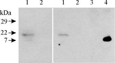
Western blot of total phagemid proteins. The left panel was detected with an HSA-HRP conjugate, i.e., the tag, and the right panel was detected with MAb 5G1. Lanes 1, phagemid containing the entire vmm gene fused to gene VIII; 2, helper phage R408; 3, whole-cell lysate of M. mycoides SC strain M223/90; and 4, whole-cell lysate of M. mycoides SC strain PG1. The scanned blots were processed with Corel Photo-Paint 9.0 and CorelDRAW 9.0.
Structure of the vmm gene.
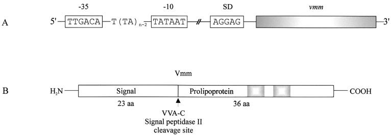
The structural features of the vmm gene region, as deduced from the DNA sequences of the type strain PG1 and the phagemid clones of strain M223/90, are outlined in Fig. 6. The vmm gene has an ORF of 177 bp, encoding 59 amino acids (aa). A typical promoter with the −35 motif TTGACA and the −10 region TATAAT is located 71 bp upstream of the start codon ATG, but there is no obvious hairpin loop structure for transcription termination 3′ of vmm. Analysis of the vmm transcript in strain PG1 by 5′ RACE and DNA sequencing showed that the transcription is initiated at the 8th nucleotide downstream of the −10 region, starting with the sequence ATCT. A Shine-Dalgarno sequence, AGGAG (68), is located 13 bp 5′ of the start codon. The vmm gene did not contain any TGA-Trp codons.
FIG. 6.
Schematic illustrations of the vmm gene with the promoter region and the Vmm precursor. (A) The vmm gene is preceded by a Shine-Dalgarno (SD) sequence and a promoter that consists of −10 and −35 regions that are identical to the consensus δ70 promoter of E. coli. A dinucleotide (TA)n repeat of variable length makes up the promoter spacer and part of the Pribnow box, and it was shown that active promoters have a total number (n) of 10 repetitions. Note that the illustration is not drawn to scale. (B) The vmm gene encodes a protein precursor of 59 aa, which has a prolipoprotein signal peptidase site, VVA-C, at positions 21 to 24, indicating that lipid modification will take place at the cysteine moiety and that the signal peptide of 23 aa will be spliced off. The shaded boxes represent two repetitive motifs of KPAD.
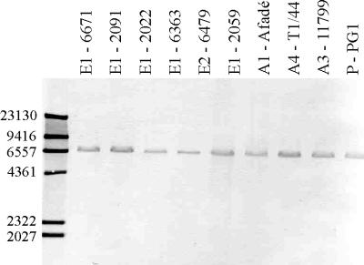
Southern blot hybridizations of HindIII-digested genomic DNA with the 5G1-insert probe showed that the vmm gene, or at least the probing region, was present in 18 tested M. mycoides SC strains that represent different geographical origins and IS1296 patterns. Ten of these are shown in Fig. 7. The M. mycoides SC strains had identical hybridization patterns with the 5G1-insert probe, characterized by a single 6,550-bp band, indicating that there is only one copy of the vmm gene and that the chromosomal location is similar. The existence of only one vmm gene was also confirmed by Southern blotting of chromosomal DNA digested with AluI from eight strains (not shown), and we know from the genome sequence database that strain PG1 has one gene. Also, the strain that did not express the Vmm possessed the gene that encodes this phase-variable surface protein. This is noteworthy because the phagemid library was coincidentally made of this strain and because the phage display assay could not have been used to select for the 5G1 epitope unless strain M223/90 possessed the vmm gene.
FIG. 7.
Southern blot showing the presence of the vmm gene in some M. mycoides SC isolates from different geographical origins. The strains also represent different IS1296 hybridization patterns as determined according to reference 21. Insertion sequence patterns are indicated before the strain designation. The Southern blots were prepared from HindIII-digested chromosomal DNA, and hybridization was performed with the 5G1-insert probe. Strain 6671 originated in Italy in 1993, strain 2091 in France in 1984, strain 2022 in France in 1984, strain 6363 in Spain in 1991, strain 6479 in Italy in 1992, strain 2059 in Spain in 1984, strain Afadé in Chad in 1968, strain T1/44 in Tanzania in 1952, and strain 11799 in Senegal in 1988. Type strain PG1 has an unknown origin. The scanned blot was processed with Powerpoint 7.0 and CorelDRAW 9.0.
Characterization of the vmm gene product.
Protein prediction analyses of the amino acid sequence recognized Vmm as being a lipoprotein where the only putative transmembrane helix is located in the signal sequence. The likeliest cleavage site for the prolipoprotein-specific signal peptidase is between aa 23 and 24, at the VVA-C motif (Fig. 6) (16, 25), leaving a mature lipoprotein of 36 aa. The Western blot of Vmm after separation on a high-resolution SDS-PAGE showed two sizes for the protein (Fig. 3), the bigger being approximately 17 kDa and a smaller peptide of almost 15 kDa. Theoretically, the signal peptide is 2.5 kDa, suggesting that the signal peptide is being spliced off. The mature protein is hydrophilic, as judged from the amino acid sequence. Characteristic repetitive regions like those for, e.g., the variable surface proteins (Vsps) of M. bovis (61) and variant lipoproteins (Vlps) of M. hyorhinis (24, 95) were not found; only a short motif of 4 aa, KPAD, was repeated twice. Similarity searches by BLASTp showed 54% identity and 79% positive matches between the Vmm signal peptide and that of spiralin of Spiroplasma citri (22). The rest of the Vmm was not similar to any protein in the databases.
The molecular basis for phase variation of Vmm.
To find the mutation that causes phase variation of Vmm, the DNA sequence of the full vmm gene with flanking regions was determined for 28 subcloned phase variants as summarized in Table 3. The table also contains data from strain M223/90 that were found when phagemid clones were sequenced. Strain M223/90, as mentioned earlier, does not express Vmm at all. All sequences were identical in the coding region, including the sequence of M223/90. The sequences were also identical in the flanking regions except for the size of the promoter spacer that separates the −35 region from the −10 region. The spacer consists of repetitive units known as (TA)n, where n is the total number of repetitions and the last two repeats were part of the −10 region (Fig. 6). Clones that express Vmm and thus have an active promoter were found to have a spacer of 17 bp in the promoter, i.e., there were 10 dinucleotide repeats. Any other number of repeats was found to disrupt the functionality of the promoter. There was, however, an exception in one revertant clone of strain B17 that was found negative by colony blotting but contained 10 TA repeats in the promoter.
TABLE 3.
Number of dinucleotide repeats in the vmm promoter for individual clones of M. mycoides SC strains B17, Afadé, T1/44, and M223/90
| Namec | Expression of Vmm | No. of (TA)n repetitions in the vmm promoter
|
|||
|---|---|---|---|---|---|
| Clone 1 | Clone 2 | Clone 3 | Clone 4 | ||
| B17 | Positive | 10 | 10 | 10 | 10 |
| B17 revertant | Negative | 12 | 10 | 12 | 12 |
| Afadé | Positive | 10 | 10 | 10 | 10 |
| Afadé revertant | Negative | 12 | 12 | 13 | 12 |
| Afadé double revertant | Positive | 10 | 10 | 10 | 10 |
| T1/44 | Negative | 12 | 12 | 12 | 12 |
| T1/44 revertant | Positive | 10 | 10 | 10 | 10 |
| M223/90 | Negativea | 6 | 7 | NDb | ND |
Strain M223/90 does not express Vmm.
ND = not determined.
Revertants and double revertants were direct lineages of the original colonies.
vmm-like genes in other species of the M. mycoides cluster.
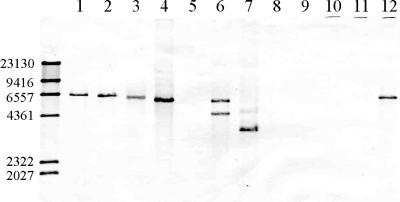
Similarity searches for the vmm with BLASTn detected a very similar gene in contig MC438 of M. capricolum. Unfortunately, this sequence was not complete, so it was not possible to fully compare the two genes. It was confirmed by Southern blot analysis with the DNA probes 5G1-insert probe and 5G1-PCR probe that, among the reference strains of the M. mycoides cluster, M. mycoides SC, M. capricolum, and M. capripneumoniae have one vmm or vmm-like gene, while Mycoplasma sp. bovine serogroup 7 may have two copies of vmm-like genes (Fig. 8). M. putrefaciens had only one barely visible band after hybridization with the oligonucleotide 5G1-insert probe, suggesting that there is one vmm-like gene that poorly hybridizes with this probe, although, when hybridized with 5G1-PCR probe, there were two clear bands (not shown). Reference strains from M. mycoides LC, M. capri, M. yeatsii, and M. cottewii seem to lack vmm-like genes, as none of the probes hybridized to DNA from these species. M. yeatsii was, however, analyzed only with the 5G1-insert probe. Although our data show that there are vmm-like genes in other mycoplasma species, they need to be further investigated and the sequences should be determined and compared to that of M. mycoides SC. Interestingly, the observations agree well with the phylogenetic relationship between these species, where M. mycoides SC forms an intermediate branch between the M. capricolum species group and the M. capri species group (74). The M. capricolum species group comprises M. capricolum, M. capripneumoniae, and Mycoplasma sp. bovine serogroup 7, while M. mycoides LC and M. capri are found in the M. capri species group. M. putrefaciens, M. yeatsii, and M. cottewii are more distantly related (43). Western blots to whole-cell lysates of the reference strains of all the M. mycoides cluster members, including M. putrefaciens, M. cottewii, and M. yeatsii, were negative for MAb 5G1, M. mycoides SC excepted. Thus, the vmm-like genes may be silent or their gene products do not contain the binding epitope for MAb 5G1.
FIG. 8.
Southern blot showing the occurrence of vmm or vmm-like genes in nine representative strains of the phylogenetic M. mycoides cluster. Genomic DNA was digested with HindIII before separation in agarose gel, and the Southern blot was hybridized with 5G1-insert probe. The samples were loaded in the following order: M. mycoides SC strain Afadé (lane 1), M. mycoides SC strain PG1 (lane 2), M. capricolum strain California Kid (lane 3), M. capricolum strain PP goat 189 (lane 4), M. putrefaciens strain KS1 (lane 5), Mycoplasma sp. bovine serogroup 7 strain PG50 (lane 6), M. capripneumoniae strain F38 (lane 7), M. mycoides LC strain Y goat (lane 8), M. capri strain PG3 (lane 9), M. yeatsii strain GHI (lane 10), M. cottewii strain VIS (lane 11), and M. mycoides SC strain 4813 (lane 12). The scanned blot was processed with Corel Photo-Paint 9.0 and CorelDRAW 9.0.
DISCUSSION
This study has shown that M. mycoides SC possesses a variable surface protein, Vmm, that undergoes high-frequency phase variation. The phase variation was caused by a dinucleotide insertion or deletion in a repetitive region of the promoter. The active vmm promoter with the TTGACA −35 region and the TATAAT −10 region separated by a 17-bp spacer is identical to the consensus promoter that is recognized by σ70-type prokaryotic sigma factors (80). Insertions or deletions of the spacer may cause changes of the promoter that affect the recognition and binding of the sigma factor. Alternatively, changes in DNA bending and the extent of supercoiling as a result of mutations in the promoter will affect the thermodynamics of DNA opening and may prevent the formation of a stable, open RNA polymerase-promoter complex and thereby disable transcription initiation. In E. coli, the promoter strength may be greatly influenced by the length of the spacer, mainly due to effects on the kinetics on the open complex formation as reviewed in reference 80. It should be noted that only a single sigma factor has been identified in mycoplasmas and that these organisms will therefore not regulate expression of any genes by alternative sigma factors (79).
Regulation strategies that are similar to the phase variation in the vmm gene, where the length of the promoter spacer controls the expression, have been described for the maa2 gene of M. arthritidis (93) and for the vlp genes of M. hyorhinis (24, 95). In both species, it is an insertion/deletion mutation in a homopolymeric nucleotide stretch that determines the length of the spacer.
When 28 selected clones of phenotypic lineages were analyzed by sequencing the vmm gene, 27 clones were consistently showing that the length of the spacer affects the Vmm expression as described above, a number that is too high to be a mere coincidence. However, there was one clone of the Vmm OFF phenotype that had the characteristic 10 (TA) repetitions that were found in all clones that express Vmm. The frequency of phase variation is high, 5 × 10⁻⁵ to 9 × 10⁻⁵ per cell per generation, and when this particular clone was checked for reversion, it was obvious that a considerable number of cells in the clone had switched phenotype. The PCR amplification may furthermore have exponentially increased the disproportion between ON- and OFF-type genes in this revertant clone of strain B17, which generated a misleading sequencing result.
Generally, it was concluded during this work that the phage display library in combination with the genome database was a very efficient means for identifying a particular protein epitope and its corresponding gene. The successful use of phage expression libraries to identify genes for mycoplasma surface proteins has been demonstrated earlier (89). There is an obvious risk of creating false epitopes in fusion proteins that can be the targets for ligands or antibodies in affinity pannings (36, 67). In this study, the affinity pannings selected five different insert variants of the same gene. The epitopes created by the fusion to gene VIII and the HSA binding region were different for the five inserts, and it is therefore unlikely that the result is an artifact from mimic epitopes. Furthermore, the fusion proteins of the phagemids were recognized by MAb 5G1 on Western blots, which confirmed identification of the true vmm gene.
The observation that there were two sizes of Vmm when separated by gradient SDS-PAGE was explained as the presence of two different states of processing of the protein, one prolipoprotein with the signal peptide still attached and one mature lipoprotein where the signal peptide was spliced off. Whether there is an aggregation of lipid-modified prolipoprotein in the cell, e.g., due to an inefficient prolipoprotein signal peptidase, needs to be further investigated (16, 42, 50). Comparison of the theoretical Vmm size as converted from the ORF (6.5 kDa including the signal peptide) to the apparent size observed by immunoblotting (17 kDa) may not seem to provide very good agreement. However, Vmm cannot be expected to migrate in the SDS-PAGE according to the standard relationship between the molecular weight and electrophoretic mobility, due to the smallness of the protein, the hydrophilic structure, and the lipid modification (6, 53, 65, 87). Moreover, the apparent size of the phage-expressed Vmm in SDS-PAGE is in accordance with the theoretical size (19 kDa for the fusion protein in Fig. 5, including the Vmm signal sequence, the tag, and protein VIII).
To study the virulence and pathogenicity mechanisms of important pathogens such as M. mycoides SC is, needless to say, crucial for the development of efficient vaccines and treatments of the disease. The surface components involved in attachment to the host tissues, in transport of metabolites or toxic substances across the membrane, and in immune evasion are key factors for pathogenicity. Although the function of Vmm in M. mycoides SC is still unknown, characterization of its structural features and the mechanism for phase variation is one step toward understanding the flexible surface of M. mycoides SC. Vmm is a small lipoprotein, and it will be important to find out if it interacts with other proteins in a complex system, thus regulating a set of processes by its phase variation. Similarly, it is of great interest to investigate if the switch of phenotype correlates to different stages of CBPP infection. It is also relevant to examine whether Vmm is a member of a large protein family, as commonly found for other mycoplasma surface proteins.
Acknowledgments
We express our sincere gratitude to Michel Solsona, Dominique Le Grand, and Marianne Persson for skillful technical assistance. We also thank Joakim Westberg for invaluable collaboration in sequencing the M. mycoides SC genome. We are furthermore grateful to Christine Citti for offering support and fruitful discussions, to Monica Rydén-Aulin at the University of Stockholm for kindly providing the E. coli suppressor strain CDJ64/Δ14, to Tapio Nikkilä for performing the electron microscopy, and to Anneli Sjöberg for determining the concentration of MAb 5G1.
This work was partly funded by grants from the Swedish Research Council for Environment, Agricultural Sciences and Spatial Planning.
REFERENCES
- 1.Abdo, E. M., J. Nicolet, R. Miserez, R. Goncalves, J. Regalla, C. Griot, A. Bensaide, M. Krampe, and J. Frey. 1998. Humoral and bronchial immune responses in cattle experimentally infected with Mycoplasma mycoides subsp. mycoides small colony type. Vet. Microbiol. 59:109-122. [DOI] [PubMed] [Google Scholar]
- 2.Altschul, S. F., T. L. Madden, A. A. Schaffer, J. Zhang, Z. Zhang, W. Miller, and D. J. Lipman. 1997. Gapped BLAST and PSI-BLAST: a new generation of protein database search programs. Nucleic Acids Res. 25:3389-3402. [DOI] [PMC free article] [PubMed] [Google Scholar]
- 3.Anonymous. 2000. OIE bulletin, vol. 112. Office International des Epizooties, Paris, France.
- 4.Anonymous. 1996. Contagious bovine pleuropneumonia, p. 85-92. In Manual of standards for diagnostic tests and vaccines, 3rd ed. Office International des Epizooties, Paris, France.
- 5.Anonymous. 2000. Reviving progressive control of CBPP in Africa, p. 1-9. In Report of the second meeting of the FAO/OIE/OAU/IAEA consultative group on contagious bovine pleuropneumonia (CBPP). Food and Agriculture Organization of the United Nations, Rome, Italy.
- 6.Banker, G. A., and C. W. Cotman. 1972. Measurement of free electrophoretic mobility and retardation coefficient of protein-sodium dodecyl sulfate complexes by gel electrophoresis. A method to validate molecular weight estimates. J. Biol. Chem. 247:5856-5861. [PubMed] [Google Scholar]
- 7.Barile, M. F., R. A. Del Giudice, and J. G. Tully. 1972. Isolation and characterization of Mycoplasma conjunctivae sp. n. from sheep and goats with keratoconjunctivitis. Infect. Immun. 5:70-76. [DOI] [PMC free article] [PubMed] [Google Scholar]
- 8.Barile, M. F., R. A. DelGiudice, T. R. Carski, C. J. Gibbs, and J. A. Morris. 1968. Isolation and characterization of Mycoplasma arginini: spec. nov. Proc. Soc. Exp. Biol. Med. 129:489-494. [DOI] [PubMed] [Google Scholar]
- 9.Belli, P., F. Poumarat, M. Perrin, D. Longchambon, and J. L. Martel. 1989. Experimental reproduction and the course of contagious bovine pleuropneumonia in a group of cattle and goats: anatomoclinical aspects. Rev. Elev. Med. Vet. Pays Trop. 42:349-356. [PubMed] [Google Scholar]
- 10.Bergonier, D., F. De Simone, P. Russo, M. Solsona, M. Lambert, and F. Poumarat. 1996. Variable expression and geographic distribution of Mycoplasma agalactiae surface epitopes demonstrated with monoclonal antibodies. FEMS Microbiol. Lett. 143:159-165. [DOI] [PubMed] [Google Scholar]
- 11.Bhugra, B., L. L. Voelker, N. Zou, H. Yu, and K. Dybvig. 1995. Mechanism of antigenic variation in Mycoplasma pulmonis: interwoven, site-specific DNA inversions. Mol. Microbiol. 18:703-714. [DOI] [PubMed] [Google Scholar]
- 12.Boesen, T., J. Emmersen, L. T. Jensen, S. A. Ladefoged, P. Thorsen, S. Birkelund, and G. Christiansen. 1998. The Mycoplasma hominis vaa gene displays a mosaic gene structure. Mol. Microbiol. 29:97-110. [DOI] [PubMed] [Google Scholar]
- 13.Boguslavsky, S., D. Menaker, I. Lysnyansky, T. Liu, S. Levisohn, R. Rosengarten, M. García, and D. Yogev. 2000. Molecular characterization of the Mycoplasma gallisepticum pvpA gene which encodes a putative variable cytadhesin protein. Infect. Immun. 68:3956-3964. [DOI] [PMC free article] [PubMed] [Google Scholar]
- 14.Bölske, G. 1988. Survey of Mycoplasma infections in cell cultures and a comparison of detection methods. Zentbl. Bakteriol. Abt. 1 Orig. A 269:331-340. [DOI] [PubMed] [Google Scholar]
- 15.Bölske, G., H. M. Msami, A. Gunnarsson, A. M. Kapaga, and P. M. Loomu. 1995. Contagious bovine pleuropneumonia in northern Tanzania, culture confirmation and serological studies. Trop. Anim. Health Prod. 27:193-201. [DOI] [PubMed] [Google Scholar]
- 16.Braun, V., and H. C. Wu. 1994. Lipoproteins, structure, function, biosynthesis and model for protein export, p. 319-341. In J.-M. Ghuysen and R. Hakenbeck (ed.), New comprehensive biochemistry, vol. 27. Bacterial cell wall. Elsevier Science B.V., Amsterdam, The Netherlands.
- 17.Brocchi, E., D. Gamba, F. Poumarat, J. L. Martel, and F. De Simone. 1993. Improvements in the diagnosis of contagious bovine pleuropneumonia through the use of monoclonal antibodies. Rev. Sci. Tech. 12:559-570. [DOI] [PubMed] [Google Scholar]
- 18.Buttery, S. H., L. C. Lloyd, and D. A. Titchen. 1976. Acute respiratory, circulatory and pathological changes in the calf after intravenous injections of the galactan from Mycoplasma mycoides subsp. mycoides. J. Med. Microbiol. 9:379-391. [DOI] [PubMed] [Google Scholar]
- 19.Carmichael, L. E., T. D. St. George, N. D. Sullivan, and N. Horsfall. 1972. Isolation, propagation, and characterization studies of an ovine Mycoplasma responsible for proliferative interstitial pneumonia. Cornell Vet. 62:654-679. [PubMed] [Google Scholar]
- 20.Cheng, X., J. Nicolet, R. Miserez, P. Kuhnert, M. Krampe, T. Pilloud, E. M. Abdo, C. Griot, and J. Frey. 1996. Characterization of the gene for an immunodominant 72 kDa lipoprotein of Mycoplasma mycoides subsp. mycoides small colony type. Microbiology 142:3515-3524. [DOI] [PubMed] [Google Scholar]
- 21.Cheng, X., J. Nicolet, F. Poumarat, J. Regalla, F. Thiaucourt, and J. Frey. 1995. Insertion element IS1296 in Mycoplasma mycoides subsp. mycoides small colony identifies a European clonal line distinct from African and Australian strains. Microbiology 141:3221-3228. [DOI] [PubMed] [Google Scholar]
- 22.Chevalier, C., C. Saillard, and J. M. Bové. 1990. Organization and nucleotide sequences of the Spiroplasma citri genes for ribosomal protein S2, elongation factor Ts, spiralin, phosphofructokinase, pyruvate kinase, and an unidentified protein. J. Bacteriol. 172:2693-2703. [DOI] [PMC free article] [PubMed] [Google Scholar]
- 23.Citti, C., and R. Rosengarten. 1997. Mycoplasma genetic variation and its implication for pathogenesis. Wien. Klin. Wochenschr. 109:562-568. [PubMed] [Google Scholar]
- 24.Citti, C., R. Watson-McKown, M. Droesse, and K. S. Wise. 2000. Gene families encoding phase- and size-variable surface lipoproteins of Mycoplasma hyorhinis. J. Bacteriol. 182:1356-1363. [DOI] [PMC free article] [PubMed] [Google Scholar]
- 25.Cleavinger, C. M., M. F. Kim, J. H. Im, and K. S. Wise. 1995. Identification of mycoplasma membrane proteins by systematic Tn phoA mutagenesis of a recombinant library. Mol. Microbiol. 18:283-293. [DOI] [PubMed] [Google Scholar]
- 26.Cottew, G. S., A. Breard, A. J. DaMassa, H. Ernø, R. H. Leach, P. C. Lefevre, A. W. Rodwell, and G. R. Smith. 1987. Taxonomy of the Mycoplasma mycoides cluster. Isr. J. Med. Sci. 23:632-635. [PubMed] [Google Scholar]
- 27.Cottew, G. S., and F. R. Yeats. 1978. Subdivision of Mycoplasma mycoides subsp. mycoides from cattle and goats into two types. Aust. Vet. J. 54:293-296. [DOI] [PubMed] [Google Scholar]
- 28.DaMassa, A. J., J. G. Tully, D. L. Rose, D. Pitcher, R. H. Leach, and G. S. Cottew. 1994. Mycoplasma auris sp. nov., Mycoplasma cottewii sp. nov., and Mycoplasma yeatsii sp. nov., new sterol-requiring mollicutes from the external ear canals of goats. Int. J. Syst. Bacteriol. 44:479-484. [DOI] [PubMed] [Google Scholar]
- 29.DelGiudice, R. A., T. R. Carski, M. F. Barile, R. M. Lemcke, and J. G. Tully. 1971. Proposal for classifying human strain navel and related simian mycoplasmas as Mycoplasma primatum sp. n. J. Bacteriol. 108:439-445. [DOI] [PMC free article] [PubMed] [Google Scholar]
- 30.Edward, D. G. 1953. Organisms of the pleuropneumonia group causing disease in goats. Vet. Rec. 65:873-874. [Google Scholar]
- 31.Edward, D. G., and E. A. Freundt. 1970. Amended nomenclature for strains related to Mycoplasma laidlawii. J. Gen. Microbiol. 62:1-2. [DOI] [PubMed] [Google Scholar]
- 32.Edward, D. G., and E. A. Freundt. 1956. The classification and nomenclature of organisms of the pleuropneumonia group. J. Gen. Microbiol. 14:197-207. [DOI] [PubMed] [Google Scholar]
- 33.Edward, D. G., and A. D. Kanarek. 1960. Organisms of the pleuropneumonia group of avian origin: their classification into species. Ann. N. Y. Acad. Sci. 79:696-702. [DOI] [PubMed] [Google Scholar]
- 34.Ernø, H., R. H. Leach, and K. J. Macowan. 1979. Further characterisation of Mycoplasma strain F38. Trop. Anim. Health Prod. 11:84.. [DOI] [PubMed] [Google Scholar]
- 35.Faxen, M., L. A. Kirsebom, and L. A. Isaksson. 1988. Is efficiency of suppressor tRNAs controlled at the level of ribosomal proofreading in vivo? J. Bacteriol. 170:3756-3760. [DOI] [PMC free article] [PubMed] [Google Scholar]
- 36.Fehrsen, J., and D. H. du Plessis. 1999. Cross-reactive epitope mimics in a fragmented-genome phage display library derived from the rickettsia, Cowdria ruminantium. Immunotechnology 4:175-184. [DOI] [PubMed] [Google Scholar]
- 37.Flitman-Tene, R., S. Levisohn, I. Lysnyansky, E. Rapoport, and D. Yogev. 2000. A chromosomal region of Mycoplasma agalactiae containing vsp-related genes undergoes in vivo rearrangement in naturally infected animals. FEMS Microbiol. Lett. 191:205-212. [DOI] [PubMed] [Google Scholar]
- 38.Glew, M. D., N. Baseggio, P. F. Markham, G. F. Browning, and I. D. Walker. 1998. Expression of the pMGA genes of Mycoplasma gallisepticum is controlled by variation in the GAA trinucleotide repeat lengths within the 5′ noncoding regions. Infect. Immun. 66:5833-5841. [DOI] [PMC free article] [PubMed] [Google Scholar]
- 39.Glew, M. D., L. Papazisi, F. Poumarat, D. Bergonier, R. Rosengarten, and C. Citti. 2000. Characterization of a multigene family undergoing high-frequency DNA rearrangements and coding for abundant variable surface proteins in Mycoplasma agalactiae. Infect. Immun. 68:4539-4548. [DOI] [PMC free article] [PubMed] [Google Scholar]
- 40.Gourlay, R. N., R. H. Leach, and C. J. Howard. 1974. Mycoplasma verecundum, a new species isolated from bovine eyes. J. Gen. Microbiol. 81:475-484. [DOI] [PubMed] [Google Scholar]
- 41.Hale, H. H., C. F. Helmboldt, W. N. Plastridge, and E. F. Stula. 1962. Bovine mastitis caused by a Mycoplasma species. Cornell Vet. 52:582-591. [PubMed] [Google Scholar]
- 42.Hayashi, S., and H. C. Wu. 1990. Lipoproteins in bacteria. J. Bioenerg. Biomembr. 22:451-471. [DOI] [PubMed] [Google Scholar]
- 43.Heldtander, M., B. Pettersson, J. G. Tully, and K.-E. Johansson. 1998. Sequences of the 16S rRNA genes and phylogeny of the goat mycoplasmas Mycoplasma adleri, Mycoplasma auris, Mycoplasma cottewii and Mycoplasma yeatsii. Int. J. Syst. Bacteriol. 48:263-268. [DOI] [PubMed] [Google Scholar]
- 44.Henderson, I. R., P. Owen, and J. P. Nataro. 1999. Molecular switches--the ON and OFF of bacterial phase variation. Mol. Microbiol. 33:919-932. [DOI] [PubMed] [Google Scholar]
- 45.Hofmann, K., P. Bucher, L. Falquet, and A. Bairoch. 1999. The PROSITE database, its status in 1999. Nucleic Acids Res. 27:215-219. [DOI] [PMC free article] [PubMed] [Google Scholar]
- 46.Hofmann, K., and W. Stoffel. 1993. TMbase--a database of membrane spanning protein segments. Biol. Chem. Hoppe-Seyler 347:166. [Google Scholar]
- 47.Howard, C. J., and R. N. Gourlay. 1981. Identification of ureaplasmas from cattle using antisera prepared in gnotobiotic calves. J. Gen. Microbiol. 126:365-369. [DOI] [PubMed] [Google Scholar]
- 48.Howard, C. J., R. N. Gourlay, and J. Collins. 1978. Serological studies with bovine ureaplasmas (T-mycoplasmas). Int. J. Syst. Bacteriol. 28:473-477. [Google Scholar]
- 49.Hudson, J. R., S. Buttery, and G. S. Cottew. 1967. Investigations into the influence of the galactan of Mycoplasma mycoides on experimental infection with that organism. J. Pathol. Bacteriol. 94:257-273. [DOI] [PubMed] [Google Scholar]
- 50.Hussain, M., Y. Ozawa, S. Ichihara, and S. Mizushima. 1982. Signal peptide digestion in Escherichia coli. Effect of protease inhibitors on hydrolysis of the cleaved signal peptide of the major outer-membrane lipoprotein. Eur. J. Biochem. 129:233-239. [DOI] [PubMed] [Google Scholar]
- 51.Jacobsson, K., and L. Frykberg. 1998. Gene VIII-based, phage-display vectors for selection against complex mixtures of ligands. BioTechniques 24:294-301. [DOI] [PubMed] [Google Scholar]
- 52.Jasper, D. E., H. Ernø, J. D. Dellinger, and C. Christiansen. 1981. Mycoplasma californicum, a new species from cows. Int. J. Syst. Bacteriol. 31:339-345. [Google Scholar]
- 53.Johansson, K.-E. 1988. Separation of antigens by analytical gel electrophoresis, p. 31-50. In O. J. Bjerrum and N. H. H. Heegaard (ed.), CRC handbook of immunoblotting of proteins, vol. 1. CRC Press, Boca Raton, Fla.
- 54.Langford, E. V., H. L. Ruhnke, and O. Onoviran. 1976. Mycoplasma canadense, a new bovine species. Int. J. Syst. Bacteriol. 26:212-219. [Google Scholar]
- 55.Leach, R. H. 1967. Comparative studies of Mycoplasma of bovine origin. Ann. N. Y. Acad. Sci. 143:305-316. [DOI] [PubMed] [Google Scholar]
- 56.Leach, R. H. 1973. Further studies on classification of bovine strains of Mycoplasmatales, with proposals for new species, Acholeplasma modicum and Mycoplasma alkalescens. J. Gen. Microbiol. 75:135-153. [DOI] [PubMed] [Google Scholar]
- 57.Le Grand, D., M. Solsona, R. Rosengarten, and F. Poumarat. 1996. Adaptive surface antigen variation in Mycoplasma bovis to the host immune response. FEMS Microbiol. Lett. 144:267-275. [DOI] [PubMed] [Google Scholar]
- 58.Lloyd, L. C., S. H. Buttery, and J. R. Hudson. 1971. The effect of the galactan and other antigens of Mycoplasma mycoides var. mycoides on experimental infection with that organism in cattle. J. Med. Microbiol. 4:425-439. [DOI] [PubMed] [Google Scholar]
- 59.Lysnyansky, I., Y. Ron, and D. Yogev. 2001. Juxtaposition of an active promoter to vsp genes via site-specific DNA inversions generates antigenic variation in Mycoplasma bovis. J. Bacteriol. 183:5698-5708. [DOI] [PMC free article] [PubMed] [Google Scholar]
- 60.Lysnyansky, I., R. Rosengarten, and D. Yogev. 1996. Phenotypic switching of variable surface lipoproteins in Mycoplasma bovis involves high-frequency chromosomal rearrangements. J. Bacteriol. 178:5395-5401. [DOI] [PMC free article] [PubMed] [Google Scholar]
- 61.Lysnyansky, I., K. Sachse, R. Rosenbusch, S. Levisohn, and D. Yogev. 1999. The vsp locus of Mycoplasma bovis: gene organization and structural features. J. Bacteriol. 181:5734-5741. [DOI] [PMC free article] [PubMed] [Google Scholar]
- 62.Maniloff, J., R. N. McElhaney, L. R. Finch, and J. B. Baseman (ed.). 1992. Mycoplasmas: molecular biology and pathogenesis. American Society for Microbiology, Washington, D.C.
- 63.Markham, P. F., M. F. Duffy, M. D. Glew, and G. F. Browning. 1999. A gene family in Mycoplasma imitans closely related to the pMGA family of Mycoplasma gallisepticum. Microbiology 145:2095-2103. [DOI] [PubMed] [Google Scholar]
- 64.Miles, R. J., R. R. Taylor, and H. Varsani. 1991. Oxygen uptake and H2O2 production by fermentative Mycoplasma spp. J. Med. Microbiol. 34:219-223. [DOI] [PubMed] [Google Scholar]
- 65.Miyake, J., S. Ochiai-Yanagi, T. Kasumi, and T. Takagi. 1978. Isolation of a membrane protein from R. rubrum chromatophores and its abnormal behavior in SDS-polyacrylamide gel electrophoresis due to a high binding capacity for SDS. J. Biochem. (Tokyo) 83:1679-1686. [DOI] [PubMed] [Google Scholar]
- 66.Muhlradt, P. F., M. Kiess, H. Meyer, R. Sussmuth, and G. Jung. 1998. Structure and specific activity of macrophage-stimulating lipopeptides from Mycoplasma hyorhinis. Infect. Immun. 66:4804-4810. [DOI] [PMC free article] [PubMed] [Google Scholar]
- 67.Murthy, K. K., I. Ekiel, S. H. Shen, and D. Banville. 1999. Fusion proteins could generate false positives in peptide phage display. BioTechniques 26:142-149. [DOI] [PubMed] [Google Scholar]
- 68.Muto, A., Y. Andachi, F. Yamao, R. Tanaka, and S. Osawa. 1992. Transcription and translation, p. 331-347. In J. Maniloff, R. N. McElhaney, L. R. Finch, and J. B. Baseman (ed.), Mycoplasmas: molecular biology and pathogenesis. American Society for Microbiology, Washington, D.C.
- 69.Nicholas, R. A., and J. B. Bashiruddin. 1995. Mycoplasma mycoides subspecies mycoides (small colony variant): the agent of contagious bovine pleuropneumonia and member of the “Mycoplasma mycoides cluster.” J. Comp. Pathol. 113:1-27. [DOI] [PubMed] [Google Scholar]
- 70.Nielsen, H., J. Engelbrecht, S. Brunak, and G. von Heijne. 1997. Identification of prokaryotic and eukaryotic signal peptides and prediction of their cleavage sites. Protein Eng. 10:1-6. [DOI] [PubMed] [Google Scholar]
- 71.Nielsen, H., and A. Krogh. 1998. Prediction of signal peptides and signal anchors by a hidden Markov model, p. 122-130. In 6th International Conference on Intelligent Systems for Molecular Biology (ISMB 6). AAAI Press, Menlo Park, Calif. [PubMed]
- 72.Nocard, R., and E. R. Roux. 1898. Le microbe de la péripneumonie. Ann. Inst. Pasteur 12:240-262. [Google Scholar]
- 73.Noormohammadi, A. H., P. F. Markham, A. Kanci, K. G. Whithear, and G. F. Browning. 2000. A novel mechanism for control of antigenic variation in the haemagglutinin gene family of Mycoplasma synoviae. Mol. Microbiol. 35:911-923. [DOI] [PubMed] [Google Scholar]
- 74.Pettersson, B., T. Leitner, M. Ronaghi, G. Bölske, M. Uhlén, and K.-E. Johansson. 1996. Phylogeny of the Mycoplasma mycoides cluster as determined by sequence analysis of the 16S rRNA genes from the two rRNA operons. J. Bacteriol. 178:4131-4142. [DOI] [PMC free article] [PubMed] [Google Scholar]
- 75.Plackett, P., S. H. Buttery, and G. S. Cottew. 1963. Carbohydrates of some Mycoplasma strains, p. 535-547. In Recent progress in microbiology. 8th International Congress for Microbiology, vol. 8. University of Toronto Press, Montreal, Canada.
- 76.Poumarat, F., D. Le Grand, M. Solsona, R. Rosengarten, and C. Citti. 1999. Vsp antigens and vsp-related DNA sequences in field isolates of Mycoplasma bovis. FEMS Microbiol. Lett. 173:103-110. [DOI] [PubMed] [Google Scholar]
- 77.Poumarat, F., B. Perrin, and D. Longchambon. 1991. Identification of ruminant mycoplasmas by dot immunobinding on membrane filtration (MF dot). Vet. Microbiol. 29:329-338. [DOI] [PubMed] [Google Scholar]
- 78.Poumarat, F., M. Solsona, and M. Boldini. 1994. Genomic, protein and antigenic variability of Mycoplasma bovis. Vet. Microbiol. 40:305-321. [DOI] [PubMed] [Google Scholar]
- 79.Razin, S., D. Yogev, and Y. Naot. 1998. Molecular biology and pathogenicity of mycoplasmas. Microbiol. Mol. Biol. Rev. 62:1094-1156. [DOI] [PMC free article] [PubMed] [Google Scholar]
- 80.Record, T. M. J., W. S. Reznikoff, M. L. Craig, K. L. McQuade, and P. L. Schlax. 1996. Escherichia coli RNA polymerase (Eσ70), promoters, and the kinetics of the steps of transcription initiation, p. 792-821. In F. C. Neidhardt, R. Curtiss III, J. L. Ingraham, E. C. C. Lin, K. B. Low, B. Magasanik, W. S. Reznikoff, M. Riley, M. Schaechter, and H. E. Umbarger (ed.), Escherichia coli and Salmonella: cellular and molecular biology, 2nd ed., vol. 1. ASM Press, Washington, D.C.
- 81.Roberts, D. H. 1964. The isolation of influenza A virus and a Mycoplasma associated with duck sinusitis. Vet. Rec. 76:470-473. [Google Scholar]
- 82.Roeder, P. L., W. N. Masiga, P. B. Rossiter, R. D. Paskin, and T. U. Obi. 1999. Dealing with animal disease emergencies in Africa: prevention and preparedness. Rev. Sci. Tech. 18:59-65. [DOI] [PubMed] [Google Scholar]
- 83.Rosengarten, R., and D. Yogev. 1996. Variant colony surface antigenic phenotypes within mycoplasma strain populations: implications for species identification and strain standardization. J. Clin. Microbiol. 34:149-158. [DOI] [PMC free article] [PubMed] [Google Scholar]
- 84.Rydén, S. M., and L. A. Isaksson. 1984. A temperature-sensitive mutant of Escherichia coli that shows enhanced misreading of UAG/A and increased efficiency for some tRNA nonsense suppressors. Mol. Gen. Genet. 193:38-45. [DOI] [PubMed] [Google Scholar]
- 85.Sambrook, J., E. F. Fritsch, and T. Maniatis. 1989. Molecular cloning: a laboratory manual, 2nd ed. Cold Spring Harbor Laboratory Press, Cold Spring Harbor, N.Y.
- 86.Shen, X., J. Gumulak, H. Yu, C. T. French, N. Zou, and K. Dybvig. 2000. Gene rearrangements in the vsa locus of Mycoplasma pulmonis. J. Bacteriol. 182:2900-2908. [DOI] [PMC free article] [PubMed] [Google Scholar]
- 87.Simons, K., and A. Helenius. 1970. Effect of sodium dodecyl sulphate on human plasma low density lipoproteins. FEBS Lett. 7:59-63. [DOI] [PubMed] [Google Scholar]
- 88.Theiss, P., A. Karpas, and K. S. Wise. 1996. Antigenic topology of the P29 surface lipoprotein of Mycoplasma fermentans: differential display of epitopes results in high-frequency phase variation. Infect. Immun. 64:1800-1809. [DOI] [PMC free article] [PubMed] [Google Scholar]
- 89.Theiss, P., and K. S. Wise. 1997. Localized frameshift mutation generates selective, high-frequency phase variation of a surface lipoprotein encoded by a mycoplasma ABC transporter operon. J. Bacteriol. 179:4013-4022. [DOI] [PMC free article] [PubMed] [Google Scholar]
- 90.Tully, J. G., M. F. Barile, D. G. Edward, T. S. Theodore, and H. Ernø. 1974. Characterization of some caprine mycoplasmas, with proposals for new species, Mycoplasma capricolum and Mycoplasma putrefaciens. J. Gen. Microbiol. 85:102-120. [DOI] [PubMed] [Google Scholar]
- 91.Tully, J. G., and S. Razin. 1969. Characteristics of a new sterol-nonrequiring Mycoplasma. J. Bacteriol. 98:970-978. [DOI] [PMC free article] [PubMed] [Google Scholar]
- 92.Vilei, E. M., E. M. Abdo, J. Nicolet, A. Botelho, R. Goncalves, and J. Frey. 2000. Genomic and antigenic differences between the European and African/Australian clusters of Mycoplasma mycoides subsp. mycoides SC. Microbiology 146:477-486. [DOI] [PubMed] [Google Scholar]
- 93.Washburn, L. R., K. E. Weaver, E. J. Weaver, W. Donelan, and S. Al-Sheboul. 1998. Molecular characterization of Mycoplasma arthritidis variable surface protein MAA2. Infect. Immun. 66:2576-2586. [DOI] [PMC free article] [PubMed] [Google Scholar]
- 94.Weisburg, W. G., J. G. Tully, D. L. Rose, J. P. Petzel, H. Oyaizu, D. Yang, L. Mandelco, J. Sechrest, T. G. Lawrence, J. Van Etten, J. Maniloff, and C. R. Woese. 1989. A phylogenetic analysis of the mycoplasmas: basis for their classification. J. Bacteriol. 171:6455-6467. [DOI] [PMC free article] [PubMed] [Google Scholar]
- 95.Yogev, D., R. Rosengarten, R. Watson-McKown, and K. S. Wise. 1991. Molecular basis of Mycoplasma surface antigenic variation: a novel set of divergent genes undergo spontaneous mutation of periodic coding regions and 5′ regulatory sequences. EMBO J. 10:4069-4079. [DOI] [PMC free article] [PubMed] [Google Scholar]
- 96.Zhang, Q., and K. S. Wise. 2001. Coupled phase-variable expression and epitope masking of selective surface lipoproteins increase surface phenotypic diversity in Mycoplasma hominis. Infect. Immun. 69:5177-5181. [DOI] [PMC free article] [PubMed] [Google Scholar]
- 97.Zhang, Q., and K. S. Wise. 1997. Localized reversible frameshift mutation in an adhesin gene confers a phase-variable adherence phenotype in mycoplasma. Mol. Microbiol. 25:859-869. [DOI] [PubMed] [Google Scholar]
- 98.Zhang, Q., and K. S. Wise. 1996. Molecular basis of size and antigenic variation of a Mycoplasma hominis adhesin encoded by divergent vaa genes. Infect. Immun. 64:2737-2744. [DOI] [PMC free article] [PubMed] [Google Scholar]
- 99.Zheng, X., L. J. Teng, H. L. Watson, J. I. Glass, A. Blanchard, and G. H. Cassell. 1995. Small repeating units within the Ureaplasma urealyticum MB antigen gene encode serovar specificity and are associated with antigen size variation. Infect. Immun. 63:891-898. [DOI] [PMC free article] [PubMed] [Google Scholar]


