

Abstract
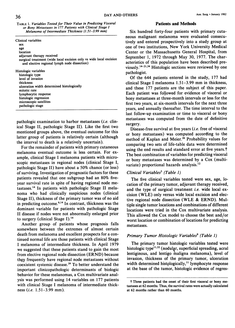
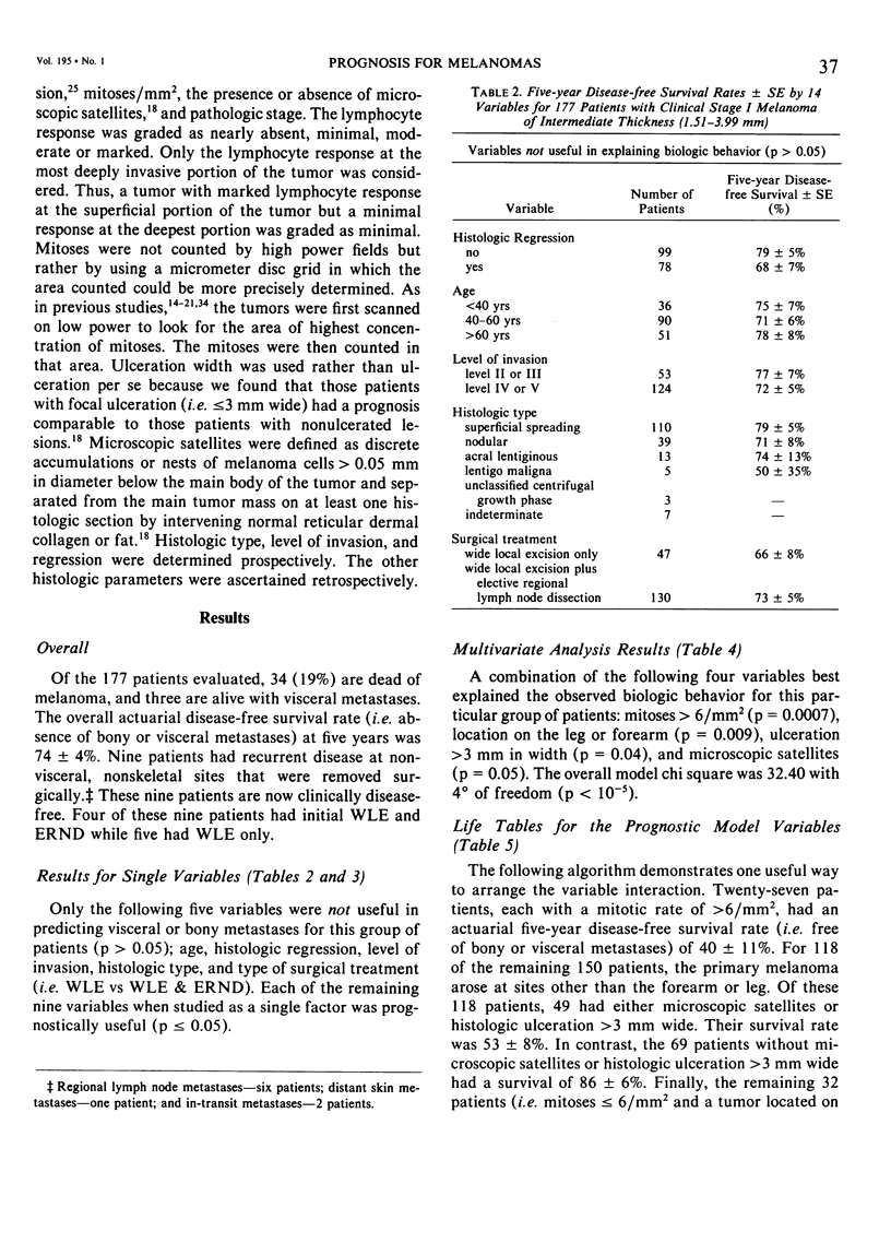
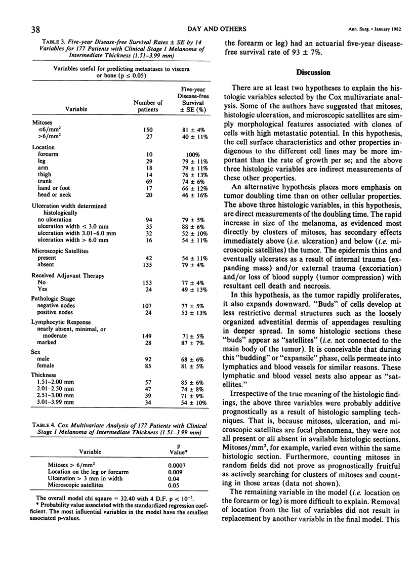
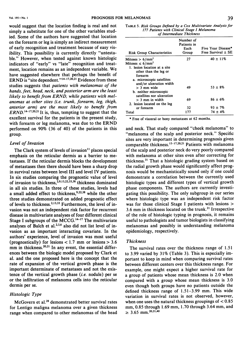
Fourteen variables were tested for their ability to predict visceral or bony metastases in 177 patients with clinical Stage I melanoma of intermediate thickness (1.51 - 3.39 mm). A Cox multivariate analysis yielded a combination of four variables that best predicted bony or visceral metastases for these patients: 1) mitoses greater than 6/min 2 (p = 0.0007), 2) location other than the forearm of leg) p = 0.009, 3) ulceration width greater than 3 mm (p = 0.04), 4) microscopic satellites (p = 0.05). The overall prognostic model chi square was 32.40 with 4 degrees of freedom (p less than 10 (-5). Combinations of the above variables were used to separate these patients into at least two risk groups. The high risk patients had at least a 35% or greater chance of developing visceral metastases within five years, while the low risk group had greater than an 85% chance of being disease free at five years. Criteria for the high risk group were as follows: 1) mitoses greater than 6/mm 2 in at least one area of the tumor, irrespective of primary tumor location, or 2) a melanoma located at some site other than the forearm or leg and histologic evidence in the primary tumor of either ulceration greater than 3 mm wide or microscopic satellites. The low risk group was defined as follows: 1) mitoses less than or equal to 6/mm 2 and a location on the leg or forearm, or 2) mitoses less than or equal to 6/mm 2 and the absence in histologic sections of the primary tumor of both microscopic satellites and ulceration greater then 3 mm wide. The number of patients in this series who did not undergo elective regional node dissection (N = 47) was probably too small to detect any benefit from this procedure. Based on survival rates from this and other studies, it is estimated that approximately 1500 patients with clinical Stage I melanoma of intermediate thickness in each arm of a randomized clinical trial would be needed to detect an increase in survival rates from elective regional node dissection.
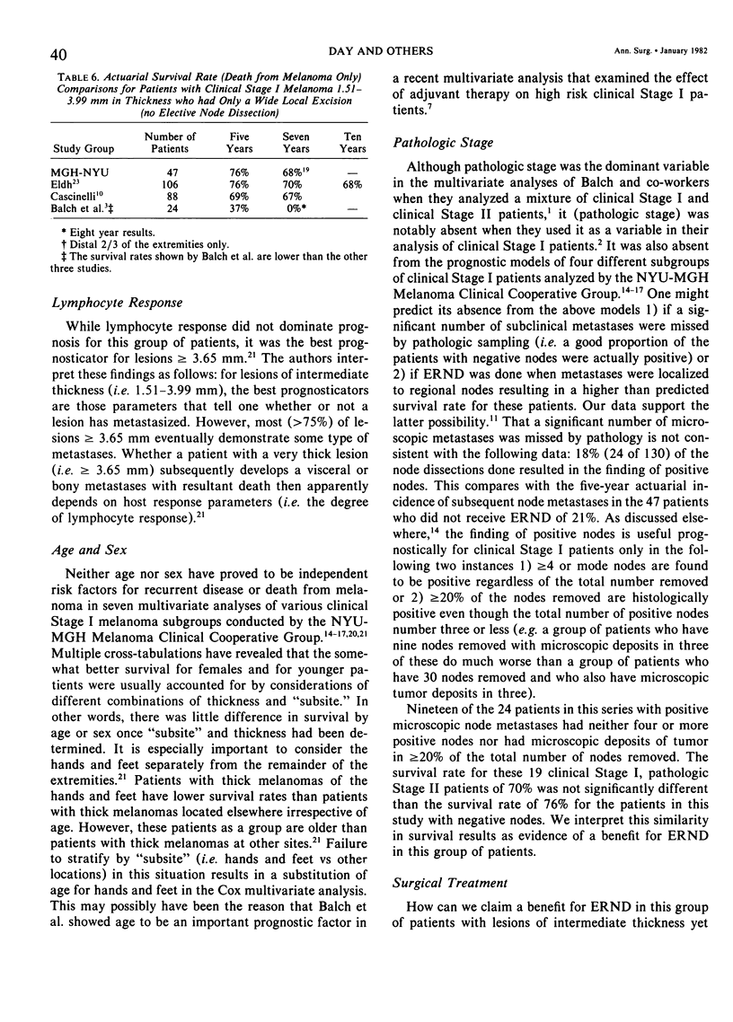
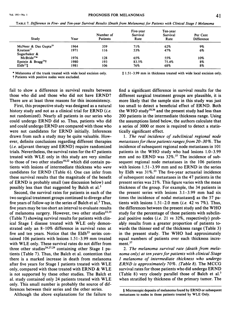
Full text
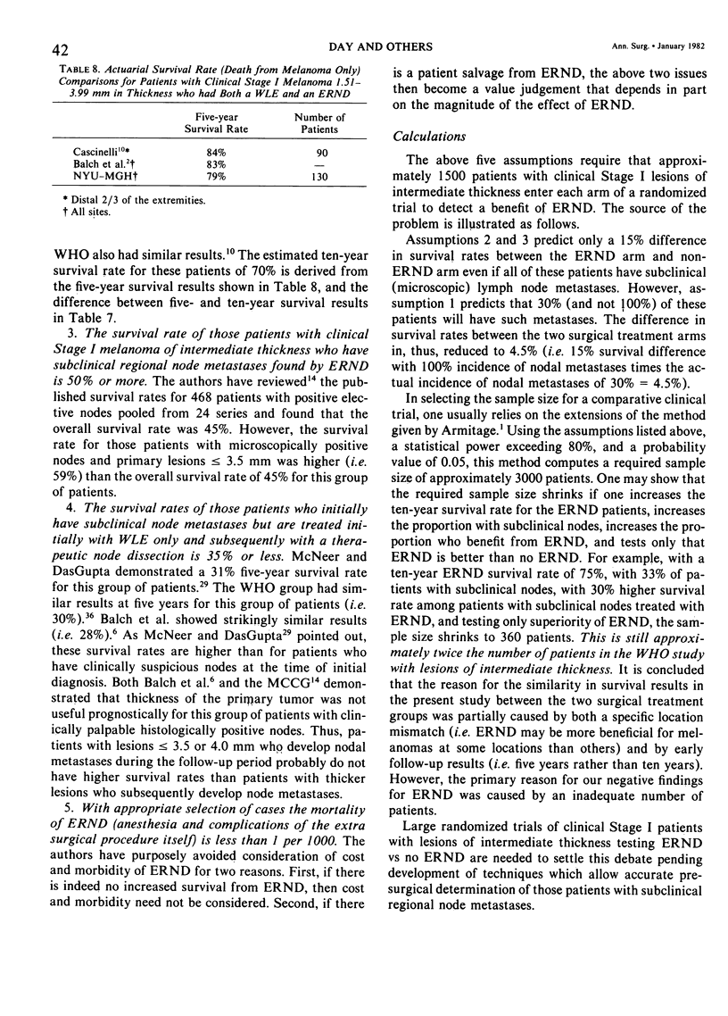
PDF
Selected References
These references are in PubMed. This may not be the complete list of references from this article.
- Balch C. M., Murad T. M., Soong S. J., Ingalls A. L., Halpern N. B., Maddox W. A. A multifactorial analysis of melanoma: prognostic histopathological features comparing Clark's and Breslow's staging methods. Ann Surg. 1978 Dec;188(6):732–742. doi: 10.1097/00000658-197812000-00004. [DOI] [PMC free article] [PubMed] [Google Scholar]
- Balch C. M., Soong S. J., Murad T. M., Ingalls A. L., Maddox W. A. A multifactorial analysis of melanoma. II. Prognostic factors in patients with stage I (localized) melanoma. Surgery. 1979 Aug;86(2):343–351. [PubMed] [Google Scholar]
- Balch C. M., Soong S. J., Murad T. M., Ingalls A. L., Maddox W. A. A multifactorial analysis of melanoma: III. Prognostic factors in melanoma patients with lymph node metastases (stage II). Ann Surg. 1981 Mar;193(3):377–388. doi: 10.1097/00000658-198103000-00023. [DOI] [PMC free article] [PubMed] [Google Scholar]
- Balch C. M. Surgical management of regional lymph nodes in cutaneous melanoma. J Am Acad Dermatol. 1980 Nov;3(5):511–524. doi: 10.1016/s0190-9622(80)80118-6. [DOI] [PubMed] [Google Scholar]
- Balch C. M., Wilkerson J. A., Murad T. M., Soong S. J., Ingalls A. L., Maddox W. A. The prognostic significance of ulceration of cutaneous melanoma. Cancer. 1980 Jun 15;45(12):3012–3017. doi: 10.1002/1097-0142(19800615)45:12<3012::aid-cncr2820451223>3.0.co;2-o. [DOI] [PubMed] [Google Scholar]
- Breslow A., Cascinelli N., van der Esch E. P., Morabito A. Stage I melanoma of the limbs: assessment of prognosis by levels of invasion and maximum thickness. Tumori. 1978 Jun 30;64(3):273–284. doi: 10.1177/030089167806400305. [DOI] [PubMed] [Google Scholar]
- Breslow A. Thickness, cross-sectional areas and depth of invasion in the prognosis of cutaneous melanoma. Ann Surg. 1970 Nov;172(5):902–908. doi: 10.1097/00000658-197011000-00017. [DOI] [PMC free article] [PubMed] [Google Scholar]
- Clark W. H., Jr, From L., Bernardino E. A., Mihm M. C. The histogenesis and biologic behavior of primary human malignant melanomas of the skin. Cancer Res. 1969 Mar;29(3):705–727. [PubMed] [Google Scholar]
- Day C. L., Jr, Lew R. A., Mihm M. C., Jr, Harris M. N., Kopf A. W., Sober A. J., Fitzpatrick T. B. The natural break points for primary-tumor thickness in clinical Stage I melanoma. N Engl J Med. 1981 Nov 5;305(19):1155–1155. doi: 10.1056/NEJM198111053051916. [DOI] [PubMed] [Google Scholar]
- Day C. L., Jr, Lew R. A., Mihm M. C., Jr, Sober A. J., Harris M. N., Kopf A. W., Fitzpatrick T. B., Harrist T. J., Golomb F. M., Postel A. A multivariate analysis of prognostic factors for melanoma patients with lesions greater than or equal to 3.65 mm in thickness. The importance of revealing alternative Cox models. Ann Surg. 1982 Jan;195(1):44–49. doi: 10.1097/00000658-198201001-00007. [DOI] [PMC free article] [PubMed] [Google Scholar]
- Day C. L., Jr, Mihm M. C., Jr, Sober A. J., Harris M. N., Kopf A. W., Fitzpatrick T. B., Lew R. A., Harrist T. J., Golomb F. M., Postel A. Prognostic factors for melanoma patients with lesions 0.76 - 1.69 mm in thickness. An appraisal of "thin" level IV lesions. Ann Surg. 1982 Jan;195(1):30–34. doi: 10.1097/00000658-198201001-00005. [DOI] [PMC free article] [PubMed] [Google Scholar]
- Day C. L., Jr, Sober A. J., Fitzpatrick T. B., Mihm M. C., Jr Prognosis in malignant melanoma. J Am Acad Dermatol. 1980 Nov;3(5):525–526. doi: 10.1016/s0190-9622(80)80119-8. [DOI] [PubMed] [Google Scholar]
- Day C. L., Jr, Sober A. J., Kopf A. W., Lew R. A., Mihm M. C., Jr, Golomb F. M., Hennessey P., Harris M. N., Gumport S. L., Raker J. W. A prognostic model for clinical stage I melanoma of the lower extremity. Location on foot as independent risk factor for recurrent disease. Surgery. 1981 May;89(5):599–603. [PubMed] [Google Scholar]
- Day C. L., Jr, Sober A. J., Kopf A. W., Lew R. A., Mihm M. C., Jr, Golomb F. M., Postel A., Hennessey P., Harris M. N., Gumport S. L. A prognostic model for clinical stage I melanoma of the trunk. Location near the midline is not an independent risk factor for recurrent disease. Am J Surg. 1981 Aug;142(2):247–251. doi: 10.1016/0002-9610(81)90286-5. [DOI] [PubMed] [Google Scholar]
- Day C. L., Jr, Sober A. J., Kopf A. W., Lew R. A., Mihm M. C., Jr, Hennessey P., Golomb F. M., Harris M. N., Gumport S. L., Raker J. W. A prognostic model for clinical stage I melanoma of the upper extremity. The importance of anatomic subsites in predicting recurrent disease. Ann Surg. 1981 Apr;193(4):436–440. doi: 10.1097/00000658-198104000-00007. [DOI] [PMC free article] [PubMed] [Google Scholar]
- Day C. L., Jr, Sober A. J., Lew R. A., Mihm M. C., Jr, Fitzpatrick T. B., Kopf A. W., Harris M. N., Gumport S. L., Raker J. W., Malt R. A. Malignant melanoma patients with positive nodes and relatively good prognoses: microstaging retains prognostic significance in clinical stage I melanoma patients with metastases to regional nodes. Cancer. 1981 Mar 1;47(5):955–962. doi: 10.1002/1097-0142(19810301)47:5<955::aid-cncr2820470523>3.0.co;2-1. [DOI] [PubMed] [Google Scholar]
- Eldh J., Boeryd B., Peterson L. E. Prognostic factors in cutaneous malignant melanoma in stage I. A clinical, morphological and multivariate analysis. Scand J Plast Reconstr Surg. 1978;12(3):243–255. doi: 10.3109/02844317809013000. [DOI] [PubMed] [Google Scholar]
- Epstein E., Bragg A. K. Curability of melanoma: a 25-year retrospective study. Cancer. 1980 Aug 15;46(4):818–821. doi: 10.1002/1097-0142(19800815)46:4<818::aid-cncr2820460429>3.0.co;2-j. [DOI] [PubMed] [Google Scholar]
- Gromet M. A., Epstein W. L., Blois M. S. The regressing thin malignant melanoma: a distinctive lesion with metastatic potential. Cancer. 1978 Nov;42(5):2282–2292. doi: 10.1002/1097-0142(197811)42:5<2282::aid-cncr2820420528>3.0.co;2-v. [DOI] [PubMed] [Google Scholar]
- MACNEER G., DASGUPTA T. PROGNOSIS IN MALIGNANT MELANOMA. Surgery. 1964 Sep;56:512–518. [PubMed] [Google Scholar]
- MUNDTH E. D., GURALNICK E. A., RAKER J. W. MALIGNANT MELANOMA: A CLINICAL STUDY OF 427 CASES. Ann Surg. 1965 Jul;162:15–28. doi: 10.1097/00000658-196507000-00003. [DOI] [PMC free article] [PubMed] [Google Scholar]
- McGovern V. J., Shaw H. M., Milton G. W., Farago G. A. Is malignant melanoma arising in a Hutchinson's melanotic freckle a separate disease entity? Histopathology. 1980 May;4(3):235–242. doi: 10.1111/j.1365-2559.1980.tb02918.x. [DOI] [PubMed] [Google Scholar]
- Schmoeckel C., Braun-Falco O. Prognostic index in malignant melanoma. Arch Dermatol. 1978 Jun;114(6):871–873. [PubMed] [Google Scholar]
- Sugarbaker E. V., McBride C. M. Melanoma of the trunk: the results of surgical excision and anatomic guidelines for predicting nodal metastasis. Surgery. 1976 Jul;80(1):22–30. [PubMed] [Google Scholar]
- Veronesi U., Adamus J., Bandiera D. C., Brennhovd I. O., Caceres E., Cascinelli N., Claudio F., Ikonopisov R. L., Javorskj V. V., Kirov S. Inefficacy of immediate node dissection in stage 1 melanoma of the limbs. N Engl J Med. 1977 Sep 22;297(12):627–630. doi: 10.1056/NEJM197709222971202. [DOI] [PubMed] [Google Scholar]
- Veronesi U., Adamus J., Bandiera D. C., Brennhovd I. O., Caceres E., Cascinelli N., Claudio F., Ikonopisov R. L., Javorskj V. V., Kirov S. Stage I melanoma of the limbs. Immediate versus delayed node dissection. Tumori. 1980 Jun 30;66(3):373–396. doi: 10.1177/030089168006600311. [DOI] [PubMed] [Google Scholar]
- Wanebo H. J., Woodruff J., Fortner J. G. Malignant melanoma of the extremities: a clinicopathologic study using levels of invasion (microstage). Cancer. 1975 Mar;35(3):666–676. doi: 10.1002/1097-0142(197503)35:3<666::aid-cncr2820350320>3.0.co;2-4. [DOI] [PubMed] [Google Scholar]