Abstract
HPr, the phosphocarrier protein of the bacterial phosphotransferase system, mediates catabolite repression of a number of operons in gram-positive bacteria. In order to participate in the regulatory process, HPr is activated by phosphorylation of a conserved serine-46 residue. To study the potential role of HPr in the regulation of Cry4A protoxin synthesis in Bacillus thuringiensis subsp. israelensis, we produced a catabolite repression-negative mutant by replacing the wild-type copy of the ptsH gene with a mutated copy in which the conserved serine residue of HPr was replaced with an alanine. HPr isolated from the mutant strain was not phosphorylated at Ser-45 by HPr kinase, but phosphorylation at His-14 was found to occur normally. The enzyme I and HPr kinase activities of the mutant were not affected. Analysis of the B. thuringiensis subsp. israelensis mutant harboring ptsH-S45A in the chromosome showed that cry4A expression was derepressed from the inhibitory effect of glucose. The mutant strain produced both cry4A and σ35 gene transcripts 4 h ahead of the parent strain, but there was no effect on σ28 synthesis. In wild-type B. thuringiensis subsp. israelensis cells, cry4A mRNA was observed from 12 h onwards, while in the mutant it appeared at 8 h and was produced for a longer period. The total amount of cry4A transcripts produced by the mutant was higher than by the parent strain. There was a 60 to 70% reduction in the sporulation efficiency of the mutant B. thuringiensis subsp. israelensis strain compared to the wild-type strain.
Biological control of dipteran pests in general and mosquitoes in particular has been a subject of primary importance for many years. Bacillus thuringiensis subsp. israelensis has been found to be the most effective microbial strain to date, possessing all the desirable properties of an ideal biocontrol agent. B. thuringiensis subsp. israelensis produces larvicidal crystal proteins in the stationary phase, concomitant with sporulation. The larvicidal activity of the parasporal crystals is attributed to the δ-endotoxin, composed of at least four major polypeptide species of about 27, 72, 128, and 135 kDa, which act synergistically in the manifestation of toxicity (27, 32). The high levels of toxin accumulation are controlled by a variety of mechanisms at the transcriptional, posttranscriptional, and posttranslational levels (2). The genes encoding different protein toxins are normally associated with large plasmids in B. thuringiensis subsp. israelensis (20, 26) and are named cry4A, cry4B, cry11A, and cytA. cry4A and cry4B code for the 135-kDa and 125-kDa protoxins, respectively, which are activated by the gut proteases in the alkaline conditions of the insect gut.
In sporulating cells of B. thuringiensis, the accumulation of large amounts of toxin proteins is achieved by expression from strong promoters associated with the gene. As in Bacillus subtilis, the developmental process is temporally and spatially regulated at the transcriptional level in B. thuringiensis by successive activation of different σ factors (6). In B. thuringiensis, the σ28 and σ35 subunits of RNA polymerase are considered functionally equivalent to EσK and EσE, respectively, of B. subtilis and have been shown to direct toxin gene transcription during the sporulation phase (1). Most cry4A gene transcription occurs by the early sporulation-specific σ35 protein, from a strong B. thuringiensis I promoter (35, 36). Later, in the mid-sporulation phase, σ28 takes over and directs transcription from the B. thuringiensis II promoter (28, 37), resulting in sustained toxin synthesis over a long period of time.
In gram-positive bacteria, catabolite repression of many catabolic operons has been shown to involve a small phosphoprotein, HPr, which is considered a key component of the signal transduction cascade representing catabolite repression. The data emerging from a number of studies (10, 12, 13, 31) point to the metabolite-activated phosphorylation of the phosphocarrier protein HPr on the conserved serine-46 as a critical step. Activation of HPr occurs under conditions favoring growth and high glycolytic activity. Once activated, the phosphorylated HPr binds to the catabolite control protein (CcpA), forming a complex with strong DNA binding affinity (12, 16). The binding of the phosphorylated serine-HPr-CcpA complex to the 14-bp cre sequence (33) associated with the target DNA leads to its transcriptional modulation (15).
Earlier we reported the effect of glucose and inorganic phosphate on Cry4A toxin synthesis (4). The modulation of toxin synthesis by both glucose and inorganic phosphate was found to occur at the mRNA level, suggesting the potential involvement of catabolite repression in the regulation. It was also observed that HPr phosphorylation and dephosphorylation in B. thuringiensis subsp. israelensis correlated with the glucose-mediated repression of Cry4A toxin synthesis (5).
In the present work, we investigated the role of HPr during glucose repression of Cry4A toxin synthesis in B. thuringiensis subsp. israelensis at the genetic level by inactivating the regulatory function of the ptsH gene, encoding the protein HPr. Since phosphorylation of the conserved serine-45 residue of HPr is primarily responsible for its regulatory catabolite repression function in B. thuringiensis subsp. israelensis (18), we first created a point mutation in the ptsH gene of B. thuringiensis subsp. israelensis by replacing the Ser-45 residue with alanine. The mutated copy of the gene was integrated into the bacterial chromosome to replace the wild-type copy by homologous recombination. We present data analyzing the effect of glucose on the synthesis of Cry4A toxin in the mutant strain.
MATERIALS AND METHODS
Bacterial strain and growth conditions.
B. thuringiensis subsp. israelensis strain HD522 (American Type Culture Collection) was grown in Luria-Bertani medium, G-medium (34), or Hogg-Jago medium (25) at 29°C as specified for different experiments. For isolation of DNA, the cultures were grown overnight in Luria-Bertani medium. The cell pellet was processed for chromosomal DNA preparation as described by Ausubel et al. (3). For estimating toxin-specific mRNA and toxin protein synthesis, the cultures were grown in G salts as described before (4). Briefly, the cells were grown with shaking for 14 to 15 h, washed once, and resuspended in starvation medium (RM) containing 50 mM Tris-HCl buffer (pH 7.5), 0.2% (NH4)2SO4, 0.008% CaCl2, 0.025% FeSO4 · 5H2O, 0.05% CuSO4 · 5H2O, 0.05% ZnSO4 · 7H2O, 0.5% MnSO4 · H2O, and 2.0% MgSO4.
DNA manipulations.
The ptsH gene from B. thuringiensis subsp. israelensis was cloned as described earlier (18). The point mutation in the ptsH gene was incorporated by using the construct pSR2 (Table 1) (18), which contained the wild-type copy of the ptsH gene. Site-directed mutagenesis was performed to replace the serine-45 residue with an alanine with the QuikChange mutagenesis kit (Stratagene). The oligonucleotide primers were 5′-GTTAACTTAAAAGCAATCATGGGCGTAATG-3′ and 5′-CATTACGCCCATGATTGCTTTTAAGTTAAC-3′. Some of the transformed colonies were sequenced to confirm the mutated residue, and one clone, SR15, was selected for further studies.
TABLE 1.
Strains and plasmids used in this study
| Plasmid or strain | Relevant characteristics | Reference |
|---|---|---|
| Plasmids | ||
| pSR2 | pGEM-T easy containing a 293-bp PCR-amplified ptsH | 18 |
| pSR15 | pGEM-T easy containing 293-bp ptsH S45A | This work |
| pSR16 | pGEM-T easy containing 293-bp ptsH S45A and kanamycin resistance cassette | This work |
| pSR18 | pGEM-T easy containing 700-bp B. thuringiensis subsp. israelensis σ33 | This work |
| pSR19 | pGEM-T easy containing 700-bp B. thuringiensis subsp. israelensis σ28 | This work |
| pDG783 | pSB118 containing kanamycin cassette at HindIII site | 14 |
| Strains | ||
| SR2 | E. coli DH5α cells harboring 293-bp ptsH gene of B. thuringiensis subsp. israelensis in pGEM-T easy | 18 |
| SR15 | E. coli DH5α cells containing 293-bp ptsH S45A gene in pGEM-T easy | This work |
| SR16 | E. coli RR1 cells containing 293-bp ptsH S45A and kanamycin cassette in pGEM-T easy | This work |
| SR17 | B. thuringiensis subsp. israelensis with ptsH S45A integrated downstream of wild-type ptsH promoter in chromosome | This work |
| E. coli RR1 | hsdS20 (r−B m+B) supE44 ara-14 proA2 rspL20(Strr) lacY1 galK2 xyl-5 mtl-1 supE44 | Promega |
| E. coli XL1-Blue | recA1 endA1 gyrA96 thi-1 hsdR17 supE44 relA1 lac [F′ proAB lacIqZΔM1515 Tn10(Tetr)] | Stratagene |
pSR15 DNA containing the S45A mutation in the ptsH gene was digested with HindIII (the site was created at the C-terminal part of the gene during cloning) for ligating a 1.5-kb kanamycin resistance cassette obtained by HindIII digestion of pDG783 (14). The ligation mix was transformed in Escherichia coli RR1 cells, and colonies were selected on plates containing ampicillin plus kanamycin. Plasmid DNA from clone SR16 was prepared by the alkaline lysis method and electroporated into B. thuringiensis subsp. israelensis cells.
Electroporation of B. thuringiensis subsp. israelensis.
B. thuringiensis subsp. israelensis cells were grown overnight in Hogg-Jago medium, subcultured, and incubated until the A600 reached 0.5. Sterile glycine was added to the growing cells at a final concentration of 8 to 12%, and the cells were grown for another hour. The cells were centrifuged and washed thrice in cold sterile EPBS (5 mM phosphate buffer [pH 8.0], 10% glycerol, 40% sorbitol), and the final pellet was resuspended in 1.0 ml of EPBS buffer. Then 200 μl of competent cells was electroporated with 1 to 5 μg of pSR16 DNA at 2 kV, 400 Ω, and 25 μF capacitance. Cold Hogg-Jago medium (800 μl) was added immediately, and the cells were incubated with shaking at 29°C for 2 h. The transformants were selected on Luria-Bertani plates containing 5 μg of kanamycin/ml.
Screening of transformants.
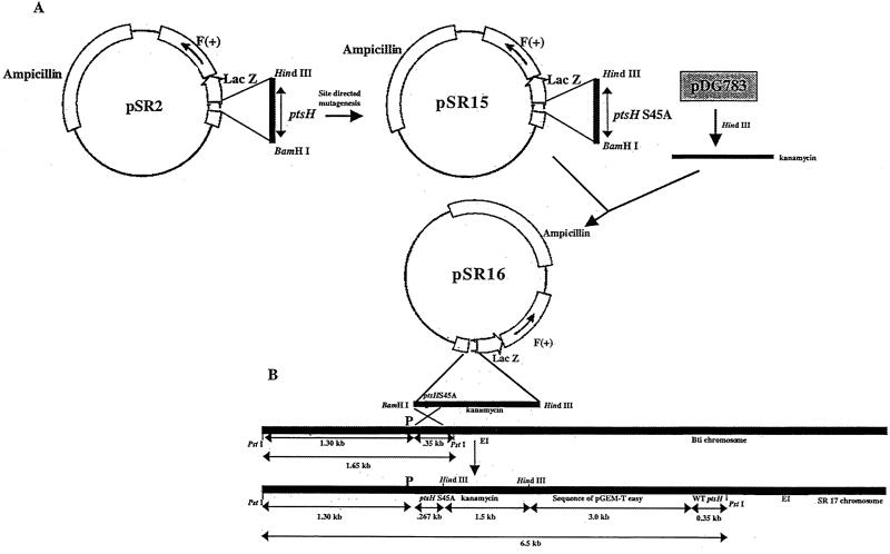
The recombinants were obtained by a single crossover event which inserted the mutated copy of the ptsH gene together with the kanamycin cassette and the vector DNA between the wild-type ptsH gene and its promoter. Genomic DNA from several kanamycin-resistant colonies was digested separately with PstI and HindIII. Southern hybridization was performed after separation on an agarose gel. The HindIII-digested DNA was probed with the 1.5-kb kanamycin gene, and the PstI-digested DNA was hybridized with a 300-bp ptsH probe. One clone, SR17, showing a positive reaction with both the kanamycin gene and the ptsH probe was selected for further studies.
To identify the point mutation in the HPr of strain SR17, it was grown overnight in Luria-Bertani medium containing 0.5% glucose. The cells were washed and resuspended in 5 mM Tris-HCl (pH 7.5) and lysed by sonication. The cell homogenate was filtered through Centricon-30, and the filtrate was dialyzed extensively against the above buffer and concentrated. The concentrated proteins containing predominantly HPr were subjected to [γ-32P]ATP- and FBP-mediated phosphorylation at Ser-45 by HPr kinase and to enzyme I (EI) and phosphoenolpyruvate (PEP)-activated phosphorylation at His-14 as described before (18). For analyzing the polar effects of insertion of ptsH on the ptsI gene, EI was prepared from SR17 cells and assayed as described earlier (18).
Growth and sporulation.
The growth pattern of the mutant strain was compared with that of the wild-type B. thuringiensis subsp. israelensis strain in Luria-Bertani medium as well as in the defined G-medium by subculturing an overnight culture in the appropriate medium. The optical density was measured at 600 nm. For determining the sporulation efficiency of the two strains, the cultures were grown in G-medium containing 0.2% glucose. Samples were removed at the designated time, serially diluted in sterile phosphate-buffered saline and subjected to heat treatment at 80°C for 15 min. Unheated and heated samples were plated in duplicate to determine the total viable and heat-resistant cell counts. The results are representative of three separate experiments.
Total RNA extraction.
Strain SR17 was grown in G-medium containing 0.2% glucose. Cells were centrifuged at 4°C after 14 to 15 h of growth and quickly resuspended in RM, which was supplemented or not with 0.5 or 1% glucose. Cultures (15 ml) were removed at different time intervals, centrifuged at 4°C, and frozen at −20°C until further use.
In another set of experiments, both the wild-type and mutant B. thuringiensis subsp. israelensis strains were grown in G-medium containing 0.2% glucose, and samples were removed from 6 h onwards. Total RNA was prepared to study the kinetics of synthesis of cry4A, σ35, and σ28 gene-specific messages in the two strains by probing with specific DNA probes. Total RNA was prepared from the cells with the RNeasy kit (Qiagen). The concentrations of RNA were calculated spectrophotometrically, and aliquots containing 2 μg of RNA were loaded. The blot was subsequently probed with a 1.7-kb DNA fragment containing the promoter and the N terminus of the cry4A gene (obtained from a cloned 3.9-kb B. thuringiensis subsp. israelensis fragment containing the cry4A gene with its promoter) and the entire σ35 and σ28 gene sequences and exposed for autoradiography.
Cloning of σ35 and σ28 genes of B. thuringiensis subsp. israelensis.
Based on the sequences of the σ35 and σ28 genes of Bacillus thuringiensis subsp. kurstaki HD1 (1), homologous sequences from B. thuringiensis subsp. israelensis were amplified by PCR and cloned, producing plasmids pSR18 and pSR19, respectively (GenBank accession numbers AY083614 and AY083615, respectively).
Uptake of 2-[14C]deoxyglucose.
One milliliter of culture (A600, 0.8) of both the wild-type and the mutant strain was centrifuged and washed with 50 mM phosphate buffer (pH 7.5) at 4°C and resuspended in the same buffer. 2-[14C]deoxyglucose was added to a final concentration of 2 mM (45 μCi/μmol) in a total volume of 500 μl. The cells were incubated at 29°C, and 25-μl samples were removed at different intervals up to 2 min and passed through a 0.2-μm filter. The filters were washed three times with 1 ml of 50 mM phosphate buffer and air dried. The filters were put in Aquasol, and the radioactivity associated with the cells was counted in a liquid scintillation counter.
Effect of glucose on Cry4A toxin synthesis.
B. thuringiensis subsp. israelensis and SR17 cells were grown in G-medium overnight at 29°C and resuspended in RM after 15 h of growth. Glucose was added to a final concentration of 0.5 and 1% to the RM. Two-milliliter samples were removed at 12 and 14 h after resuspension, and aliquots containing equal amounts of proteins (22) were solubilized in loading dye and resolved by sodium dodecyl sulfate-10% polyacrylamide gel electrophoresis (SDS-PAGE). The proteins were visualized by Coomassie blue staining. For Western blotting, antibodies were raised against Cry4A protoxin isolated from the recombinant B. thuringiensis subsp. israelensis strain 4Q2-81(pHT606) containing the cry4A protoxin gene only (8).
RESULTS
Preparation of HPr-S45A mutant.
The PCR-amplified nicked circular product containing the S45A mutation in the ptsH gene was obtained at 60°C. It was transformed into E. coli to produce clone SR15, which contained the desired mutation.
The plasmid DNA obtained from clone SR16, a derivative of pSR15 (Fig. 1), contained the 1.5-kb kanamycin cassette at the HindIII site at the 3′ end of the ptsH gene. Electroporation of the pSR16 DNA into B. thuringiensis subsp. israelensis occurred at a rather low frequency (5 × 10⁻⁷). In the Southern blot, a 6.5-kb fragment in the PstI-digested genomic DNA of SR17 reacted with the 300-bp ptsH probe, which was absent in the wild-type strain (data not presented). Hybridization of the HindIII-digested genomic DNA with the kanamycin gene sequence yielded two bands at 1.5 and 4.5 kb, and both the bands were absent in the wild-type B. thuringiensis subsp. israelensis DNA (data not shown), indicating that the kanamycin cassette had integrated in the B. thuringiensis subsp. israelensis chromosome at the designated location.
FIG. 1.
Construction of B. thuringiensis subsp. israelensis mutant SR17, containing the ptsH S45A allele in the chromosome. (A) The wild-type promoterless ptsH gene in pSR2 was mutagenized by reverse PCR with primers containing the mutated residue. A kanamycin resistance cassette was incorporated at the HindIII site of pSR15, producing pSR16. (B) Homologous recombination of pSR16 by single crossing over at the ptsH locus resulted in integration of the ptsH S45A allele downstream of the wild-type ptsH promoter, rendering the wild-type (wt) ptsH gene nonfunctional. The size of the PstI-digested fragment containing the wild-type ptsH gene increased from 1.65 to 6.5 kb due to duplication of the gene and addition of the vector DNA and kanamycin cassette between two ptsH copies.
Identification of point mutation.
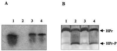
In contrast to the wild-type protein, no phosphorylation was observed when the same amount of HPr prepared from strain SR17 was subjected to phosphorylation at Ser-45 by HPr kinase and [γ-32P]ATP (Fig. 2A, lanes 1 and 2). As expected, there was no effect on the EI-mediated phosphorylation at His-14 (Fig. 2B). The above result demonstrated that in the mutant, the wild-type copy of the ptsH gene is totally silent, and the HPr produced by the latter is the altered HPr-S45A only. The mutant strain was able to produce HPr kinase normally, as shown in Fig. 2A, lanes 3 and 4, in which HPr kinases prepared from the wild-type strain and the mutant strain were used to phosphorylate recombinant B. thuringiensis subsp. israelensis HPr at Ser-45. Furthermore, partially purified cell homogenate from strain SR17, used as a source of EI, was able to transfer the phosphoryl group to recombinant B. thuringiensis subsp. israelensis HPr as efficiently as the wild-type strain (data not shown). This indicated that there was no polar effect on the expression of the ptsI gene, and the second copy of the ptsH gene is not expressed.
FIG. 2.
Phosphorylation pattern of HPr from wild-type B. thuringiensis subsp. israelensis and ptsH S45A mutant strain SR17. (A) Autoradiogram showing phosphorylation of HPr by HPr kinase and [γ-32P]ATP at serine-45. Lane 1, HPr from B. thuringiensis subsp. israelensis phosphorylated by HPr kinase from B. thuringiensis subsp. israelensis; lane 2, HPr from SR17 phosphorylated by HPr kinase from SR17; lanes 3 and 4, B. thuringiensis subsp. israelensis recombinant HPr phosphorylated by HPr kinase from B. thuringiensis subsp. israelensis and SR17, respectively. (B) Western blot of nondenaturing polyacrylamide gel, showing phosphorylation of HPr by PEP and recombinant EI from B. subtilis at histidine-14. Lane 1, HPr from B. thuringiensis subsp. israelensis; lane 2, HPr from B. thuringiensis subsp. israelensis phosphorylated by EI and PEP; lane 3, HPr from SR17; lane 4, HPr from SR17 phosphorylated by EI and PEP.
Growth, sporulation, and 2-[14C]deoxyglucose uptake.
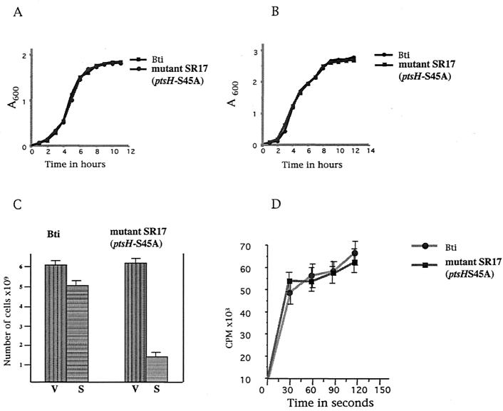
The growth kinetics of the two strains in G-medium and in Luria-Bertani medium were identical (Fig. 3A and B). The cultures grew exponentially up to 6 to 7 h and entered the stationary phase after 8 h. Sporulation efficiency was determined after 20 h, and the time of appearance of heat-resistant spores varied in different experiments and occurred between 18 and 24 h. The mutant strain showed a 60 to 70% reduction in the formation of heat-resistant spores. The sporulation efficiency in the latter was 20 to 30%, compared to more than 80% observed in the wild-type strain after 24 h (Fig. 3C). The rate of glucose uptake by both strains was similar, as shown in Fig. 3D. The rate calculated in the initial 30 s was almost the same in both cases.
FIG. 3.
Growth and sporulation characteristics of ptsH mutant strain SR17. (A) Growth patterns of wild-type B. thuringiensis subsp. israelensis (Bti) and ptsH mutant strain SR17 in G-medium. (B) Growth patterns of wild-type B. thuringiensis subsp. israelensis and ptsH mutant strain SR17 in Luria-Bertani medium. (C) Comparison of sporulation efficiencies of the two strains in G-medium. The formation of heat-resistant cells was monitored after growing the strains in G-medium for 20 h. V, total viable cell counts; S, heat-resistant cell counts. (D) Uptake of 2-[14C]deoxyglucose by the wild-type and mutant strains. Cells in the mid-exponential phase were resuspended in the presence of 2-[14C]deoxyglucose, and the incorporation of radioactivity by the cells was monitored by liquid scintillation counting. The mean values of three independent experiments have been plotted, and the standard deviations have been plotted as error bars.
Effect of glucose on cry4A gene expression.
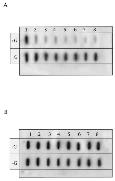
cry4A-specific mRNA levels were reduced in the presence of glucose in the wild-type but not in the mutant strain (Fig. 4A and B). The concentration of cry4A transcripts was considerably higher at the time of resuspension (0 h) in the mutant strain than in the wild-type (Fig. 4A and B, lane 1). cry4A mRNA could be detected till 5 h after resuspension in both cases. In the parent strain, the transcript levels were drastically reduced within 15 min of addition of glucose, but there was no effect in the mutant cells (Fig. 4A, lane 2).
FIG. 4.
Effect of glucose on cry4A-specific mRNA in B. thuringiensis subsp. israelensis and the ptsH S45A mutant. Both strains were grown in G-medium for 15 h and resuspended in RM with or without 0.5% glucose. Then 15-ml samples were removed at different times, and total RNA was prepared and probed with a 1.7-kb cry4A-specific DNA probe. (A) Total RNA from B. thuringiensis subsp. israelensis. (B) Total RNA from ptsH mutant SR17 (ptsH S45A). Lanes 1 to 8, samples collected at 0 h, 15 min, 30 min, 1 h, 2 h, 3 h, 4 h, and 5 h, respectively.
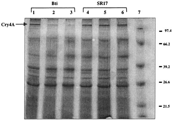
Cry4A protein synthesis was monitored in the cultures after resuspension in starvation medium with or without glucose. Similar to its effect on the mRNA levels, a corresponding effect could be observed on the synthesis of Cry4A protoxin. When the cells were collected 12 to 14 h after resuspension and total proteins were subjected to SDS-PAGE, the response of the two strains was markedly different in the presence of glucose. Glucose at 0.5 and 1% (Fig. 5, lanes 2 and 3) inhibited protoxin synthesis in the wild-type strain, but the mutant was totally insensitive to glucose (Fig. 5, lanes 4, 5, and 6). The location of the Cry4A protein was determined by blotting the separated proteins with Cry4A-specific antibodies (data not shown). Other toxin or nontoxin proteins apparently remained unaffected.
FIG. 5.
Protein profiles of wild-type B. thuringiensis subsp. israelensis (Bti) and the ptsH S45A mutant, showing the effect of glucose on Cry4A synthesis. Both strains were grown in G-medium for 15 h and resuspended in RM containing 0, 0.5, or 1% glucose. Aliquots of cells were removed 12 h after resuspension, and the proteins were solubilized in SDS sample buffer and resolved on 10% polyacrylamide gels. Lanes 1 and 4, no glucose; lanes 2 and 5, 0.5% glucose; lanes 3 and 6, 1% glucose; lane 7, molecular size markers, indicated at the right in kilodaltons.
Kinetics of cry4A, σ35, and σ28 mRNA synthesis.
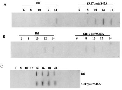
The two strains differed in the time of appearance as well as the level of cry4A mRNA (Fig. 6A). In the wild-type B. thuringiensis subsp. israelensis, the mRNA could be detected from 12 to 14 h of growth, and its levels were lower than that obtained in the mutant strain, in which the transcripts could be detected from 8 h onwards (Fig. 6A). In both strains, the cry4A transcripts persisted beyond the 14th hour of growth.
FIG. 6.
mRNA profiles of σ35, σ28, and cry4A in B. thuringiensis subsp. israelensis (Bti) and SR17 (ptsH S45A) mutant during growth. Both strains were grown in G-medium, and cell samples were removed at different times. Total RNA was prepared from these cells and probed with cry4A-, σ35-, or σ28-specific probes to determine the transcript level. (A) Profile of cry4A-specific mRNA. (B) Profile of σ35-specific mRNA. (C) Profile of σ28-specific mRNA. Slots 1 to 8, samples removed at 6 h, 8 h, 10 h, 12 h, 14 h, 16 h, 18 h, and 20 h, respectively.
Since σ35 is the major sigma factor involved in transcription of the cry4A gene during sporulation, its profile was determined to see if the loss in activity of the ptsH gene had any effect on the levels of σ35 protein (Fig. 6B). σ35-specific mRNA was detectable between 12 and 14 h in the wild-type strain; the timing matched the appearance of cry4A mRNA (Fig. 6A and B). In the mutant strain, σ35 transcripts were visible much earlier, at 8 h, and remained until 12 h, coinciding with the synthesis of cry4A mRNA. There was no difference in the levels of the σ35 transcripts produced in the two strains (Fig. 6B). These results indicate that σ35 gene transcription is also regulated by a ptsH-mediated repression in B. thuringiensis subsp. israelensis. The time of appearance and concentration of σ28 were not affected in the ptsH mutant (Fig. 6C), indicating the insensitivity of σ28 to ptsH-mediated regulation.
DISCUSSION
The results described in this study demonstrate that phosphorylation of HPr at serine-45 mediates glucose repression of Cry4A protoxin synthesis in B. thuringiensis subsp. israelensis. Glucose catabolite repression is considered a global regulatory mechanism, controlling the activity of many catabolic operons (9, 12, 13, 31) in response to carbon source availability in bacteria. We created a catabolite repression-negative ptsH mutant of B. thuringiensis subsp. israelensis, in which an increase in cry4A gene transcription has occurred at the expense of the developmental process. Bryan et al. described regulation of the σE-dependent mmg operon by glucose catabolite repression in B. subtilis (7), providing the rationale for such a regulation in maintaining the optimum metabolic state of the mother cell during endospore development. Another EσE-driven operon, the glycogen operon, which is repressed by glucose in sporulating cells of B. subtilis, also supports the need for metabolic control of the stationary-phase phenomenon (19).
In the light of the remarkable conservation of sigma subunits between B. thuringiensis and B. subtilis, the temporal regulation of sporulation in B. thuringiensis through the cascade of sigma factors is believed to be closely similar to that of B. subtilis (1, 21, 23, 24). Homologs of several important transcription factors described in B. subtilis have been identified in B. thuringiensis also (1). In B. thuringiensis subsp. israelensis, the total Cry4A protein produced is the sum of three different promoter activities. In the stationary phase, σH-containing RNA polymerase initiates low-level transcription (28), followed by σ35 in the mid-sporulation phase (35), and finally σ28-directed transcription occurs from a weak promoter in the late sporulation stages (37). The expression of the EσE precursor in B. subtilis starts just before septation by σA-containing RNA polymerase in conjunction with Spo0A (11, 17, 29), a protein belonging to the family of global response regulator proteins (30).
In the event of a similar mechanism operating in B. thuringiensis subsp. israelensis, an additional control through ptsH on either of these genes active in the vegetative phase would serve as a switch linked to the metabolic state of the cell. In the wild-type B. thuringiensis subsp. israelensis, cry4A mRNA was detected between 12 and 14 h, with the simultaneous induction of σ35 mRNA. Very low transcription of cry4A occurred before that, indicating the predominance of the σ35-directed synthesis of cry4A transcription in that period, while σH promoter activity remained very low. Significantly, in the ptsH mutant, σ35 transcripts appeared 4 h earlier than in the wild type, at 8 h of growth, with concomitant synthesis of cry4A mRNA.
Derepression of both genes in the ptsH mutant strongly supports the key role of ptsH-mediated regulation of Cry4A toxin synthesis. The regulation is apparently exerted through primary modulation of the σ35 gene or the proteins responsible for its transcription, i.e., σA or Spo0A (20, 28). It is necessary to mention here that two potential cre sequences at −488 and +63 nucleotides were found in the upstream and coding regions of the σ35 gene of B. thuringiensis subsp. kurstaki (1). In the later periods (12 to 14 h), the cry4A gene is apparently transcribed by both σ35 and σ28 in the ptsH mutant, as shown by the σ28 profile.
As a consequence of early induction of Eσ35 in the mutant strain, an enhancement in sporulation efficiency parallel to toxin synthesis was expected. However, contrary to our expectations, sporulation was inhibited in the mutant in comparison to the wild-type strain. Derepression of negative regulators of sporulation from ptsH-mediated catabolite repression in the catabolite repression-negative strain could be one of the reasons for this. Alternatively, the untimely induction of Eσ35 protein in cells metabolically not ready to start the developmental process could be another reason for aborted spore development, and this is supported by our kinetics data on the induction of cry4A and the sigma factor genes. The third reason could be titration of Eσ35 by the high-throughput cry gene promoters in the absence of the appropriate sporulation machinery, leading to increased transcription of the cry4A gene at the expense of the developmental process. The reduced sporulation efficiency of the ptsH mutant observed in this study reinforces the basic argument that catabolite repression of the toxin gene in the early stages is necessary for providing adequate levels of the sigma factors to the developmental machinery.
Interestingly, the concentration of cry4A transcripts remained high till 14 h of growth in the mutant, when transcription occurred mainly by a σ28-controlled promoter (Fig. 6A and C). However, our resuspension experiments with cells of similar age (15 h old) showed that glucose repressed cry4A transcription in the wild-type B. thuringiensis subsp. israelensis cells but not in the ptsH mutant. Considering the fact that the ptsH mutation had no effect on the σ28 profile, the insensitivity of the ptsH mutant to glucose in this period also suggests an additional ptsH-mediated control on the toxin gene, which is probably required for finer regulation of toxin synthesis.
We have identified several putative cre sequences (33) in the upstream region and also within the coding sequence of the cry4A gene (Table 2). Studies are in progress to verify their role in regulation. The presence of a functional cre sequence downstream of the promoters or within the open reading frame could be expected to result in regulation of cry4A gene transcription dependent on the concentration of carbon metabolites.
TABLE 2.
Putative cre sequences present in cry4A
| cre (position) | Sequence |
|---|---|
| Consensus | TG(A/T)∗A∗CG∗T∗(T/A)CA |
| −6 to −19 | 5′-AAATTACGAAT ACT-3′ |
| +246 to +259 | 5′-TGGTTTCGGGT TCA-3′ |
| +591 to +604 | 5′-TGATTGCGATT ACT-3′ |
| +3504 to +3517 | 5′-TTATATCGAAA GCA-3′ |
In conclusion, the analysis of cry4A gene transcription in a ptsH mutant of B. thuringiensis subsp. israelensis demonstrates that protoxin synthesis is indeed controlled by ptsH-mediated glucose catabolite repression. The primary target of the regulatory process could not be ascertained in this study. Significantly, the ptsH mutant strain of B. thuringiensis subsp. israelensis has the potential to produce larger amounts of toxin than the wild-type strain over a longer period of time. This study demonstrates for the first time the key role of HPr in the modulation of Cry4A toxin expression in B. thuringiensis subsp. israelensis.
Acknowledgments
We thank Armelle Delecluse and D. R. Zeigler for strain 4Q2-81(pHT606) and vector pDG783, respectively.
Sharik R. Khan was supported by a University Grants Commission Government of India Fellowship.
REFERENCES
- 1.Adams, L. F., K. L. Brown, and H. R. Whiteley. 1991. Molecular cloning and characterization of two genes encoding sigma factors that direct transcription from a Bacillus thuringiensis crystal protein gene promoter. J. Bacteriol. 173:3846-3854. [DOI] [PMC free article] [PubMed] [Google Scholar]
- 2.Agaisse, H., and D. Lereclus. 1995. How does Bacillus thuringiensis produce so much insecticidal crystal protein? J. Bacteriol. 177:6027-6032. [DOI] [PMC free article] [PubMed] [Google Scholar]
- 3.Ausubel, F. M., R. Brent, R. E. Kingston, D. D. Moore, J. G. Seidman, J. A. Smith, and K. Struhl. 1989. Current protocols in molecular biology, vol. I and II. John Wiley and Sons, New York, N.Y.
- 4.Banerjee-Bhatnagar, N. 1998. Modulation of CryIVA toxin protein expression by glucose in Bacillus thuringiensis israelensis. Biochem. Biophys. Res. Commun. 252:402-406. [DOI] [PubMed] [Google Scholar]
- 5.Banerjee-Bhatnagar, N. 1999. Inorganic phosphate regulates CryIVA protoxin expression in Bacillus thuringiensis israelensis. Biochem. Biophys. Res. Commun. 262:359-364. [DOI] [PubMed] [Google Scholar]
- 6.Baum, J. A., and T. Malvar. 1995. Regulation of insecticidal crystal protein production in Bacillus thuringiensis. Mol. Microbiol. 18:1-12. [DOI] [PubMed] [Google Scholar]
- 7.Bryan, E. M., B. W. Beall, and C. P. Moran, Jr. 1996. A σE-dependent operon subject to catabolite repression during sporulation in Bacillus subtilis. J. Bacteriol. 178:4778-4786. [DOI] [PMC free article] [PubMed] [Google Scholar]
- 8.Delecluse, A., S. Poncet, A. Klier, and G. Rapoport. 1993. Expression of cry4A and cry4B genes, independently or in combination, in a crystal-negative strain of Bacillus thuringiensis subsp. israelensis. Appl. Environ. Microbiol. 59:3922-3927. [DOI] [PMC free article] [PubMed]
- 9.Deutscher, J., J. Reizer, C. Fischer, A. Galinier, M. H. Saier, Jr., and M. Steinmetz. 1994. Loss of protein kinase-catalyzed phosphorylation of HPr, a phosphocarrier protein of the phosphotransferase system, by mutation of the ptsH gene confers catabolite repression resistance to several catabolic genes of Bacillus subtilis. J. Bacteriol. 176:3336-3344. [DOI] [PMC free article] [PubMed] [Google Scholar]
- 10.Deutscher, J., E. Kuster, U. Bergstedt, V. Charrier, and W. Hillen. 1995. Protein kinase-dependent HPr/CcpA interaction links glycolytic activity to carbon catabolite repression in Gram-positive bacteria. Mol. Microbiol. 15:1049-1053. [DOI] [PubMed] [Google Scholar]
- 11.Errington, J. 1993. Bacillus subtilis sporulation: regulation of gene expression and control of morphogenesis. Microbiol. Rev. 57:1-33. [DOI] [PMC free article] [PubMed] [Google Scholar]
- 12.Fujita, Y., Y. Miwa, A. Galinier, and J. Deutscher. 1995. Specific recognition of the Bacillus subtilis gnt cis-acting catabolite-responsive element by a protein complex formed between CcpA and seryl-phosphorylated HPr. Mol. Microbiol. 17:953-960. [DOI] [PubMed] [Google Scholar]
- 13.Galinier, A., J. Deutscher, and I. Martin-Verstraete. 1999. Phosphorylation of either Crh or HPr mediates binding of CcpA to the Bacillus subtilis xyn cre and catabolite repression of xyn operon. J. Mol. Biol. 286:307-314. [DOI] [PubMed] [Google Scholar]
- 14.Guerout-Fleury, A.-M., K. Shazand, N. Frandsen, and P. Stragier. 1995. Antibiotic resistance cassettes for Bacillus subtilis. Gene 167:335-336. [DOI] [PubMed] [Google Scholar]
- 15.Hueck, C. J., and W. Hillen. 1995. Catabolite repression in Bacillus subtilis: a global regulatory mechanism for the Gram-positive bacteria? Mol. Microbiol. 15:395-401. [DOI] [PubMed] [Google Scholar]
- 16.Jones, E. B., V. Dossonnet, E. Kuster, W. Hillen, J. Deutscher, and R. E. Klevit. 1997. Binding of the catabolite repressor protein CcpA to its DNA target is regulated phosphorylation of its corepressor HPr. J. Biol. Chem. 272:26530-26535. [DOI] [PubMed] [Google Scholar]
- 17.Kenney, T. J., K. York, P. Youngman, and C. P. Moran, Jr. 1989. Genetic evidence that RNA polymerase associated with σA factor uses a sporulation specific promoter in Bacillus subtilis. Proc. Natl. Acad. Sci. USA 86:9109-9113. [DOI] [PMC free article] [PubMed] [Google Scholar]
- 18.Khan, S. R., J. Deutscher, R. A. Vishwakarma, V. Monedero, and N. Banerjee-Bhatnagar. 2001. The ptsH gene from Bacillus thuringiensis israelensis. Characterization of a new phosphorylation site on the protein HPr. Eur. J. Biochem. 268:521-530. [DOI] [PubMed] [Google Scholar]
- 19.Kiel, J. A. K. W., J. M. Boels, G. Beldman, and G. Venema. 1994. Glycogen in Bacillus subtilis: molecular characterization of an operon encoding enzymes involved in glycogen biosynthesis and degradation. Mol. Microbiol. 11:203-211. [DOI] [PubMed] [Google Scholar]
- 20.Lereclus, D., A. Delecluse, and M.-M. Lecadet. 1993. Diversity of Bacillus thuringiensis toxins and genes, p. 37-69. In P. Entwistle, J. S. Cory, M. J. Bailey, and S. Higgs (ed.), Bacillus thuringiensis, an environmental biopesticide: theory and practice. John Wiley and Sons, Ltd., Chichester, United Kingdom.
- 21.Lereclus, D., H. Agaisse, M. Gominet, and J. Chaufaux. 1995. Overproduction of encapsulated insecticidal crystal proteins in a Bacillus thuringiensis spo0A mutant. Bio/Technology 13:67-71. [DOI] [PubMed] [Google Scholar]
- 22.Lowry, O. H., N. J. Rosebrough, A. L. Farr, and R. J. Randall. 1951. Protein measurement with the Folin phenol reagent. J. Biol. Chem. 193:265-275. [PubMed] [Google Scholar]
- 23.Malvar, T., C. Gawron-Burke, and J. A. Baum. 1994. Overexpression of Bacillus thuringiensis HknA, a histidine protein kinase homolog, bypasses early Spo− mutations that result in CryIIIA overproduction. J. Bacteriol. 176:4742-4749. [DOI] [PMC free article] [PubMed]
- 24.Malvar, T., and J. A. Baum. 1994. Tn5401 disruption of spo0F gene, identified by direct chromosomal sequencing, results in CryIIIA overproduction in Bacillus thuringiensis. J. Bacteriol. 176:4750-4753. [DOI] [PMC free article] [PubMed] [Google Scholar]
- 25.Marciset, O., and B. Mollet. 1994. Multifactorial experimental designs for optimizing transformation: electroporation of Streptococcus thermophilus. Biotechnol. Bioeng. 43:490-496. [DOI] [PubMed] [Google Scholar]
- 26.Margalith, Y., and E. Ben-Dov. 2000. Biological control by Bacillus thuringiensis subsp. israelensis, p. 243-301. In Insect pest management: techniques for environmental protection. CRC Press, Boca Raton, Fla.
- 27.Poncet, S., A. Delecluse, A. Klier, and G. Rappoport. 1995. Evaluation of synergistic interactions among the CryIVA, CryIVB, and CryIVD toxic components of B. thuringiensis subsp. israelensis crystals. J. Invertebr. Pathol. 66:131-135. [Google Scholar]
- 28.Poncet, S., E. Dervyn, A. Klier, and G. Rappoport. 1997. Spo0A represses transcription of the cry toxin genes in Bacillus thuringiensis. Microbiology 143:2743-2751. [DOI] [PubMed] [Google Scholar]
- 29.Satola, S., P. A. Kirchman, and C. P. Moran, Jr. 1991. Spo0A binds to a promoter used by σA RNA polymerase during sporulation in Bacillus subtilis. Proc. Natl. Acad. Sci. USA 88:4533-4537. [DOI] [PMC free article] [PubMed] [Google Scholar]
- 30.Stock, J. B., A. J. Ninfa, and A. M. Stock. 1989. Protein phosphorylation and regulation of adaptive response in bacteria. Microbiol. Rev. 53:450-490. [DOI] [PMC free article] [PubMed] [Google Scholar]
- 31.Stulke, J., I. Martin-Verstraete, V. E. Charrier, A. Klier, J. Deutscher, and G. Rappoport. 1995. HPr protein of the phosphotransferase system links induction and catabolite repression of the Bacillus subtilis levanase operon. J. Bacteriol. 177:6928-6936. [DOI] [PMC free article] [PubMed] [Google Scholar]
- 32.Tabashnik, B. E. 1992. Evaluation of synergism among Bacillus thuringiensis toxins. Appl. Environ. Microbiol. 58:3343-3346. [DOI] [PMC free article] [PubMed] [Google Scholar]
- 33.Weickert, M. J., and G. H. Chambliss. 1990. Site directed mutagenesis of a catabolite repression operator sequence in Bacillus subtilis. Proc. Natl. Acad. Sci. USA 87:6238-6242. [DOI] [PMC free article] [PubMed] [Google Scholar]
- 34.Wu, D., X. L. Cao, Y. Y. Bai, and A. I. Aronson. 1991. Sequence of an operon containing a novel δ-endotoxin gene from Bacillus thuringiensis. FEMS Microbiol. Lett. 81:32-36. [DOI] [PubMed] [Google Scholar]
- 35.Yoshisue, H., T. Fukada, K.-I. Yoshida, K. Sen, S.-I. Kurosawa, H. Sakai, and T. Komano. 1993. Transcriptional regulation of Bacillus thuringiensis subsp. israelensis mosquito larvicidal crystal gene cryIVA. J. Bacteriol. 175:2750-2753. [DOI] [PMC free article] [PubMed] [Google Scholar]
- 36.Yoshisue, H., K. Ihara, T. Nishimoto, H. Sakai, and T. Komano. 1995. Expression of the genes for insecticidal proteins in Bacillus thuringiensis cryIVA not cryIVB is transcribed by RNA polymerase containing σH and that containing σE. FEMS Microbiol. Lett. 127:65-72. [DOI] [PubMed] [Google Scholar]
- 37.Yoshisue, H., H. Sakai, K. Sen, M. Yamagiwa, and T. Komano. 1997. Identification of a second transcriptional start site for the insecticidal protein gene cryIVA of Bacillus thuringiensis subsp. israelensis. Gene 185:251-252. [DOI] [PubMed] [Google Scholar]


