

Abstract
1. The enzyme horseradish peroxidase (HRP) was injected into single axons that innervated hair follicle receptors to study the morphology of their collaterals in the dorsal horn of the cord. The axons were impaled near the dorsal root entrance zone in the lumbosacral spinal cord of anaesthetized cats and HRP injected by passing current through the intra-axonal micro-electrode. The morphology was revealed by subsequent histochemistry.
2. Thirteen hair-follicle afferent fibres were stained including six that innervated tylotrichs (type T hair follicle afferent units) and one that innervated guard hairs (type G unit). The remaining six axons were not classified according to hair type, but, on the basis of their axonal conduction velocities, would have been either type G or T.
3. Eleven axons could be traced back into the dorsal roots. Eight of these, upon entering the cord, turned and ran towards the brain. They did not divide into rostral and caudal branches. Three of the eleven did divide and gave rise to both rostral and caudal branches.
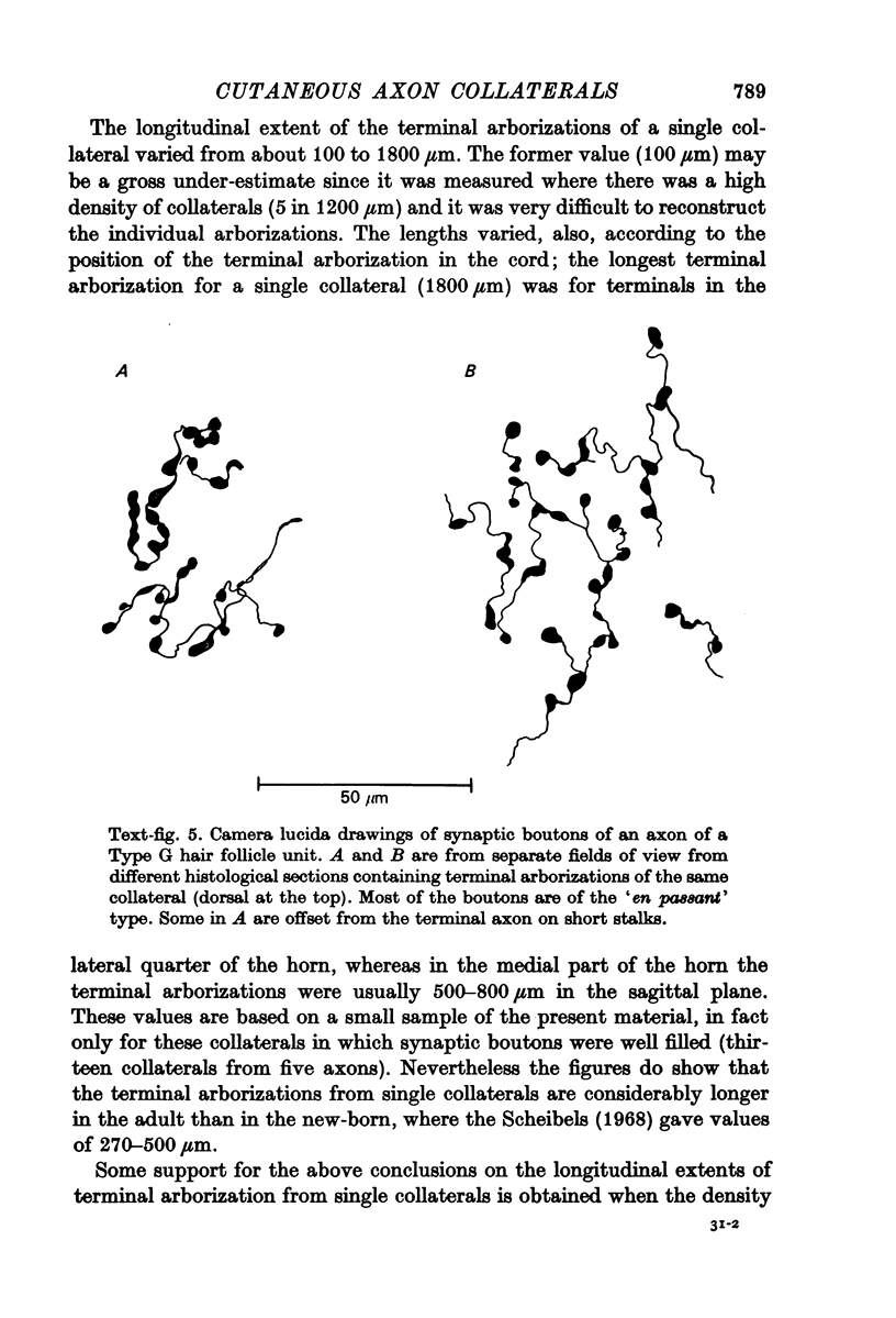
4. Sixty-three collaterals were given off the thirteen stained axons. All well-filled collaterals had a strikingly similar morphology. They descended through laminae I-III of the dorsal horn into the deeper parts of lamina IV or into lamina V, before turning and ascending back into superficial lamina IV and lamina III where they branched profusely to give rise to their terminal arborizations. Terminal boutons, most commonly of the `en passant' type, were numerous in lamina III, but were also seen in the dorsal part of lamina IV and in ventral lamina II. None were observed in dorsal lamina II or near the junction of the grey and white matter (lamina I) or in lamina V.
5. The terminal arborizations of collaterals from a single hair follicle afferent fibre were in line with one another in the longitudinal axis of the cord. In the better-stained preparations the terminal arborizations of adjacent collaterals from a single axon formed a continuous longitudinal column through the dorsal horn. There was a gradual shift of the column of arborizations from lateral to medial as the more rostral collaterals were given off.
6. The hair-follicle afferent fibre collaterals are now identified as the `collaterales grosses et profondes de la substance de Rolando' of Ramón y Cajal (1909) which give rise to the `flame-shaped arbors' of Scheibel & Scheibel (1968).
7. The importance of the longitudinal organization of the terminal arborizations for an understanding of the topographical properties of dorsal horn neurones is discussed.
Full text
PDF


Images in this article


Selected References
These references are in PubMed. This may not be the complete list of references from this article.
- Brown A. G. Cutaneous axons and sensory neurones in the spinal cord. Br Med Bull. 1977 May;33(2):109–112. doi: 10.1093/oxfordjournals.bmb.a071409. [DOI] [PubMed] [Google Scholar]
- Brown A. G., House C. R., Rose P. K., Snow P. J. The morphology of spinocervical tract neurones in the cat. J Physiol. 1976 Sep;260(3):719–738. doi: 10.1113/jphysiol.1976.sp011540. [DOI] [PMC free article] [PubMed] [Google Scholar]
- Brown A. G., Iggo A. A quantitative study of cutaneous receptors and afferent fibres in the cat and rabbit. J Physiol. 1967 Dec;193(3):707–733. doi: 10.1113/jphysiol.1967.sp008390. [DOI] [PMC free article] [PubMed] [Google Scholar]
- Brown A. G., Rose P. K., Snow P. J. The morphology of spinocervical tract neurones revealed by intracellular injection of horseradish peroxidase. J Physiol. 1977 Sep;270(3):747–764. doi: 10.1113/jphysiol.1977.sp011980. [DOI] [PMC free article] [PubMed] [Google Scholar]
- Brown A. G., Rose R. K., Snow P. J. The morphology of identified curtaeous afferent fibre collaterals in the spinal cord [proceedings]. J Physiol. 1976 Dec;263(1):132P–134P. [PubMed] [Google Scholar]
- Bryan R. N., Trevino D. L., Coulter J. D., Willis W. D. Location and somatotopic organization of the cells of origin of the spino-cervical tract. Exp Brain Res. 1973 Apr 30;17(2):177–189. doi: 10.1007/BF00235027. [DOI] [PubMed] [Google Scholar]
- Czarkowska J., Jankowska E., Sybirska E. Axonal projections of spinal interneurones excited by group I afferents in the cat, revealed by intracellular staining with horseradish peroxidase. Brain Res. 1976 Dec 10;118(1):115–118. doi: 10.1016/0006-8993(76)90844-1. [DOI] [PubMed] [Google Scholar]
- Czarkowska J., Jankowska E., Sybirska E. Diameter and internodal length of axons of spinal interneurones excited by group I afferents in the cat. Brain Res. 1976 Dec 10;118(1):119–122. doi: 10.1016/0006-8993(76)90845-3. [DOI] [PubMed] [Google Scholar]
- Graybiel A. M., Devor M. A microelectrophoretic delivery technique for use with horseradish peroxidase. Brain Res. 1974 Mar 15;68(1):167–173. doi: 10.1016/0006-8993(74)90541-1. [DOI] [PubMed] [Google Scholar]
- Iles J. F. Central terminations of muscle afferents on motoneurones in the cat spinal cord. J Physiol. 1976 Oct;262(1):91–117. doi: 10.1113/jphysiol.1976.sp011587. [DOI] [PMC free article] [PubMed] [Google Scholar]
- Jankowska E., Rastad J., Westman J. Intracellular application of horseradish peroxidase and its light and electron microscopical appearance in spinocervical tract cells. Brain Res. 1976 Apr 9;105(3):557–562. doi: 10.1016/0006-8993(76)90603-x. [DOI] [PubMed] [Google Scholar]
- Kirkwood P. A., Sears T. A. Monosynaptic excitation of motoneurones from secondary endings of muscle spindles. Nature. 1974 Nov 15;252(5480):243–244. doi: 10.1038/252243a0. [DOI] [PubMed] [Google Scholar]
- Kitai S. T., Kocsis J. D., Preston R. J., Sugimori M. Monosynaptic inputs to caudate neurons identified by intracellular injection of horseradish peroxidase. Brain Res. 1976 Jun 18;109(3):601–606. doi: 10.1016/0006-8993(76)90039-1. [DOI] [PubMed] [Google Scholar]
- McCrea R. A., Bishop G. A., Kitai S. T. Intracellular staining of Purkinje cells and their axons with horseradish peroxidase. Brain Res. 1976 Dec 10;118(1):132–136. doi: 10.1016/0006-8993(76)90847-7. [DOI] [PubMed] [Google Scholar]
- Ralston H. J., 3rd The fine structure of neurons in the dorsal horn of the cat spinal cord. J Comp Neurol. 1968 Feb;132(2):275–302. doi: 10.1002/cne.901320205. [DOI] [PubMed] [Google Scholar]
- Scheibel M. E., Scheibel A. B. Terminal axonal patterns in cat spinal cord. II. The dorsal horn. Brain Res. 1968 Jun;9(1):32–58. doi: 10.1016/0006-8993(68)90256-4. [DOI] [PubMed] [Google Scholar]
- Snow P. J., Rose P. K., Brown A. G. Tracing axons and axon collaterals of spinal neurons using intracellular injection of horseradish peroxidase. Science. 1976 Jan 23;191(4224):312–313. doi: 10.1126/science.54936. [DOI] [PubMed] [Google Scholar]
- Stauffer E. K., Watt D. G., Taylor A., Reinking R. M., Stuart D. G. Analysis of muscle receptor connections by spike-triggered averaging. 2. Spindle group II afferents. J Neurophysiol. 1976 Nov;39(6):1393–1402. doi: 10.1152/jn.1976.39.6.1393. [DOI] [PubMed] [Google Scholar]
- WALL P. D. Cord cells responding to touch, damage, and temperature of skin. J Neurophysiol. 1960 Mar;23:197–210. doi: 10.1152/jn.1960.23.2.197. [DOI] [PubMed] [Google Scholar]
- Wall P. D., Werman R. The physiology and anatomy of long ranging afferent fibres within the spinal cord. J Physiol. 1976 Feb;255(2):321–334. doi: 10.1113/jphysiol.1976.sp011282. [DOI] [PMC free article] [PubMed] [Google Scholar]