Abstract
The soil bacterium Rhizobium lupini H13-3 has complex right-handed flagellar filaments with unusual ridged, grooved surfaces. Clockwise (CW) rotation propels the cells forward, and course changes (tumbling) result from changes in filament speed instead of the more common change in direction of rotation. In view of these novelties, fluorescence labeling was used to analyze the behavior of single flagellar filaments during swimming and tumbling, leading to a model for directional changes in R. lupini. Also, flagellar filaments were investigated for helical conformational changes, which have not been previously shown for complex filaments. During full-speed CW rotation, the flagellar filaments form a propulsive bundle that pushes the cell on a straight path. Tumbling is caused by asynchronous deceleration and stops of individual filaments, resulting in dissociation of the propulsive bundle. R. lupini tumbles were not accompanied by helical conformational changes as are tumbles in other organisms including enteric bacteria. However, when pH was experimentally changed, four different polymorphic forms were observed. At a physiological pH of 7, normal flagellar helices were characterized by a pitch angle of 30°, a pitch of 1.36 μm, and a helical diameter of 0.50 μm. As pH increased from 9 to 11, the helices transformed from normal to semicoiled to straight. As pH decreased from 5 to 3, the helices transformed from normal to curly to straight. Transient conformational changes were also noted at high viscosity, suggesting that the R. lupini flagellar filament may adapt to high loads in viscous environments (soil) by assuming hydrodynamically favorable conformations.
Motile bacteria respond to environmental changes by biasing their overall movement in a favorable direction. They can move on solid surfaces as performed in swarming, gliding, or twitching motility, or they can swim in liquid (19). Free-swimming bacteria are propelled by helical flagellar filaments connected to the basal body by a flexible hook and driven by a flagellar rotary motor (for reviews see references 1, 16, 20, and 30). There are two types of flagellar filaments, plain and complex. These have been distinguished by their physical properties and their appearance by electron microscopy. The plain flagellar filaments of organisms such as Escherichia coli, Salmonella spp., and Rhodobacter sphaeroides have a smooth surface structure, whereas the complex filaments of soil bacteria, such as Rhizobium lupini H13-3 and Sinorhizobium meliloti, exhibit a prominent helical surface pattern of alternating ridges and grooves (14, 18, 25, 28). Plain filaments can assume distinct polymorphic forms with different helical patterns (5, 15). Enterobacteria with plain filaments switch flagellar rotation from counterclockwise (CCW) to clockwise (CW), which induces a helical transition from left-handedness to right-handedness. A cell tumbles when at least one filament switches from CCW to CW rotation. Concomitantly, the right-handed helix transforms to semicoiled and curly (11, 17, 29). The photosynthetic bacterium R. sphaeroides has a single plain filament that rotates only in the CW mode, and these cells change direction by stopping and restarting rotation of the filament (2), which exists in three distinct polymorphic forms: a helical structure while rotating, a straight form during fast rotation, and a coiled structure when at rest (3).
Whereas the E. coli filament consists of a single type of flagellin, the complex R. lupini filament consists of three related flagellin subunits that are assembled as functional heterodimers (23). These filaments are more stable and do not switch handedness due to interflagellin bonds that lock the filament in a right-handed helical conformation (7). They are also suitable for propulsion in viscous media, which is a necessary adaptation for soil bacteria (9). Complex filaments are able to rotate only in the CW direction (10). Directional changes of swimming S. meliloti cells were shown previously to be a consequence of variable rotary speeds of individual flagella and of differences in rotary speed among the four to six flagella of a single cell (21, 24). No structural polymorphism has previously been observed related to the modulation of flagellar rotary speed in complex filaments.
In an effort to understand the role of individual complex filaments in controlling swimming behavior, the technique of fluorescence labeling developed by Turner et al. (29) was used to visualize swimming cells of R. lupini and their flagellar filaments in real time. This work also provided the first evidence of polymorphic transitions in complex filaments artificially induced by variations of pH and by rotation at high viscosity.
MATERIALS AND METHODS
Fluorescence labeling of cells and filaments.
R. lupini H13-3 strains RU12/001 (a spontaneous streptomycin-resistant derivative of wild-type H13-3), RU12/003 (ΔflaB), and RU12/004 (ΔflaD) (23) were grown in TYC (0.5% tryptone, 0.3% yeast extract, 0.13% CaCl2 · 6H2O [pH 7.0])-streptomycin (600 mg/liter) at 30°C for 2 days. Motile cells were obtained by diluting cells from a stationarily grown TYC culture in 10 ml of RB (9) to an optical density at 600 nm of 0.05, layering them on Bromfield agar plates (27), and incubating them at room temperature for 16 to 20 h to an optical density at 600 nm of approximately 0.2 to 0.5. Bacterial cells from two plates (15 ml of culture) were washed three times at room temperature and were concentrated on a 0.45-μm-pore-size HV Durapore filter to about 1 ml. Cells were gently resuspended in 10 ml of labeling buffer (10 mM KPi [pH 8.0], 1 mM NaCl, 0.4 mM CaCl2, 25 μM EDTA, 0.025% [wt/vol] Tween 20). The detergent was added to prevent cells from sticking to the filter and to each other. The final suspension (0.3 ml) contained bacteria at about 50-fold their original concentration.
One vial of Cy3 monofunctional succinimidyl ester (Amersham Pharmacia) was dissolved in 100 μl of labeling buffer and added to the bacterial suspension. Sodium bicarbonate (25 μl [1 M]) was added to maintain a pH of 8.0. The suspension was incubated at room temperature on a shaker platform (100 rpm) for 75 to 90 min. Bacteria were washed free of dye by filtration as described above and were resuspended in motility buffer (10 mM KPi [pH 7.1], 1 mM NaCl, 1 mM CaCl2, 25 μM EDTA, 0.05% [wt/vol] Tween 20).
Immobilization of filaments.
Five microliters of anticyanine monoclonal antibody (1:200; Rockland, Gilbertsville, Pa.) was spread on a coverslip cleaned with KOH-saturated 95% ethanol. Equal volumes of cells and 1% glutaraldehyde were mixed and incubated for 2 min before 10 μl of the mixture was added to the antibody solution on the coverslip. Cells were allowed to settle for 1 h in a humidity chamber before image acquisition.
Observation of filament polymorphisms.
Buffers between pH 2 and 5 were prepared with 0.2 M citric acid and 0.2 M Na2HPO4, and buffers between pH 8 and 11 were prepared with 0.2 M Tris and 0.2 M glycine essentially as described previously (26) to yield final concentrations of 100 mM buffer either without NaCl or with 1 M NaCl. Equal volumes of buffer and cells were mixed and incubated for 5 min before image acquisition. A 20% (wt/vol) Ficoll 400 (Sigma Chemical Co., St. Louis, Mo.) stock solution in motility buffer was mixed with the cell suspension at a ratio of 19:1, and rotating filaments were observed as described below.
Tethering of cells.
A glass coverslip was supported over a glass slide at its edges by two other coverslips greased with Apiezon L (Fisher). Two milliliters of motile cells was sheared 20 times (10), washed once with motility buffer, and resuspended in 200 μl of motility buffer. Twenty microliters of the cell suspension was mixed with 2 μl of 0.5 M mannitol and 1 μl of purified antiflagellin polyclonal antibody (1:50) (23). Cells were added to the space between the coverslip and the slide. The slide was inverted, incubated for 30 min in a humidity chamber, and rinsed with several volumes of motility buffer.
Image acquisition and analysis.
Cells and filaments were observed with a fluorescence microscope illuminated with an argon-ion laser with the setup of Turner et al. (29). Speeds of swimming cells were determined from the distance traveled per video field, and deflections of the trajectory generated by a tumble were noted.
RESULTS
Fluorescence labeling.
Flagellins are three-domain proteins, with the N terminus being required for export, both the N- and C-terminal domains being involved in filament assembly, and the central domain comprising portions exposed at the surface of the filaments (22). The outermost subdomain of the flagellin FliC from E. coli contains at least three lysine residues, which are sufficient to brightly label flagellar filaments with amino-specific dyes, such as Alexa Fluor, containing succinimidyl ester-reactive groups (29). While the plain filaments from enterobacteria consist of one type of flagellin monomers, the complex filaments of R. lupini H13-3 are composed of three related flagellin subunits (23). The central domain of these flagellin subunits contains 12 basic amino acid residues with primary amino groups, which allowed bright labeling with the orange-fluorescing cyanine Cy3, a monofunctional succinimidyl ester specific for free amino groups, in a procedure adapted from the work of Turner et al. (29). Fluorescently labeled cells were highly motile for several hours and retained motility even after storage at room temperature overnight. Attempts to label flagellar filaments of the closely related soil bacteria S. meliloti and Agrobacterium tumefaciens were unsuccessful, a difficulty perhaps related to the inaccessibility of primary amino groups.
Observation of swimming cells.
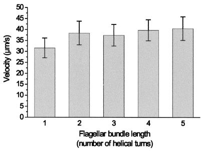
To confirm that the fluorescent dye does not impair swimming proficiency, the swimming speeds of 158 labeled R. lupini cells were determined from frame to frame with the ruler tool provided by the Scion Image program. The average instant velocity was 37.9 ± 5.4 μm/s, and this value is consistent with the 37.3 ± 3.4 μm/s determined by computerized motion analysis with a Hobson Bactracker (23). Therefore, the flagellar filaments of R. lupini tolerate the chemical modification without reduction in function. Since complex flagellar filaments are rather fragile (14, 28), it was important to see whether flagellar length has an influence on swimming speed. Flagellar bundle lengths of the 158 cells were determined as before. The histogram shown in Fig. 1 correlates five classes of flagellar bundle lengths to swimming velocity. As seen, no significant differences in speed were observed among cells possessing flagellar filaments of two to five helical turns in length. However, cells possessing flagellar filaments of one helical turn in length swam 19% slower than others, suggesting that a very short filament length does affect swimming speed.
FIG. 1.
Swimming velocity of R. lupini as a function of the flagellar filament length. The filament length was measured as the number of helical turns. Swimming speeds were determined from frame to frame with the ruler tool provided by the Scion Image program. A total of 158 cells were analyzed: 16 cells with one helical turn, 47 with two helical turns, 48 with three helical turns, 35 with four helical turns, and 12 with five helical turns. Error bars depict standard deviations of the means.
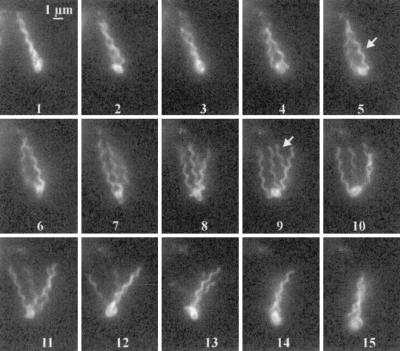
Swimming R. lupini cells alternate between straight runs and brief tumbles. When the flagellar bundle falls apart (causing a tumble), the cell body orients in a new direction, whereupon the flagellar bundle re-forms, and the cell swims straight in the new direction. This pattern is reminiscent of swimming E. coli bacteria that switch the flagellar rotation from CCW to CW (17, 29). However, given that R. lupini flagella rotate exclusively in the CW mode and that tumbling is brought about by differential modulation of the rotary speed of single flagella, it was of great interest to see how single flagellar filaments of a swimming cell behave. Figure 2 shows a typical swimming pattern of an R. lupini cell with four flagellar filaments in successive fields over a time span of 0.25 s. All filaments form a bundle, and the cell body is propelled forward along a straight path (field 1) until the bundle falls apart in a procession from proximal to distal, yielding two filament pairs that have completely separated (fields 2 to 5). The pairs fall apart into four individual filaments (fields 7 to 11); subsequently, single filaments rejoin to form a bundle (fields 12 to 15) that drives the cell into a new direction.
FIG. 2.
A series of 15 successive frames at 60 Hz (deinterlaced, total time span of 0.25 s) showing an R. lupini cell with four flagellar filaments. The bundle disintegrates (frames 2 to 5) when a single flagellar filament slows down (in the focal plane and marked by an arrow in frame 5), and it falls apart (frames 7 to 11) when one flagellar filament comes to a stop (in the focal plane and marked by an arrow in frame 9), thus causing a reorientation of the swimming cell. A flagellar bundle is re-formed (frames 12 to 15) when all flagellar filaments resume the same maximum rotary speed. The cell was labeled with Cy3 and illuminated by an argon-ion laser with a strobe (the technique used for all subsequent figures).
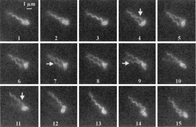
What causes the flagellar bundle to fall apart? The filaments of R. lupini are locked in right-handed helicity by interflagellin bonds (28), and also, the flagellar motor never reverses its CW sense of rotation. It is most likely that, as the rotary speed of individual flagella decreases at different rates, the flagella of a bundle will jam unless they separate into single filaments. The method used to analyze flagellar motion distinguishes only extremely decelerated filaments (motionless or stopped), which are difficult to observe in a cell with several filaments. Therefore, cells with only one or two filaments were analyzed in the following. Figure 3 shows an analysis of the filament movement and swimming behavior of a biflagellated cell. Both filaments rotate together in a bundle (fields 1 to 3). The cell body is stopped in its forward movement (field 4), and the two filaments come apart from proximal to distal until they are completely separated (field 6). The first filament stops rotation (arrow in field 4), and then the second filament stops (arrow in field 7). Both filaments resumed rotation immediately thereafter (arrows, fields 9 and 11). The bundle re-forms, and the cell body starts a forward movement in a different direction than before (fields 12 to 15). No difference in filament shape between rotating and stopping flagella has been observed (Table 1).
FIG. 3.
A series of 15 successive frames at 60 Hz (total time span of 0.25 s) showing an R. lupini cell with two flagellar filaments swimming and tumbling. The bundle disintegrates (frames 4 to 9) when one flagellar filament (marked by an arrow in frame 4) slows down and comes to a stop, followed by the halting of the second filament (marked by an arrow in frame 7). The second filament resumes rotation (marked by an arrow in frame 9) followed by the first filament (marked by an arrow in frame 11). A bundle is re-formed, when both filaments rotate at the same speed, and the cell moves on in a new direction (frames 12 to 15). Criteria for an active filament are (i) its displacement in successive frames and (ii) an occasionally blurred image, slightly out of the focal plane.
TABLE 1.
Geometrical parameters of flagellar filaments attached to wild-type and two mutant strains of R. lupinia
| Strain | Pitch angle (°) | Pitch (μm) | Diam (μm) | nb |
|---|---|---|---|---|
| Wild type (swimming)c | 30.7 ± 1.2 | 1.36 ± 0.03 | 0.50 ± 0.03 | 104 |
| Wild type (immobilized) | 29.9 ± 1.6 | 1.36 ± 0.04 | 0.50 ± 0.02 | 111 |
| ΔflaB strain (immobilized) | 27.6 ± 2.4 | 1.36 ± 0.04 | 0.49 ± 0.03 | 51 |
| ΔflaD strain (immobilized) | 27.9 ± 2.7 | 1.36 ± 0.05 | 0.50 ± 0.04 | 54 |
Images were analyzed with Scion Image Beta 4.02 (29).
Number of flagellar filaments analyzed.
Assessed during a tumble.
The criteria for an active filament are (i) its displacement in successive frames and (ii) an occasionally blurred image slightly out of the focal plane. By contrast, a still filament can be clearly visualized for the entire time that it is positioned in the focal plane. Intermittent stops, as the ultimate slowing down of flagellar motor rotation, lead to the observed changes in swimming direction. A single “pausing” filament(s) causes the bundle to come apart. Consequently, a brief tumble of the cell occurs, accompanied with a reorientation of the cell body. When the pausing filament(s) starts rotating again, the bundle consolidates, and the cell moves in the new direction.
Unidirectional CW rotation and rotary speed modulation have been demonstrated previously for S. meliloti (10, 27), a close relative of R. lupini H13-3 (23). In this study, the mode of rotation of the R. lupini flagellar motor has been directly assessed by the tethered cell assay. Four cells tethered by a single flagellum were observed for 2 min at 60 Hz. The flagellar motors rotated solely in the CW mode and never reversed their sense of rotation but slowed down to brief intermittent stops of 0.09 ± 0.02 s every 0.97 ± 0.6 s (data not shown). In a swimming cell of R. lupini, these brief rotational stops of individual flagella (as the ultimate of slowing down) force the bundle to fall apart, leading to a directional change.
Determinants of swimming direction.
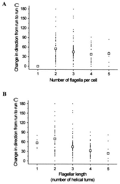
What determines the degree of directional changes after a tumble? Three parameters were analyzed for their impact on the swimming pattern: the flagellar number per cell, the flagellar filament length, and the duration of a tumble. The degree of directional change was analyzed by using the angle tool provided by the Scion Image program, and it is shown as a function of flagellar number per cell (averaging about 2.9 ± 0.8) in Fig. 4A. In 160 cells tested, the average angle of reorientation was 46 ± 39°. Interestingly, reorientation of cells with two and three filaments ranged between 0 and 180°, whereas it was reduced in cells with four and five filaments to between 0 and 90°. Hence, a higher number of filaments per cell narrows the degree of directional change. This result is consistent with the following working model (see also references 12 and 29): a single uncoordinated filament has a smaller impact on the integrity of a bundle of four than on a bundle of two filaments. In this model, cells with one flagellar filament cannot change direction, a result consistent with experimental observations (Fig. 4). Pausing of flagellar filament rotation on a cell with a single filament results in an immediate halt of the cell body due to low mass and lack of inertia. When the pausing filament starts rotating again, the single filament drives the cell in the same direction as before.
FIG. 4.
Change in direction from run to run plotted as a function of total number of flagellar filaments per cell (A) and as a function of flagellar length measured as the number of helical turns (B). A total of 160 cells were analyzed. Each point represents an individual cell. Open squares, mean directional change.
Next, the influence of flagellar length on directional change was analyzed. Filament lengths were determined for 160 swimming cells undergoing a tumble reaction. The degree of directional changes (determined as before) was plotted as a function of filament lengths in Fig. 4B (average number of helical turns, 3.1. ± 0.9). Maximum changes of direction were observed when cells had filaments of between two and three helical turns (0 to 180° directional change), whereas cells with longer filaments (four and five helical turns) exhibited directional changes from 0 to 90°. Due to mechanical reasons, bundles formed by longer filaments take a longer time to separate, resulting in a smaller directional change of the cell. Cells with very short filaments (one helical turn) exhibit small changes (0 to 80°). Very short filaments are apparently less capable than longer filaments of pushing the cell body in a new direction.
Finally, the influence of tumble duration on directional change was analyzed. In 160 cells tested, the duration of a tumble varied from 0.02 s (equaling one frame) to 0.87 s with an average of 0.21 ± 0.12 s. No causal relationship between the duration of reorientation and deviation of the swimming path was observed (data not shown).
Thus, the swimming behavior of an R. lupini cell can be described as follows: when all flagellar filaments turn at the same speed, they form a bundle that propels the cell on a straight path. When single filaments slow down or stop, the bundle comes apart in a progression from proximal to distal, which leads to a short tumble. The cell reorients to a degree dependent on filament number and length. When the pausing filament resumes rotation and joins with the other flagella to turn at the same speed, the bundle consolidates and the cell is driven in a new direction.
Flagellar filament shape.
Unlike plain flagella from enteric bacteria (29), no polymorphic transitions between different helical conformations have been observed in complex filaments. In an attempt to determine whether the complex flagella of R. lupini ever undergo conformational changes, I first analyzed the normal geometrical parameters of these filaments and then exposed the bacteria to changes in pH and ion concentration that have been previously shown to affect flagellar helical stability (13). Because single flagellar filaments are concealed in a bundle, cells were deenergized and their filaments were immobilized on the coverslip (with anti-Cy3 monoclonal antibody). These stationary filaments were all expected to have a normal waveform (29). However, if waveform transitions are required for tumbling, I would expect to see a filament change in cells caught midtumble. For higher accuracy, images of immobilized filaments were averaged digitally over 30 video frames (60 fields). These data are listed in Table 1. Both data sets result in the same normal waveform, indicating that waveform transition is not required for tumbling. The sole conformation was characterized by an average pitch angle of 30°, a pitch of 1.36 μm, and a helical diameter of 0.50 μm.
Alterations of the helical waveform may also be caused by genetic changes in the flagellin composition of the complex R. lupini filaments. Of the three flagellin monomers, FlaA, FlaB, and FlaD, the secondary species FlaB and FlaD are not absolutely needed for motility, although flaB and flaD knockout mutants exhibit a reduction of motility by 10 and 25%, respectively (23). When assessed as described above, the geometrical parameters of these mutant filaments were found to be very similar to those of the wild type (Table 1). Therefore, unlike plain filaments in bacteria such as E. coli, the right-handed complex filaments of R. lupini do not undergo conformational changes during tumbles, and it is solely the difference in rotary speed that drives the filaments of a bundle apart. However, this does not exclude a conformational change under conditions different from those found in a neutral aqueous environment.
Polymorphic transitions induced by changes in pH and ionic strength.
Changes in pH or ion concentration are known to affect the structure of E. coli (13) and R. sphaeroides (26) flagellar filaments, so I asked whether changes in these conditions also induce conformational alterations in the R. lupini flagellar filament. These conditions may represent an unnatural environment for soil bacteria but may reveal properties of the helical polymer.
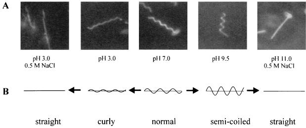
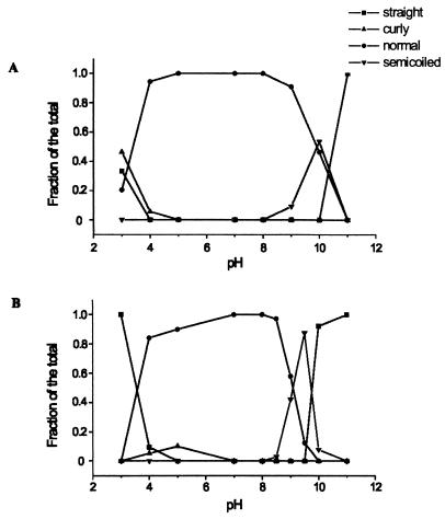
Accordingly, R. lupini flagellar filaments were examined at various pH values and salt concentrations. Fluorescently labeled flagellated cells were incubated for 5 min in appropriately adjusted buffer before images were taken. The following four forms of the right-handed R. lupini filament were identified at various pH values and ion concentrations: normal, semicoiled, curly, and straight. The helical parameters of each are listed in Table 2. Examples are illustrated and the filament contours are drawn schematically in Fig. 5B. It should be noted that the R. lupini waveforms are defined by their morphological appearance and are not related to the specifications of the E. coli filament forms. The normal waveform has a pitch angle of 28°, a pitch of 1.35 μm, and a diameter of 0.50 μm (in close agreement with values listed in Table 1). The semicoiled waveform observed at pH 8 to 10 had twice the pitch angle, a pitch decreased by about 20%, and a diameter increased by about 50%. The curly waveform was induced at pH 3 to 4 and had a pitch angle decreased by about 50%, a slightly reduced pitch, and only half the normal diameter. At extreme pH values, pH 3 and 11, respectively, straight filaments formed. When acidity was further increased to pH 2, filaments disintegrated and eventually depolymerized (data not shown). The frequencies at which the different waveforms appeared in the pH range from 2 to 11 are plotted in Fig. 6A. These experiments showed that an R. lupini complex flagellar filament will transform sequentially from normal to curly to straight when shifted from neutral to low pH and from normal to semicoiled to straight when shifted from neutral to high pH. The same transitions were observed in the presence of 0.5 M NaCl, which enhanced the pH effect (Fig. 6B).
TABLE 2.
Geometrical parameters of the four polymorphic waveforms of R. lupini filaments observed at different pH values (pH 3 to 11) and in the presence of 0.5 M NaCl as shown in Fig. 6 a
| Waveform | Pitch angle (°) | Pitch (μm) | Diam (μm) | nb |
|---|---|---|---|---|
| Normal | 28.0 ± 1.9 | 1.35 ± 0.03 | 0.50 ± 0.03 | 351 |
| Semicoiled | 51.8 ± 3.4 | 1.11 ± 0.05 | 0.77 ± 0.04 | 137 |
| Curly | 16.7 ± 1.8 | 1.27 ± 0.06 | 0.27 ± 0.04 | 36 |
| Straight | 0 | ∞ | 0 |
Images were analyzed with Scion Image Beta 4.02 (29).
Number of flagellar filaments analyzed.
FIG. 5.
Polymorphic forms of R. lupini flagellar filaments induced by changes of pH and ion concentration. (A) Examples of each conformation are shown that were optimally observed at the conditions indicated below. (B) The graphical representations of four different waveforms were drawn with the parameters of Table 2, each with a contour length of 8 μm. Arrows indicate the directions of conformational transition.
FIG. 6.
Dependence of polymorphic waveforms on pH and salt concentration. Percentages of a waveform observed at a given pH were averaged (53 individual data on average). Data were collected in the absence of NaCl (A) or in the presence of 0.5 M NaCl (B) on cells incubated in buffers of given pH.
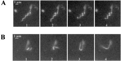
In rare cases, it was possible to observe the transition from one form to another. Such an event is recorded in Fig. 7A, where a transition from normal to semicoiled was captured in a time span of 1.48 s. A cell paralyzed by a high pH and salt concentration exhibits one filament with three normal helical turns. The first, third, and second turns are transformed from normal to semicoiled, respectively (fields 2 to 4; the third helical turn is slightly out of the focal plane). Transitions do not follow an ordered pattern along the filament but may start at any site and then expand in both directions. A transition from semicoiled to straight was recorded within 0.28 s, as shown in Fig. 7B. The semicoiled filament unwinds into a hairpin-like structure (field 3), before it straightens completely (field 4 and the following [data not shown]). In all observed cases, the filament straightens similarly to the uncoiling of a twisted elastic band, with the intermediate formation of a hairpin structure. The observed changes of pitch angle, pitch, and diameter may be caused by the interference of pH (and salt) with weak inter- and intrasubunit bonds. However, a change of handedness was never observed.
FIG. 7.
Time-lapse exposures of two polymorphic transitions. (A) Transition from normal to semicoiled at pH 9.5 and 0.5 M NaCl recorded at 0.5-s intervals. (B) Transition from semicoiled to straight at pH 10 and 0.5 M NaCl recorded at 0.1-s intervals.
Polymorphic transitions of rotating filaments induced by viscosity.
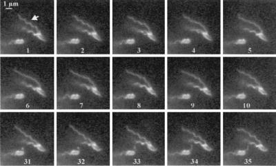
In E. coli flagella, polymorphic transitions have been induced not only by switching the senses of rotation but also by viscous media (8, 13), leading me to conjecture that artificially induced transitions of wave morphology might also be observed in filaments rotating on a cell. Accordingly, the viscosity of the medium was increased by adding Ficoll 400, and the filaments were analyzed. At a final concentration of 19% Ficoll, corresponding to a viscosity of about 20 cP (as extrapolated from Fig. 3 of reference 6), all cells stuck to the glass of the coverslip, but their flagellar filaments were still rotating. Under these conditions of high load, I observed polymorphic transitions in rotating complex R. lupini flagella. A typical example is shown in Fig. 8. The rotating filament in focus undergoes a transition from normal to curly that proceeds from proximal to distal (fields 1 to 6). This curly conformation is then reversed to normal, a transition that again proceeds from proximal to distal (fields 7 to 35). Also, transitions from normal to straight to curly and back to normal have been observed (data not shown). The propagation of a conformational transition is expected to occur from proximal to distal when torsionally induced. The ability of a complex filament to react to high viscous drag by a change of conformation reveals a higher stability needed under extreme conditions, such as high viscosity in the soil habitat.
FIG. 8.
Polymorphic transitions of a complex flagellar filament rotating at high viscosity. The figure shows an R. lupini cell with two flagella rotating in 19% Ficoll 400, stuck to the coverslip. One filament (arrow) undergoes transitions from normal (field 1) to curly (field 6) and back to normal (field 32). Successive fields are shown at 60 Hz, and the total time span was 0.60 s. No changes were observed between fields 10 and 31.
DISCUSSION
The complex flagella of R. lupini rotate solely in the CW sense without reversals, but they vary their rotary speed as a means to direct changes in the swimming path. Here, I have used fluorescence labeling to visualize single rotating filaments of R. lupini to study their behavior during translational motion and tumbles of single cells. This technique permitted the recording of individual swimming cells in real time at a resolution of 60 Hz with a fluorescence microscope with laser illumination with a strobe and equipped with a charge-coupled device camera. Until now, their motion had been monitored only by differential interference contrast microscopy, which requires that filaments be observed close to a glass surface or indirectly by a tethering assay. Therefore, the use of this labeling technique opens new perspectives for the analysis of complex flagella. When all filaments rotated at high speed, they formed a bundle that drove the swimming cell on a straight track. Tumbles and change of swimming direction were initiated when one or more flagellar filaments slowed down or came to a stop and the bundle fell apart. The model of swimming R. lupini in Fig. 9 summarizes the results obtained in this study as well as those of previously published observations (24, 27). No change of the normal helical conformation (pitch angle, 30°; pitch, 1.36 μm; diameter, 0.5 μm) was observed under these swimming conditions. However, different conformations of the complex flagellar filaments appeared at high and low pH and when rotating at high viscosity. Under these conditions, normal, semicoiled, curly, and straight waveforms were observed and may play important roles in vivo.
FIG. 9.
Flagellar rotation and swimming patterns of R. lupini. Decreasing and increasing the rotary speed of individual flagella directs the swimming pattern of R. lupini. (1) Full-speed CW rotation causes the right-handed helical filaments to form a bundle that drives the cell forward. (2) When the CW rotation of individual flagella declines at different rates or comes to a transient stop, the flagellar bundle flies apart and the cell tumbles and assumes a new direction. (3) The coordinated increase of rotary speed of all flagella induces bundle formation and (4) straight translatory swimming movement.
In an effort to shed new light on the behavior of complex filaments, I examined swimming and tumbling cells for flagellar movement. During a tumble of a swimming R. lupini cell, individual flagellar filaments were observed to come to a complete stop. The deflection of the cell body during a tumble is dependent on the number and length of the flagellar filaments possessed by the cell. The longer the filaments or the more flagella per cell, the smaller the deflection (Fig. 4). The influence of a factor like Brownian motion is negligible, because it is assumed to be a slow and inefficient mechanism for redirecting the swimming path in water and even less efficient in the relatively viscous soil environment given the short average time for a stop.
Polymorphic transitions of the helical filament do not need to occur during changes in swimming speed in R. lupini, although they can be induced artificially by pH and viscosity. The complex R. lupini filament can exist in four distinct forms, which are defined as normal, semicoiled, curly, and straight, very similar to the E. coli filament waveforms discovered by Asakura (4). (The existence of more than one curly form, as described for E. coli, cannot be excluded, but resolution of the low-amplitude helix was hampered by the fairly low fluorescence intensity of the dye at low pH.)
It may be noteworthy that a 1- to 2-μm straight portion of the filaments of swimming cells at the proximal end was occasionally observed (similar to Fig. 7A). Its significance is unclear, but its appearance might be caused by the mechanical forces applied during the labeling and washing procedure. In E. coli, transitions from normal to semicoiled to curly occur when the flagellar motor switches from CCW to CW rotation (17, 29). The CW rotation of the left-handed filaments places them under torsional load and initiates polymorphic transitions to right-handed helical waveforms as the key to tumbling motion (29). Variances in coiling can be produced by two different packing interactions of flagellin called L and R (22). In contrast, the complex filament of R. lupini is locked in its right-handed state (7). Helical perturbations in the structural organization of complex filaments imply a high stability suitable for propulsion in viscous media. While the inner tubes of the concentric structure are nearly indistinguishable from the enterobacterial plain filament, the external layer mainly contributes to the stable structure of the complex filament in addition to strong intersubunit connectivity. Upon pH changes, release and re-forming of intra and/or intermonomer bonds due to changes in the protonation state might occur in this region, leading to changes in waveform but not in handedness.
In addition to the irreversible changes of waveforms induced by pH, reversible changes of rotating complex filaments are induced under conditions of high viscous load. Rotating R. lupini flagellar filaments transformed from their normal into curly and straight waveforms and transformed back to normal. The transformations propagated from the base to the tip, as expected when torsionally driven. It is difficult to speculate on the biological significance of these transitions. The complex filaments yield to a high load in a viscous environment by assuming a curly or even straight shape. This still permits rotation, though less efficient propulsion of the cell, whereas a filament with the normal helical waveform would come to a stop because of high frictional forces.
The soil bacterium R. lupini has evolved a stable, unidirectional rotating flagellar filament, optimally adapted to the viscous environment. In conjunction, R. lupini had developed a novel swimming mode composed of straight-swimming periods—when all filaments rotate at the same maximum speed in a bundle—and tumbles—when single filaments change their rotational speed and the bundle comes apart. These two different swimming modes are the basis for an optimal chemotactic response toward environmental stimuli.
Acknowledgments
I am grateful to Howard Berg and Linda Turner for valuable discussions and for their hospitality.
This work was supported by grant Scha914/1-1 from the Deutsche Forschungsgemeinschaft and by the Rowland Institute for Science.
REFERENCES
- 1.Aizawa, S. I., C. S. Harwood, and R. J. Kadner. 2000. Signaling components in bacterial locomotion and sensory reception. J. Bacteriol. 182:1459-1471. [DOI] [PMC free article] [PubMed] [Google Scholar]
- 2.Armitage, J. P., and R. M. Macnab. 1987. Unidirectional, intermittent rotation of the flagellum of Rhodobacter sphaeroides. J. Bacteriol. 169:514-518. [DOI] [PMC free article] [PubMed] [Google Scholar]
- 3.Armitage, J. P., T. P. Pitta, M. A. Vigeant, H. L. Packer, and R. M. Ford. 1999. Transformations in flagellar structure of Rhodobacter sphaeroides and possible relationship to changes in swimming speed. J. Bacteriol. 181:4825-4833. [DOI] [PMC free article] [PubMed] [Google Scholar]
- 4.Asakura, S. 1970. Polymorphism of bacterial flagella. Tanpakushitsu Kakusan Koso 15:1372-1381. [PubMed] [Google Scholar]
- 5.Calladine, C. R. 1978. Change in waveform in bacterial flagella: the role of mechanics at the molecular level. J. Mol. Biol. 118:457-479. [Google Scholar]
- 6.Chen, X., and H. C. Berg. 2000. Torque-speed relationship of the flagellar rotary motor of Escherichia coli. Biophys. J. 78:1036-1041. [DOI] [PMC free article] [PubMed] [Google Scholar]
- 7.Cohen-Krausz, S., and S. Trachtenberg. 1998. Helical perturbations of the flagellar filament: Rhizobium lupini H13-3 at 13 Å resolution. J. Struct. Biol. 122:267-282. [DOI] [PubMed] [Google Scholar]
- 8.Fahrner, K. A., S. M. Block, S. Krishnaswamy, J. S. Parkinson, and H. C. Berg. 1994. A mutant hook-associated protein (HAP3) facilitates torsionally induced transformations of the flagellar filament of Escherichia coli. J. Mol. Biol. 238:173-186. [DOI] [PubMed] [Google Scholar]
- 9.Götz, R., N. Limmer, K. Ober, and R. Schmitt. 1982. Motility and chemotaxis in two strains of Rhizobium with complex flagella. J. Gen. Microbiol. 128:789-798. [Google Scholar]
- 10.Götz, R., and R. Schmitt. 1987. Rhizobium meliloti swims by unidirectional, intermittent rotation of right-handed flagellar helices. J. Bacteriol. 169:3146-3150. [DOI] [PMC free article] [PubMed] [Google Scholar]
- 11.Hotani, H. 1982. Micro-video study of moving bacterial flagellar filaments. III. Cyclic transformation induced by mechanical force. J. Mol. Biol. 156:791-806. [DOI] [PubMed] [Google Scholar]
- 12.Ishihara, A., J. E. Segall, S. M. Block, and H. C. Berg. 1983. Coordination of flagella on filamentous cells of Escherichia coli. J. Bacteriol. 155:228-237. [DOI] [PMC free article] [PubMed] [Google Scholar]
- 13.Kamiya, R., H. Hotani, and S. Asakura. 1982. Polymorphic transition in bacterial flagella. Symp. Soc. Exp. Biol. 35:53-76. [PubMed] [Google Scholar]
- 14.Krupski, G., R. Götz, K. Ober, E. Pleier, and R. Schmitt. 1985. Structure of complex flagellar filaments in Rhizobium meliloti. J. Bacteriol. 162:361-366. [DOI] [PMC free article] [PubMed] [Google Scholar]
- 15.Kuwajima, G., J. Asaka, T. Fujiwara, T. Fujiwara, K. Node, and E. Kondo. 1986. Nucleotide sequence of the hag gene encoding flagellin of Escherichia coli. J. Bacteriol. 168:1479-1483. [DOI] [PMC free article] [PubMed] [Google Scholar]
- 16.Macnab, R. M. 1996. Flagella and motility, p. 123-145. In F. C. Neidhardt, R. Curtiss III, J. L. Ingraham, E. C. C. Lin, K. B. Low, B. Magasanik, W. S. Reznikoff, M. Riley, M. Schaechter, and H. E. Umbarger (ed.), Escherichia coli and Salmonella: cellular and molecular biology, 2nd ed., vol. 1. ASM Press, Washington, D.C.
- 17.Macnab, R. M., and M. K. Ornston. 1977. Normal-to-curly flagellar transitions and their role in bacterial tumbling. Stabilization of an alternative quaternary structure by mechanical force. J. Mol. Biol. 112:1-30. [DOI] [PubMed] [Google Scholar]
- 18.Maruyama, M., G. Lodderstaedt, and R. Schmitt. 1978. Purification and biochemical properties of complex flagella isolated from Rhizobium lupini H13-3. Biochim. Biophys. Acta 535:110-124. [DOI] [PubMed] [Google Scholar]
- 19.McBride, M. J. 2001. Bacterial gliding motility: multiple mechanisms for cell movement over surfaces. Annu. Rev. Microbiol. 55:49-75. [DOI] [PubMed] [Google Scholar]
- 20.Namba, K., and F. Vonderviszt. 1997. Molecular architecture of bacterial flagellum. Q. Rev. Biophys. 30:1-65. [DOI] [PubMed] [Google Scholar]
- 21.Platzer, J., W. Sterr, M. Hausmann, and R. Schmitt. 1997. Three genes of a motility operon and their role in flagellar rotary speed variation in Rhizobium meliloti. J. Bacteriol. 179:6391-6399. [DOI] [PMC free article] [PubMed] [Google Scholar]
- 22.Samatey, F. A., K. Imada, S. Nagashima, F. Vonderviszt, T. Kumasaka, M. Yamamoto, and K. Namba. 2001. Structure of the bacterial flagellar protofilament and implications for a switch for supercoiling. Nature 410:331-337. [DOI] [PubMed] [Google Scholar]
- 23.Scharf, B., H. Schuster-Wolff-Bühring, R. Rachel, and R. Schmitt. 2001. Mutational analysis of Rhizobium lupini H13-3 and Sinorhizobium meliloti flagellin genes: importance of flagellin A for flagellar filament structure and transcriptional regulation. J. Bacteriol. 183:5334-5342. [DOI] [PMC free article] [PubMed] [Google Scholar]
- 24.Schmitt, R. 2002. Sinorhizobial chemotaxis: a departure from the enterobacterial paradigm. Microbiology 148:627-631. [DOI] [PubMed] [Google Scholar]
- 25.Schmitt, R., I. Bamberger, G. Acker, and F. Mayer. 1974. Feinstrukturanalyse der komplexen Geißeln von Rhizobium lupini. Arch. Microbiol. 100:145-162. [Google Scholar]
- 26.Shah, D. S., T. Perehinec, S. M. Stevens, S. I. Aizawa, and R. E. Sockett. 2000. The flagellar filament of Rhodobacter sphaeroides: pH-induced polymorphic transitions and analysis of the fliC gene. J. Bacteriol. 182:5218-5224. [DOI] [PMC free article] [PubMed] [Google Scholar]
- 27.Sourjik, V., and R. Schmitt. 1996. Different roles of CheY1 and CheY2 in the chemotaxis of Rhizobium meliloti. Mol. Microbiol. 22:427-436. [DOI] [PubMed] [Google Scholar]
- 28.Trachtenberg, S., D. J. DeRosier, S. Aizawa, and R. M. Macnab. 1986. Pairwise perturbation of flagellin subunits. The structural basis for the differences between plain and complex bacterial flagellar filaments. J. Mol. Biol. 190:569-576. [DOI] [PubMed] [Google Scholar]
- 29.Turner, L., W. S. Ryu, and H. C. Berg. 2000. Real-time imaging of fluorescent flagellar filaments. J. Bacteriol. 182:2793-2801. [DOI] [PMC free article] [PubMed] [Google Scholar]
- 30.Wilson, D. R., and T. J. Beveridge. 1992. Bacterial flagellar filaments and their component flagellins. Can. J. Microbiol. 39:451-472. [DOI] [PubMed] [Google Scholar]


