Abstract
Objective:
To determine the population-based incidence, anatomic distribution, and survival rates of gastrointestinal carcinoid tumors.
Background:
Carcinoid tumors arise from neuroendocrine cells and may develop in almost any organ. Many textbooks and articles represent single institution studies and report varying incidence rates, anatomic distribution of tumors, and patient survival rates. Population-based statistics remain largely unknown.
Methods:
Data was obtained from the National Cancer Institute Surveillance, Epidemiology, and End Results program (1973 to 1997). Incidence rates, distribution, and 5-year survival rates were analyzed. Multivariate Cox regression was used to identify predictors of survival using age, race/ethnicity, gender, and tumor characteristics (size, lymph node status, and stage).
Results:
Of the 11,427 cases analyzed, the average age was 60.9 years, and 54.2% were female. The overall incidence rates for carcinoid tumors have increased significantly over the past 25 years, although rates for some sites have decreased (eg, appendix). The gastrointestinal tract accounted for 54.5% of the tumors. Within the gastrointestinal tract, the small intestine was the most common site (44.7%), followed by the rectum (19.6%), appendix (16.7%), colon (10.6%), and stomach (7.2%). The 5-year survival rates for the most common gastrointestinal sites were stomach (75.1%), small intestine (76.1%), appendix (76.3%), and rectum (87.5%).
Conclusions:
Using national, population-based cancer registry data, this study demonstrates that (1) incidence rates for carcinoid tumors have changed, (2) the most common gastrointestinal site is not the appendix (as is often quoted), but the small intestine, followed in frequency by the rectum, and (3) survival rates differ between individual anatomic sites.
An updated, population-based evaluation of carcinoid tumors was performed using a nationwide database (n = 11,427). Incidence rates have increased for many carcinoid tumors, and the small intestine is the most common site for these tumors to develop in the gastrointestinal tract.
Carcinoid tumors are the most frequently occurring neuroendocrine tumors of the gastrointestinal tract and have been an area of continual interest in the field of general surgery. These tumors are derived predominately from enterochromaffin or Kulchitsky's cells and have diverse pathologic findings that typically correspond to the site of origin and hormone-secreting ability.1 Despite advances in the diagnosis and treatment of these tumors, several aspects remain unknown, and 3 of these areas are the focus of this report.
First, the true incidence rate for carcinoid tumors is currently unclear. Although reports have estimated rates to be 1 per 100,000 individuals, other studies have found carcinoids in approximately 1% of necropsies.2,3 As many previous research studies reflect single institution data, the actual incidence rates (population-based) for carcinoid tumors overall and per individual anatomic sites are unknown.
Second, the reported anatomic distribution of primary carcinoid tumors varies depending on the source of data quoted. Although, many textbooks cite the appendix as the most common site in the gastrointestinal tract, they disagree widely as to the actual percentage. For example, Schwartz states that the appendix accounts for 46% of gastrointestinal carcinoids, Greenfield comments that it accounts for >50%, and Cameron states that as many as 85% are attributed to the appendix.4–6
The majority of these articles describe data derived from the 1960s.7 While some more contemporary articles support the idea that the appendix is the most common site, there are several studies identifying other locations as being more frequent. Jetmore et al found that the rectum was the most frequent site for gastrointestinal carcinoid tumors, accounting for 55% of the tumors treated at their facility between the years 1958 and 1990.8 These inconsistencies regarding the incidence rates for the anatomic locations may be due to the relatively rare occurrence of this tumor as well the influence of patient selection and specific institutional biases. As such, this issue remains unresolved.
Finally, while it has been established that survival rates differ widely on the basis of the origin of the carcinoid tumor, most of this work has also been obtained from single institutional experiences. To date, the overall survival rates in the population for individual anatomic sites remain unknown.
This study examines a nationwide population-based cancer registry to address 3 specific aims: (1) to measure the incidence of carcinoids over the past 25 years, (2) to determine the distribution of these tumor throughout the gastrointestinal tract and note trends over time, and (3) to determine survival rates for the most common tumor sites.
METHODS
All patients diagnosed with carcinoid tumor and recorded by the Surveillance, Epidemiology and End Results (SEER) national cancer registry between the years 1973 and 1997 were analyzed. The diagnosis of carcinoid was based on ICD-9 histology codes (including 8240–8246). Carcinoid tumors classified as in situ were excluded from the analyses (accounting for 0.1% of cases). This national cancer database collects patient records from selected tumor registry sites across the United States and currently represents 14% of the population. At each registry, trained coordinators report information on all tumors. Cases are identified through pathology records and detailed data are submitted into the national entry system.9
Patient Demographics and Cancer-Specific Data
Demographics reported for each patient included age, gender, race, and year of diagnosis. Site of tumor, tumor stage, and surgical treatment were also recorded. After separating tumor origin into gastrointestinal and nongastrointestinal categories, descriptive statistics were performed, followed by analyses of individual sites (ie, stomach, small intestine, appendix, colon, and rectum). Cecal tumors were defined to be appendiceal on the basis of coding practices. We wanted to capture all appendiceal tumors that may have involved the cecum. Stage at presentation was determined for each site of location (ie, where the tumor occurred).
Incidence and Distribution
Age-Adjusted Incidence Rates Were Determined Using Population Estimates
To determine the incidence rate, the number of cancer cases reported in the SEER database (numerator) as defined by the registry sites was divided by the total number of people in those geographic areas as reported by the Census Bureau (denominator). Overall incidence rates were then age-adjusted to the standard 2000 population.
Using this method, overall incidence rates and rates by site of origin of the tumor were determined. To evaluate the change in incidence from 1973 to 1997, the percent change (PC) and estimated annual percent change (EAPC), as determined by weighted least squares, were calculated. These above calculations were performed within SEER*Stat 4.0 (Information Management Services, Inc, Silver Spring, MD).
The distribution of carcinoids throughout the body was determined for the years 1973 to 1997. The trends over time for each location of gastrointestinal carcinoid tumors was also analyzed by dividing the study period into 3 groups on the basis of year of diagnosis: 1973 to 1979, 1980 to 1989, and 1990 to 1997.
Survival
Age-adjusted, 5-year survival rates were calculated for each location and grouped by tumor stage at presentation (localized, regional, and distant). Both cancer-specific (ie, death attributed to carcinoid) and relative survival (ie, death from all causes) rates were determined. As mentioned, tumors classified as cecal were categorized as appendiceal for the analysis. To evaluate a narrower definition of appendiceal carcinoids, a subanalysis of survival was performed exclusively on tumors coded as appendiceal alone (excluding cecal coded tumors).
Multivariate Cox regression was used to identify tumor characteristics (tumor size, lymph node status, and disease stage) and patient factors (age and race) that predict survival (ie, hazard of dying). Multivariate logistic regression was used to identify predictors of lymph node involvement. All statistical analyses were completed using SAS statistical package 8.01 (SAS Institute, Cary, NC).
RESULTS
Demographics
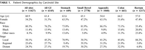
A total of 11,427 patient records were obtained for analysis. Patient demographics and tumor stage data are provided in Table 1. The average age of patients was 60.9 years. Patients with appendiceal tumors were the youngest, and those with small intestine tumors were the oldest. Of the group overall, 54.2% were female, and approximately 80% were white. At presentation, slightly more than 50% of the patients were found to have localized disease, and the remainder was divided evenly between regional and distant stage disease.
TABLE 1. Patient Demographics by Carcinoid Site
Incidence of Carcinoids
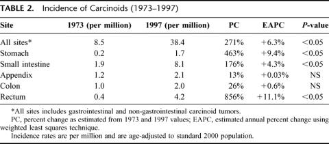
The overall incidence of carcinoid tumors was 38.4 per one million individuals in the year 1997 (age-adjusted), as determined from the population-based data (see Table 2). In comparison, the incidence rate for 1973 was only 8.5 (per million). This increase translates into +6.3% EAPC (P < 0.05). The incidence of carcinoids found in the rectum, stomach, and small intestine increased significantly over the time period (P < 0.05), while the incidences of tumors in the appendix and colon have remained constant (P = NS). Incidence rates for black patients were 2-fold higher than for white patients; however, rates for both racial groups increased incrementally at a similar pace over the 25-year time period.
TABLE 2. Incidence of Carcinoids (1973–1997)
Distribution of Carcinoids
Carcinoids were distributed widely throughout the body (eg, gastrointestinal tract, breast, genital-urinary system, respiratory, and head and neck regions). The majority occur in the gastrointestinal tract (54.5%) followed in decreasing frequency by lung and bronchus (30.1%), pancreas (2.3%), gynecologic/ovarian (1.2%), biliary (1.1%), and head and neck (0.4%). The remaining sites included such areas as soft tissue, genital/urinary, etc. (9.7%).
Within the gastrointestinal tract, carcinoids occur predominately in the small intestine (44.7%) followed in decreasing frequency by the rectum (19.6%), appendix (16.7%), colon (10.6%), and stomach (7.2%); see Table 3. Within the small intestine, carcinoid tumors are distributed predominately in the ileum (>50%), less frequently in the jejunum, and rarely in the duodenum. Within the colon, tumors develop most commonly in the sigmoid (35.7%), followed in decreasing frequency by rectosigmoid junction (26.7%), ascending colon (20.8%), transverse colon (9.2%), and descending colon (9.0%).
TABLE 3. Distribution of Gastrointestinal Carcinoids by Decade
Along with the increase in the incidence rates of rectal, stomach, and small intestinal carcinoids, there has been a concomitant decrease in the percentage of tumors found in the appendix (relative to the other sites) over the past 25 years (Table 3). More specifically, 31.8% of all carcinoids were found in the appendix in 1973 to 1979, compared with 12.0% in 1990 to 1997.
Survival
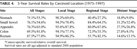
The overall cancer-specific 5-year survival for all sites was 69.7% (relative survival, 56.2%). Survival rates for carcinoid tumors varied significantly depending on their site of origin and tumor stage. Table 4 shows the survival per stage for the various sites in the gastrointestinal tract. Cancer-specific survival rates were high for all localized tumors (regardless of stage). Rates were variable for regional stage disease depending on the tumor site. Regional appendiceal tumors had better 5-year survival compared with stomach tumors (80.0% versus 40.4%, P < 0.05). Overall, rectal carcinoids had the highest 5-year survival (87.5%), which is likely the result of the high proportion of these patients who present with local stage disease. Relative 5-year survival rates are also provided in Table 4.
TABLE 4. 5-Year Survival Rates by Carcinoid Location (1973–1997)
Subanalysis for survival rates was completed using a narrower definition of appendiceal tumors: excluding those coded as cecal in origin. For this classification, the appendiceal origin had a 5-year cancer-specific survival of 88.6% for all stages, 96.2% for localized stage, and 86.7% for regional stage.
Predictors of Lymph Node Involvement and Mortality
Logistic regression revealed that increased tumor size was associated with a greater likelihood of lymph node involvement (odds ratio = 1.90; CI, 1.58 to 2.28; P < 0.0001), even when controlling for age, gender, race, and location in gastrointestinal tract.
Gender and race/ethnicity were both found to be predictors associated with a higher hazard of dying. Specifically, male patients, controlling for the other factors, had a hazard ratio of 1.30 of dying compared with female patients (CI, 1.12 to 1.52; P = 0.0006). Black patients had a hazard ratio of 1.53 compared with white patients (CI, 1.23 to 1.89; P = 0.0001), whereas Hispanic patients had 0.87 hazard ratio, which was not statistically significant (CI, 0.50 to 1.52; P = 0.63), controlling for age, gender, and tumor stage.
DISCUSSION
This analysis of over 11,000 patients with carcinoid tumors is the largest epidemiological study to date. We determined that the overall incidence of carcinoid tumors has markedly increased and most recently was 38.5 per million persons (in 1997). To put this into perspective with other cancers, the incidence for carcinoids is similar to esophageal (48.1 cases per million persons) and gastric cancer (85.6 cases per million persons).10
The overall incidence of carcinoids has increased at an annual rate of 6.3%. This rise has occurred more rapidly than for other common cancers such as breast (+2.1% EAPC) and lung (+1.0% EAPC). In comparison, both colorectal and pancreatic cancer have remained fairly constant (−0.5% and −0.3% EAPC, respectively). Surprisingly, differential changes in incidence have occurred on the basis of carcinoid tumor location. While the incidence rate of appendiceal carcinoids has remained relatively constant, those for rectal, stomach, and small intestine carcinoids have increased.
As carcinoid tumors are relatively uncommon, previous evidence has been primarily limited to anecdotal or single-institution experiences. Most authors reference an article from 1975 by Godwin, which was the most comprehensive analysis of carcinoid tumor epidemiology at the time.4 They studied 2837 cases reported by the End Results Group (ERG) and the Third National Cancer Survey (TNCS) programs of the National Cancer Institute between 1950 and 1971. Their results were based on data from the 1950s and 1960s. They found that the majority of carcinoids occurred in the appendix (43.9 to 35.5%), followed by the rectum (15.4 to 12.3%), and small intestine (13.8 to 10.8%). This analysis by Godwin appears to have remained the gold standard for decades.
We found evidence that is contrary to conventional wisdom; the appendix does not appear to be the most common site for gastrointestinal carcinoids. Similar results have been reported; however, these findings have yet to be accepted into the surgical culture.5,11–12 Additional confirmation from other sources (ie, other large tumor registries or studies from other countries) is warranted.
The etiology for this possible shift in distribution can only be speculated at this time. While it is possible that the aging of population may play a role, this influence should have been minimized, as our determination for incidence rates was age-adjusted. However, the quality of this age-adjustment will be dependent on the Census population determinants. In addition, the age-adjustment may not account for the entire potential impact of aging on the incidence of carcinoids. We speculate that the explanation for the observed change in the distribution of carcinoids is likely to be multifactorial and could include factors such as lengthened life expectancy, improved technology and diagnosis, changes in diet, and environmental exposures.
Regarding survival, our study confirms that rates for carcinoid tumors are generally favorable, but they vary by tumor site and stage. Local stage tumors have relatively high survival rates, regardless of site of origin. Survival rates are substantially lower for those with regional disease, particularly for stomach, rectal, and small intestine tumors. Tumor size and lymph node status were important predictors of survival, as has been demonstrated by others.12–18
Racial disparities were apparent for carcinoids, as has been seen for other solid tumors. In our study, blacks patients had a higher incidence of carcinoid tumors and poorer survival (even when controlling for age and tumor stage). These findings may be partially due to factors like access to care, socioeconomic status, and presence of comorbidities. Future studies are needed to further examine these relationships to work towards minimizing disparities in care.
The SEER database allows for a longitudinal examination of population-based cancer data, but it has limitations. SEER lacks comprehensive outpatient data (receipt of radiation and chemotherapy). While the primary therapy for carcinoid is surgical resection, chemotherapy, which is used for regional and distant disease, likely affects survival. We were unable to determine the effect of chemotherapy on survival using this database. Another potential limitation involves the accuracy of the data, as coding errors and inability to follow-up patients may exist. However, the validity of SEER as a comprehensive data source has been well established. Incidence rates, mortality rates, and tumor distributions have all been confirmed by other methods to be accurate and valid.19–20
Use of these data has advantages, including its multigeographic areas, wide representation of racial groups, large sample size, and broad spectrum of age groups. Due to the relatively uncommon occurrence of carcinoid tumors, this large dataset offers unique insights that single institutional series may lack.
In summary, our work shows that the distribution of carcinoid tumors appears to have changed significantly over the 25-year study period. The incidence rate for appendiceal carcinoids has remained stable, while the rates for small intestine, rectum, and stomach have increased. The relative distribution of carcinoids in these organs has changed. The appendix may no longer be the most common site for gastrointestinal carcinoids to develop; the small intestine and rectum appear to be the more frequent sites. We have also clarified the population-based survival rates for each carcinoid location in the body. This work disputes currently held beliefs about the distribution of carcinoids and highlights the role of population-based data registries in determining incidence rates, distribution, and survival rates, particularly for diseases that are relatively less common. One favorite question heard on surgical wards and found on the in-service training examination is: what is the most common site for carcinoids to occur? The answer may have changed.
Footnotes
Support provided by the Robert Wood Johnson Clinical Scholars Program, UCLA, Los Angeles, California.
Reprints: Melinda A Maggard, MD, UCLA School of Medicine, Department of Surgery, CHS, Rm 72-215, 10833 Le Conte Ave, Los Angeles, CA 90095. E-mail: mmaggard@mednet.ucla.edu.
REFERENCES
- 1.Creutzfeldt W. Carcinoid tumors: development of our knowledge. World J Surg. 1996;20:126–131. [DOI] [PubMed] [Google Scholar]
- 2.Caplin ME, Buscombe JR, Hilson AJ, et al. Carcinoid tumour. The Lancet. 1998;352:799–805. [DOI] [PubMed] [Google Scholar]
- 3.Hodgson HJ. Carcinoid tumors and the carcinoid syndrome. In: Bonchier IA, Allan RN, Hodgson HJ, et al., eds. Gastroenterology: Clinical Science and Practice. London: WB Sanders; 1992:643–658. [Google Scholar]
- 4.Schwartz SI. Appendix. In: Schwartz SI, Shires GT, Spencer FC, eds. Principles of Surgery, 6th Edition. New York: McGraw-Hill, Inc, Health Provisions Division; 1994:1307–1318.
- 5.Campbell KA. Small bowel tumors. In: Cameron JL, ed. Current Surgical Therapy, 7th Edition. Philadelphia: Mosby, Inc; 2001:139–149. [Google Scholar]
- 6.Liu CD, McFadden DW. Acute abdomen and appendix. In: Greenfield LJ, Mulholland MW, Oldham KT, et al., eds. Surgery: Scientific Principles and Practice. Philadelphia: Lippincott-Raven; 1997:1246–1261. [Google Scholar]
- 7.Godwin JD. Carcinoid tumors. An analysis of 2837 cases. Cancer. 1975;36:560–569. [DOI] [PubMed] [Google Scholar]
- 8.Jetmore AB, Ray JE, Gathright JB Jr, et al. Rectal carcinoids: the most frequent carcinoid tumor. Dis Colon Rectum. 1992;35:717–725. [DOI] [PubMed] [Google Scholar]
- 9.Brooks JM, Chrischilles E, Scott S, et al. Information gained from linking SEER Cancer Registry Data to state-level hospital discharge abstracts. Surveillance, epidemiology, and end results. Med Care. 2000;38:1131–1140. [DOI] [PubMed] [Google Scholar]
- 10.SEER Cancer Statistics Review 1973–1999. National Cancer Institute. Available at: http://seer.cancer.gov. Accessed September 20, 2002.
- 11.Modlin IM, Sandor A. An analysis of 8305 cases of carcinoid tumors. Cancer. 1997;79:813–829. [DOI] [PubMed] [Google Scholar]
- 12.Lauffer JM, Zhang T, Modlin IM. Review article: current status of gastrointestinal carcinoids. Aliment Pharmacol Ther. 1999;13:271–287. [DOI] [PubMed] [Google Scholar]
- 13.Stinner B, Kisker O, Ziele A, et al. Surgical management for carcinoid tumors of small bowel, appendix, colon and rectum. World J Surg. 1996;20:183–188. [DOI] [PubMed] [Google Scholar]
- 14.Talamonti MS, Goetz LH, Rao S, et al. Primary cancers of the small bowel: analysis of prognostic factors and results of surgical management. Arch Surg. 2002;137:564–570; discussion 570–571. [DOI] [PubMed]
- 15.Soreide JA, van Heerden JA, Thompson GB, et al. Gastrointestinal carcinoid tumors: long-term prognosis for surgically treated patients. World J Surg. 2000;24:1431–1436. [DOI] [PubMed] [Google Scholar]
- 16.Marshall JB, Bodnarchuk G. Carcinoid tumors of the gut. Our experience over three decades and review of the literature. J Clin Gastroenterol. 1993;16:123–129. [PubMed] [Google Scholar]
- 17.Ballantyne GH, Savoca PE, Flannery JT, et al. Incidence and mortality of carcinoids of the colon. Data from the Connecticut Tumor Registry. Cancer. 1992;69:2400–2405. [DOI] [PubMed] [Google Scholar]
- 18.Eller R, Frazee R, Roberts J. Gastrointestinal carcinoid tumors. Am Surg. 1991;57:434–437. [PubMed] [Google Scholar]
- 19.Merrill RM, Capocaccia R, Feuer EJ, et al. Cancer prevalence estimates based on tumour registry data in the Surveillance, Epidemiology, and End Results (SEER) Program. Int J Epidemiol. 2000;29:197–207. [DOI] [PubMed] [Google Scholar]
- 20.Gamel JW, Vogel RL. Non-parametric comparison of relative versus cause-specific survival in Surveillance, Epidemiology and End Results (SEER) programme breast cancer patients. Stat Methods Med Res. 2001;10:339–352. [DOI] [PubMed] [Google Scholar]


