Abstract
Ebola virus, a prime example of an emerging pathogen, causes fatal hemorrhagic fever in humans and in nonhuman primates. Identification of major determinants of Ebola virus pathogenicity has been hampered by the lack of effective strategies for experimental mutagenesis. Here we exploit a reverse genetics system that allows the generation of Ebola virus from cloned cDNA to engineer a mutant Ebola virus with an altered furin recognition motif in the glycoprotein (GP). When expressed in cells, the GP of the wild type, but not of the mutant, virus was cleaved into GP1 and GP2. Although posttranslational furin-mediated cleavage of GP was thought to be an essential step in Ebola virus infection, generation of a viable mutant Ebola virus lacking a furin recognition motif in the GP cleavage site demonstrates that GP cleavage is not essential for replication of Ebola virus in cell culture.
Ebola virus, a member of the family Filoviridae, is a nonsegmented negative-strand RNA virus and is one of the most lethal human pathogens recognized to date (2, 16). Ebola virus infection manifests as a typical viral hemorrhagic fever, presenting a much-needed model to study virus-induced mechanisms leading to coagulation disorders and vascular instability. For Ebola virus infection, cleavage of the glycoprotein (GP) is thought to be an important determinant of viral pathogenicity (3). The Ebola virus GP contains a highly conserved consensus motif for the subtilisin-like endoprotease furin, and previous studies demonstrated GP cleavage by this protease (21). However, studies of murine leukemia virus (25) or vesicular stomatitis virus (5), pseudotyped with mutant Ebola virus GPs lacking the furin recognition motif at the cleavage site, showed that GP cleavage by furin was not essential for infectivity of the pseudotyped viruses. In many viruses, GP cleavage by furin and related endoproteases is essential for their infectivity. Thus, the significance of GP cleavage for the Ebola virus life cycle remains in question.
Reverse genetics systems, i.e., systems for the generation of infectious virus entirely from cloned cDNA, provide excellent tools for deciphering factors that determine viral pathogenicity. Recent completion of the genomic sequence (22), as well as the establishment of an artificial replication system to identify the viral proteins required for transcription and replication (12), have presented the opportunity to devise a reverse genetics system for Ebola virus research. To directly address the requirement for GP cleavage by furin for Ebola virus infectivity, we established a reverse genetics system for the generation of Ebola virus from cloned cDNA. We used this system to generate a mutant virus with an altered furin cleavage motif in GP, and we studied the growth characteristics of the resultant virus in cell culture.
Generation of transfectant Ebola virus.
To generate a cDNA clone encoding the entire genome of Ebola virus (Zaire species, strain Mayinga), we reverse transcribed viral RNA with Thermo Script Reverse Transcriptase (Gibco/BRL, Rockville, Md.) and amplified it by PCR with Pfu Turbo (Stratagene, La Jolla, Calif.). The resulting cDNA fragments were cloned in a Bluescript vector or its derivatives. A consensus sequence was determined and compared to a reference sequence (GenBank accession number AF086833). An A insertion was found between nucleotides 9,744 and 9,745, which was also detected in a partial Ebola virus genomic sequence (GenBank accession number L11365). In addition, an A insertion was found between nucleotides 18,495 and 18,496, and an A-to-T replacement was detected at position 18,226. The latter two changes have also been reported for a functional Ebola virus minigenome (12). We assembled a full-length Ebola virus cDNA construct in a modified pTM1 vector (11), using conventional cloning techniques. Sequence analysis of the resulting full-length clone proved that no mutations had occurred during cloning procedures in E. coli.
Negative-sense RNA viruses have been generated from constructs encoding either the negative-sense viral RNA (vRNA) or the positive-sense complementary RNA (cRNA) (9, 13, 14, 17, 18), albeit with higher efficiencies from the latter (1, 6). We therefore generated cDNA constructs encoding the entire viral genome, flanked by the T7 RNA polymerase promoter and a ribozyme, in both positive-sense and negative-sense orientations (Fig. 1A). To achieve efficient transcription, we used the wild-type T7 RNA polymerase promoter, which yields transcripts with an additional G at the 5′ end, resulting in pTM-T7G-Ebo-Rib and pTM-Rib-Ebo-GT7 (Fig. 1A).
FIG. 1.
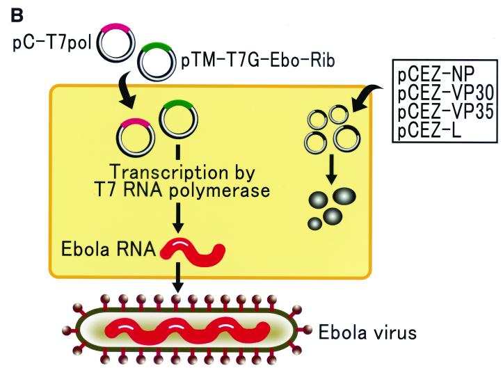
Generation of Ebola virus entirely from cloned cDNA. (A) Schematic diagram of cDNA plasmids for Ebola virus cRNA or vRNA synthesis and their efficiencies for virus generation. T7 and Rib indicate T7 RNA polymerase promoter and ribozyme sequences. G designates a guanine nucleotide inserted between the promoter and the Ebola virus cDNA. Synthesis of positive-sense Ebola virus cRNA is represented by “Ebola,” while the inverse lettering denotes synthesis of negative-sense vRNA. To determine the efficiency of virus generation, we cotransfected Vero E6 cells with protein expression plasmids and the plasmid for Ebola virus vRNA or cRNA synthesis. Four days after transfection, we measured the efficiency of virus generation by determining the dose required to infect 50% of tissue culture cells (TCID50) per ml of supernatant. The data are representative results from three independent experiments. Experiments for the generation of Ebola virus as well as the characterization of recombinant Ebola virus were carried out in the BSL4 facility at the Canadian Science Centre for Human and Animal Health, Winnipeg, Canada. (B) Schematic diagram of Ebola virus generation. Cells were transfected with plasmids for the expression of the Ebola virus NP, L, VP30, and VP35 proteins, and with the plasmid for Ebola virus cRNA or vRNA synthesis, controlled by T7 RNA polymerase promoter and ribozyme sequences. T7 RNA polymerase was provided by cotransfection of cells with pC-T7Pol.
The generation of negative-sense RNA viruses requires viral proteins and genomic RNA for replication and transcription. For Ebola virus, the proteins necessary for replication and transcription include the RNA-dependent RNA polymerase L, the nucleoprotein (NP), and two additional auxiliary proteins (VP30 and VP35) (12). To generate constructs for the expression of Ebola viral proteins, we amplified the respective cDNA fragments by PCR, sequenced the products, and then cloned them into the eukaryotic expression vector pCAGGS/MCS (controlled by the chicken β-actin promoter) (8, 15), resulting in four plasmids (pCEZ-NP, pCEZ-VP30, pCEZ-VP35, and pCEZ-L).
To generate Ebola virus, we transfected 5 × 10⁵ Vero E6 (African green monkey kidney) cells with 1 μg of the respective plasmid for Ebola virus vRNA or cRNA synthesis and with the following amounts of protein expression plasmids: 1 μg of pCEZ-NP, 0.3 μg of pCEZ-VP30, 0.5 μg of pCEZ-VP35, and 2 μg of pCEZ-L (Fig. 1B). To drive the transcription of viral RNA from the T7 RNA polymerase promoter, we cotransfected cells with 1 μg of an expression plasmid for this enzyme (pC-T7pol). Four days later, supernatants were collected and used to infect fresh Vero E6 cells. When examined at 6 to 8 days postinfection, the cells showed cytopathic effects, indicating the generation of infectious Ebola virus entirely from cloned cDNA. We were able to produce Ebola virus from constructs encoding either negative-sense vRNA or positive-sense cRNA (Fig. 1A). To determine the efficiency of virus generation, supernatants of transfected cells were collected 4 days after transfection, and the titer of virus in the supernatant was determined in Vero E6 cells. The efficiencies of virus generation from negative-sense vRNA or positive-sense cRNA were comparable, resulting in the generation of 102 50% tissue culture infective doses (TCID50) per ml of supernatant.
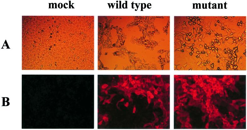
A subsequent passage of the virus in Vero E6 cells was performed to confirm the authenticity of the replicating agent. The first signs of a cytopathic effect were observed at 48 h postinfection and became more prominent during the following days (Fig. 2A). Indirect immunofluorescence assays with a rabbit antiserum to Ebola virus GP/secreted GP (sGP) demonstrated the presence of these Ebola virus GPs (Fig. 2B). None of the negative controls (untreated cells or cells transfected with the full-length cDNA construct or the protein expression plasmids alone) showed cytopathic effects or reacted with the anti-GP/sGP antiserum.
FIG. 2.
Replication of Ebola virus in cell culture. Vero E6 cells were infected at a multiplicity of infection of 10−2 with either wild-type Ebola virus generated from plasmids or Ebola virus with an altered furin recognition sequence in its GP. Six days later, cells were observed under a light microscope (A). For an immunofluorescence assay (B), 3 days after infection, cells were fixed with 2% paraformaldehyde in phosphate-buffered saline, followed by inactivation by gamma irradiation (2 Mrads). Cells were permeabilized with 0.1% Triton X-100 in phosphate-buffered saline for 15 min, washed three times with phosphate-buffered saline, and incubated for 1 h at room temperature with an anti-Ebola virus Zaire rabbit antiserum (1:100 dilution in phosphate-buffered saline). After three washes with phosphate-buffered saline, Cy3-labeled anti-rabbit (1:500) conjugate (Rockland, Gilbertsville, Pa.) was added for 1 h at room temperature. The cells were then washed with phosphate-buffered saline, mounted, and analyzed using an Axioplan 2 microscope (Zeiss).
GP cleavage by furin is not essential for viral replication in tissue culture.
The availability of a method for generating Ebola virus mutants greatly increases opportunities to dissect mechanisms of viral pathogenesis. For many viruses, posttranslational cleavage of membrane glycoproteins by host proteolytic enzymes, including subtilisin-like proteases such as furin, is a prerequisite for fusion between the viral envelope and cellular membranes and therefore an important step in pathogenesis (7). The Ebola virus GP is cleaved by furin or furin-like proteases at a highly conserved sequence motif (R-X-K/R-R; X, any amino acid) (21). Since the amino acid sequence of the GP of the Reston species, the least pathogenic of all Ebola virus subtypes in humans, deviates from the optimal furin recognition sequence, GP cleavage has been thought to be an important determinant of Ebola virus pathogenicity (3).
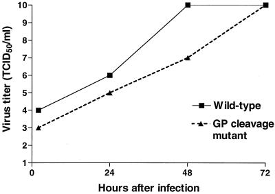
We therefore studied the effect of an altered furin recognition motif on Ebola virus replication, modifying pTM-T7G-Ebo-Rib by replacing the multibasic furin recognition site (RRTRR at amino acid positions 497 to 501 of the GP) with 497-AGTAA-501. The modified plasmid, designated pTM-T7G-Ebo-Rib-Cl(−), was transfected into Vero E6 cells, together with protein expression plasmids for the NP, VP30, VP35, and L proteins and for T7 RNA polymerase. Fresh Vero E6 cells were subsequently incubated with supernatants derived from the transfected cells. Six days later, we observed cytopathic effects in these cells. Indirect immunofluorescence assays with antiserum to Zaire Ebola virus GP/sGP verified virus replication (Fig. 2). Growth curves in Vero E6 cells demonstrated that although the mutant virus grew slightly more slowly than the wild-type virus (Fig. 3), it reached 1010 TCID50/ml at 3 days postinfection.
FIG. 3.
Replication kinetics of wild-type Ebola virus and its GP cleavage mutant. Vero E6 cells were infected with the wild-type or cleavage site mutant at a multiplicity of infection of 10−2. Supernatants were harvested at 2, 24, 48, and 72 h postinfection. The TCID50 was determined by infecting Vero E6 cells with 10-fold dilutions of the supernatants obtained at the above-mentioned time points.
To confirm the presence of mutations in the GP cleavage motif, we passaged wild-type and mutant viruses three times in Vero E6 cells, extracted RNA from virions, and performed reverse transcriptase PCR with primers spanning the altered furin recognition motif. Direct sequencing of the PCR products confirmed the retention of mutations in the GP cleavage site (data not shown).
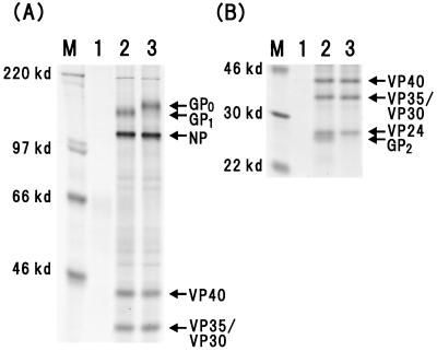
Figure 4 shows the results of experiments testing the cleavability of the Ebola virus mutant GP lacking a furin recognition motif. Virions derived from labeled Vero E6 cells infected with wild-type or mutant virus were lysed, and viral proteins were detected by immunoprecipitation using a horse antiserum to Zaire Ebola virus. For wild-type virus, both cleavage products GP1 (140 kDa) and GP2 (26 kDa) were detected (Fig. 4A and B, lanes 2). By contrast, alteration of the furin recognition sequence abolished the generation of GP1 and GP2, and only the precursor, GP0, was detected (Fig. 4A and B, lanes 3), confirming that furin or related proteases are the major host cell proteases for GP cleavage. These results indicate that the furin recognition motif at the Ebola GP cleavage site is dispensable for replication of the virus in cell culture.
FIG. 4.
Comparison of GP cleavage between wild-type and mutant Ebola virus. Vero E6 cells were infected at a multiplicity of infection of 10−2 and incubated until a cytopathic effect was observed. After the medium was removed, the cells were washed once with methionine- and cysteine-free DMEM and labeled for 24 h in 2 ml of methionine- and cysteine-free Dulbecco’s modified Eagle’s medium containing 2% dialyzed fetal calf serum and 10 μCi of protein labeling mix (NEN, Mississauga, Canada)/ml. The supernatants were then clarified by centrifugation (1,000 × g for 5 min at 4°C). An equal volume of 2× RIPA buffer (2% Triton X-100, 2% sodium deoxycholate, 0.2% sodium dodecyl sulfate, 0.3 M NaCl, 40 mM Tris-HCl [pH 7.7], 20 mM EDTA [pH 8.0], 0.4 U of aprotinin/ml, 2 mM phenylmethylsulfonyl fluoride, 20 mM iodoacetamide) was added to the supernatants, and the solutions were subsequently inactivated by gamma irradiation (2 Mrad). Aliquots of the inactivated labeled material were mixed with an anti-Ebola virus Zaire horse serum and incubated at 4°C overnight. The immune complexes were mixed with 30 μl of protein G sepharose for 3 h at 4°C with rotation. After three washes with RIPA buffer, the immunoprecipitated proteins were recovered by boiling them in 1× RIPA buffer. Labeled proteins were separated on 8% (A) and 15% (B) sodium dodecyl sulfate-polyacrylamide gels under reducing conditions. M, molecular mass marker; lanes 1, mock-infected Vero E6 cells; lanes 2, wild-type Ebola virus generated from plasmids; lanes 3, GP cleavage mutant virus.
Marburg and Ebola viruses have been difficult to study because they must be handled in high-containment facilities. Furthermore, effective methods of experimental mutagenesis were lacking until recently, when Volchkov et al. reported the generation of Ebola virus from cDNA (23). These limitations have restricted the development of antiviral drugs and vaccines, although reports of potentially useful experimental vaccines (4, 19, 20, 26) and antibody-mediated treatments (10, 24) are beginning to emerge. Here, we also established a reverse genetics system, which enables one to generate Ebola virus mutants entirely from cloned cDNA, opening a new era of filovirus research.
We exploited reverse genetics to determine the significance of the furin cleavage motif in the GP protein for Ebola virus replication. For many viruses in the Orthomyxoviridae and Paramyxoviridae families, GP cleavage by furin and other host cell proteases is absolutely required for their infectivity and thus determines the extent of viral pathogenicity (7). In contrast, findings with viruses pseudotyped with Ebola GPs as well as the present results demonstrate that GP cleavage is dispensable for replication of Ebola virus, at least in cell culture. The furin cleavage motif is highly conserved among all Ebola GP sequences determined thus far, and its conservation suggests a role in the viral life cycle. Hence, GP cleavage by furin is not critical for Ebola virus replication in the cells tested in this study, but it may be required for Ebola virus replication in vivo and/or in its natural reservoir. Further studies with animal models will be needed to establish the role of GP cleavage in Ebola virus replication and pathogenicity.
T7 RNA polymerase-based reverse genetics systems rely on the expression of this enzyme within the transfected cells. To this end, two approaches have been explored (reviewed in references 9, 13 and 17). T7 RNA polymerase has been provided from recombinant vaccinia virus or from stable cell lines constitutively expressing this enzyme. The former approach leaves investigators with the task of separating the artificially generated recombinant virus from vaccinia virus. On the other hand, cell lines expressing T7 RNA polymerase may produce insufficient amounts to efficiently transcribe the viral genome. In contrast to Volchkov et al. (23), who used a BHK-21 cell line stably expressing T7 RNA polymerase, we use an entirely plasmid-based system by achieving T7 RNA polymerase expression under control of the strong chicken β-actin promoter. This approach allows us to produce 102 PFU of virus per ml of culture supernatant. Expression of T7 RNA polymerase from plasmids may therefore be an alternative for the generation of other nonsegmented, negative-sense RNA viruses, thereby circumventing restrains encountered with the established systems.
The reverse genetics systems for the generation of Ebola virus can be used to identify key regulatory elements and structure-function relationships in the viral life cycle, and it will now allow investigators to study mechanisms of filovirus pathogenicity in animal models. Ultimately, the system will promote the development of new vaccines, including replication-deficient viruses.
Acknowledgments
We thank John Gilbert for editing the manuscript, Daryl Dick, Michael Garbutt, Krisna Wells, and Martha McGregor for excellent technical assistance, and Yuko Kawaoka for illustrations. Plasmid pC-T7Pol was kindly provided by Toru Takimoto (St. Jude Children’s Research Hospital, Memphis, Tenn.), and the horse antiserum to Zaire species was kindly provided by Aleksandr Chepurnov (Russian National Research Center of Virology and Biotechnology, Koltosovo, Russia). We also thank Anthony Sanchez for a rabbit antiserum to GP/sGP and the Department of Virology at the University of Marburg, Marburg, Germany (Director, H.-D. Klenk) for constructs encoding Ebola virus NP, VP30, VP35, and L genes.
This work was supported by National Institute of Allergy and Infectious Diseases Public Health Service research grants to Y.K., by Grants-in-Aid awarded by the Ministry of Education, Culture, Sports, Science and Technology and by the Ministry of Health, Labor and Welfare, Japan, to Y.K., and by research grants from the Canadian Institutes of Health Research (MOP-43921) and the European Union (INCO project no. ERBIC 18 CT98 0382) to H.F.
REFERENCES
- 1.Durbin, A. P., S. L. Hall, J. W. Siew, S. S. Whitehead, P. L. Collins, and B. R. Murphy. 1997. Recovery of infectious human parainfluenza virus type 3 from cDNA. Virology 235:323–332. [DOI] [PubMed] [Google Scholar]
- 2.Feldmann, H., A. Sanchez, and H. D. Klenk. 1998. Filoviruses, p. 651–664. In Topley and Wilson’s microbiology and microbial infections, 9th ed. Edward Arnold, London, United Kingdom.12649037
- 3.Feldmann, H., V. E. Volchkov, V. A. Volchkova, and H. D. Klenk. 1999. The glycoproteins of Marburg and Ebola virus and their potential role in pathogenesis. Arch. Virol. Suppl. 15:159–196. [DOI] [PubMed] [Google Scholar]
- 4.Hevey, M., D. Negley, P. Pushko, J. Smith, and A. Schmaljohn. 1998. Marburg virus vaccines based upon alphavirus replicons protect guinea pigs and nonhuman primates. Virology 251:28–37. [DOI] [PubMed] [Google Scholar]
- 5.Ito, H., S. Watanabe, A. Takada, and Y. Kawaoka. 2001. Ebola virus glycoprotein: proteolytic processing, acylation, cell tropism, and detection of neutralizing antibodies. J. Virol. 75:1576–1580. [DOI] [PMC free article] [PubMed] [Google Scholar]
- 6.Kato, A., Y. Sakai, T. Shioda, T. Kondo, M. Nakanishi, and Y. Nagai. 1996. Initiation of Sendai virus multiplication from transfected cDNA or RNA with negative or positive sense. Genes Cells 1:569–579. [DOI] [PubMed] [Google Scholar]
- 7.Klenk, H. D., and W. Garten. 1994. Host cell proteases controlling viral pathogenicity. Trends Microbiol. 2:39–43. [DOI] [PubMed] [Google Scholar]
- 8.Kobasa, D., M. E. Rodgers, K. Wells, and Y. Kawaoka. 1997. Neuraminidase hemadsorption activity, conserved in avian influenza A viruses, does not influence viral replication in ducks. J. Virol. 71:6706–6713. [DOI] [PMC free article] [PubMed] [Google Scholar]
- 9.Marriott, A. C., and A. J. Easton. 1999. Reverse genetics of the Paramyxoviridae. Adv. Virus Res. 53:321–340. [PubMed] [Google Scholar]
- 10.Maruyama, T., L. L. Rodriguez, P. B. Jahrling, A. Sanchez, A. S. Khan, S. T. Nichol, C. J. Peters, P. W. H. I. Parren, and D. R. Burton. 1999. Ebola virus can be effectively neutralized by antibody produced in natural human infection. J. Virol. 73:6024–6030. [DOI] [PMC free article] [PubMed] [Google Scholar]
- 11.Moss, B., O. Elroy-Stein, T. Mizukami, W. A. Alexander, and T. R. Fuerst. 1990. Product review. New mammalian expression vectors. Nature 348:91–92. [DOI] [PubMed] [Google Scholar]
- 12.Mühlberger, E., M. Weik, V. E. Volchkov, H.-D. Klenk, and S. Becker. 1999. Comparison of the transcription and replication strategies of Marburg virus and Ebola virus by using artificial replication systems. J. Virol. 73:2333–2342. [DOI] [PMC free article] [PubMed] [Google Scholar]
- 13.Nagai, Y., and A. Kato. 1999. Paramyxovirus reverse genetics is coming of age. Microbiol. Immunol. 43:613–624. [DOI] [PubMed] [Google Scholar]
- 14.Neumann, G., T. Watanabe, H. Ito, S. Watanabe, H. Goto, P. Gao, M. Hughes, D. R. Perez, R. Donis, E. Hoffmann, G. Hobom, and Y. Kawaoka. 1999. Generation of influenza A viruses entirely from cloned cDNAs. Proc. Natl. Acad. Sci. USA 96:9345–9350. [DOI] [PMC free article] [PubMed] [Google Scholar]
- 15.Niwa, H., K. Yamamura, and J. Miyazaki. 1991. Efficient selection for high-expression transfectants by a novel eukaryotic vector. Gene 108:193–200. [DOI] [PubMed] [Google Scholar]
- 16.Peters, C. J., A. Sanchez, P. E. Rollin, T. G. Ksiazek, and F. A. Murphy. 1996. Filoviridae: Marburg and Ebola viruses, p. 1161–1176. In B. N. Fields, D. M. Knipe, and P. M. Howley (ed.), Fields virology, 3rd ed. Lippincott-Raven Publishers, Philadelphia, Pa.
- 17.Roberts, A., and J. K. Rose. 1999. Redesign and genetic dissection of the rhabdoviruses. Adv. Virus Res. 53:301–319. [DOI] [PubMed] [Google Scholar]
- 18.Schnell, M. J., T. Mebatsion, and K. K. Conzelmann. 1994. Infectious rabies viruses from cloned cDNA. EMBO J. 13:4195–4203. [DOI] [PMC free article] [PubMed] [Google Scholar]
- 19.Sullivan, N. J., A. Sanchez, P. E. Rollin, Z. Yang, and G. J. Nabel. 2000. Development of a preventative vaccine for Ebola virus infection in primates. Nature 408:605–609. [DOI] [PubMed] [Google Scholar]
- 20.Vanderzanden, L., M. Bray, D. Fuller, T. Roberts, D. Custer, K. Spik, P. Jahrling, J. Huggins, A. Schmaljohn, and C. Schmaljohn. 1998. DNA vaccines expressing either the GP or NP genes of Ebola virus protect mice from lethal challenge. Virology 246:134–144. [DOI] [PubMed] [Google Scholar]
- 21.Volchkov, V. E., H. Feldmann, V. A. Volchkova, and H. D. Klenk. 1998. Processing of the Ebola virus glycoprotein by the proprotein convertase furin. Proc. Natl. Acad. Sci. USA 95:5762–5767. [DOI] [PMC free article] [PubMed] [Google Scholar]
- 22.Volchkov, V. E., V. A. Volchkova, A. A. Chepurnov, V. M. Blinov, O. Dolnik, S. V. Netesov, and H. Feldmann. 1999. Characterization of the L gene and 5′ trailer region of Ebola virus. J. Gen. Virol. 80:355–362. [DOI] [PubMed] [Google Scholar]
- 23.Volchkov, V. E., V. A. Volchkova, E. Muhlberger, L. V. Kolesnikova, M. Weik, O. Dolnik, and H. D. Klenk. 2001. Recovery of infectious Ebola virus from complementary DNA: RNA editing of the GP gene and viral cytotoxicity. Science 291:1965–1969. [DOI] [PubMed] [Google Scholar]
- 24.Wilson, J. A., M. Hevey, R. Bakken, S. Guest, M. Bray, A. L. Schmaljohn, and M. K. Hart. 2000. Epitopes involved in antibody-mediated protection from Ebola virus. Science 287:1664–1666. [DOI] [PubMed] [Google Scholar]
- 25.Wool-Lewis, R. J., and P. Bates. 1999. Endoproteolytic processing of the Ebola virus envelope glycoprotein: cleavage is not required for function. J. Virol. 73:1419–1426. [DOI] [PMC free article] [PubMed] [Google Scholar]
- 26.Xu, L., A. Sanchez, Z. Yang, S. R. Zaki, E. G. Nabel., S. T. Nichol, and G. J. Nabel. 1998. Immunization for Ebola virus infection. Nat. Med. 5:373–374. [DOI] [PubMed] [Google Scholar]


