

Abstract
1. A method is described for recording in vivo the action potentials of afferent and efferent fibres in whole nerves supplying the rabbit's uterus.
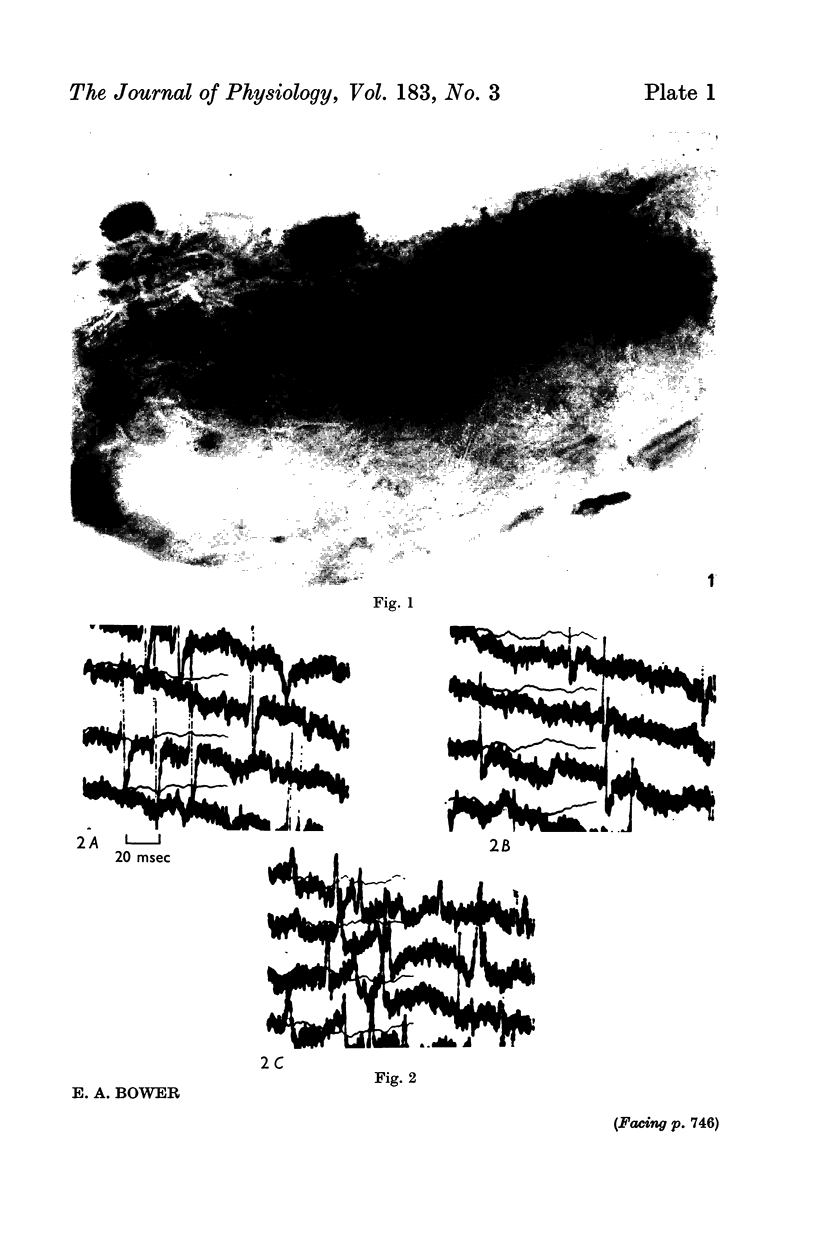
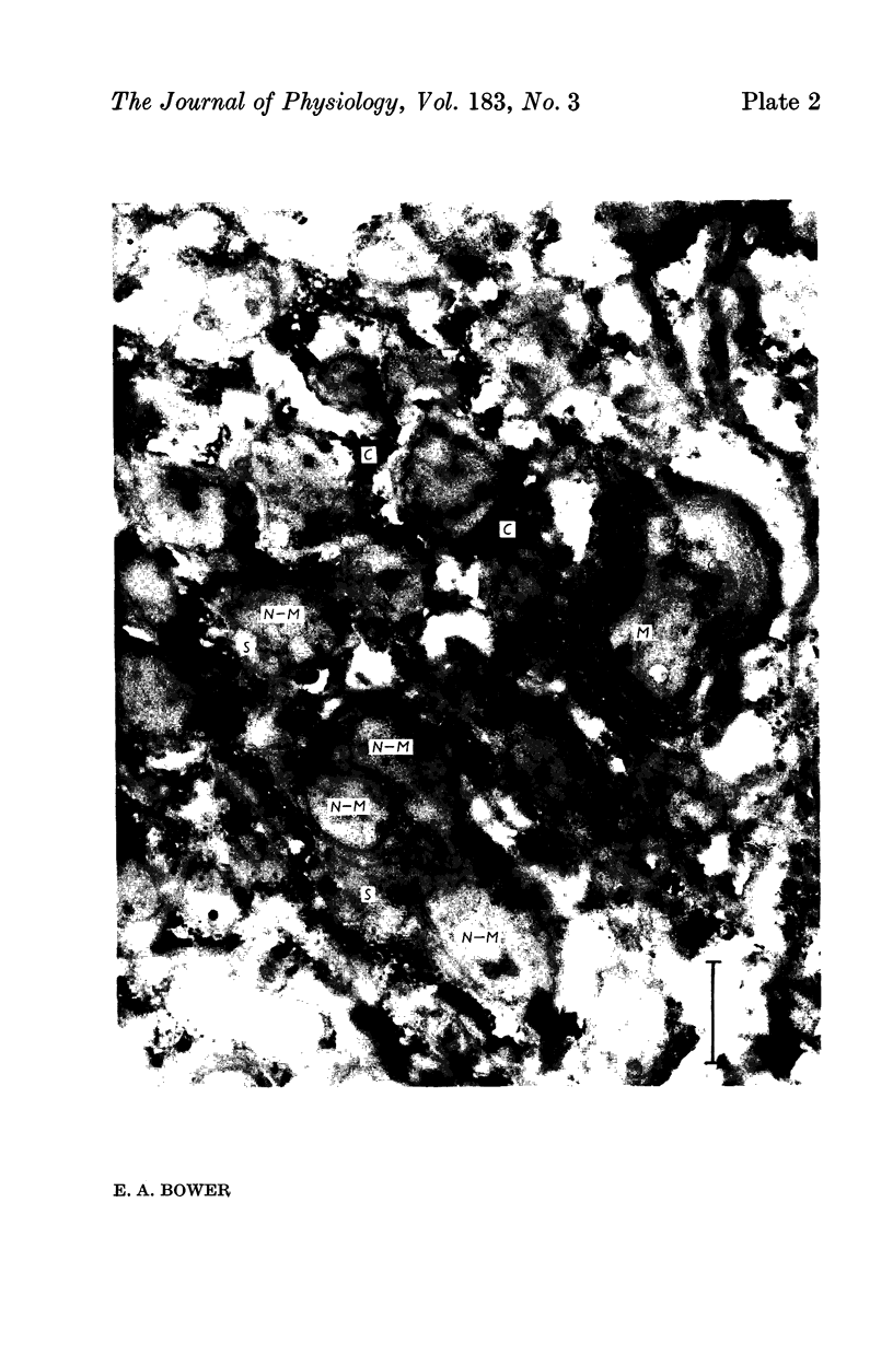
2. Examination of these nerves under the light microscope and the electron microscope showed them to be composed almost entirely of non-myelinated fibres.
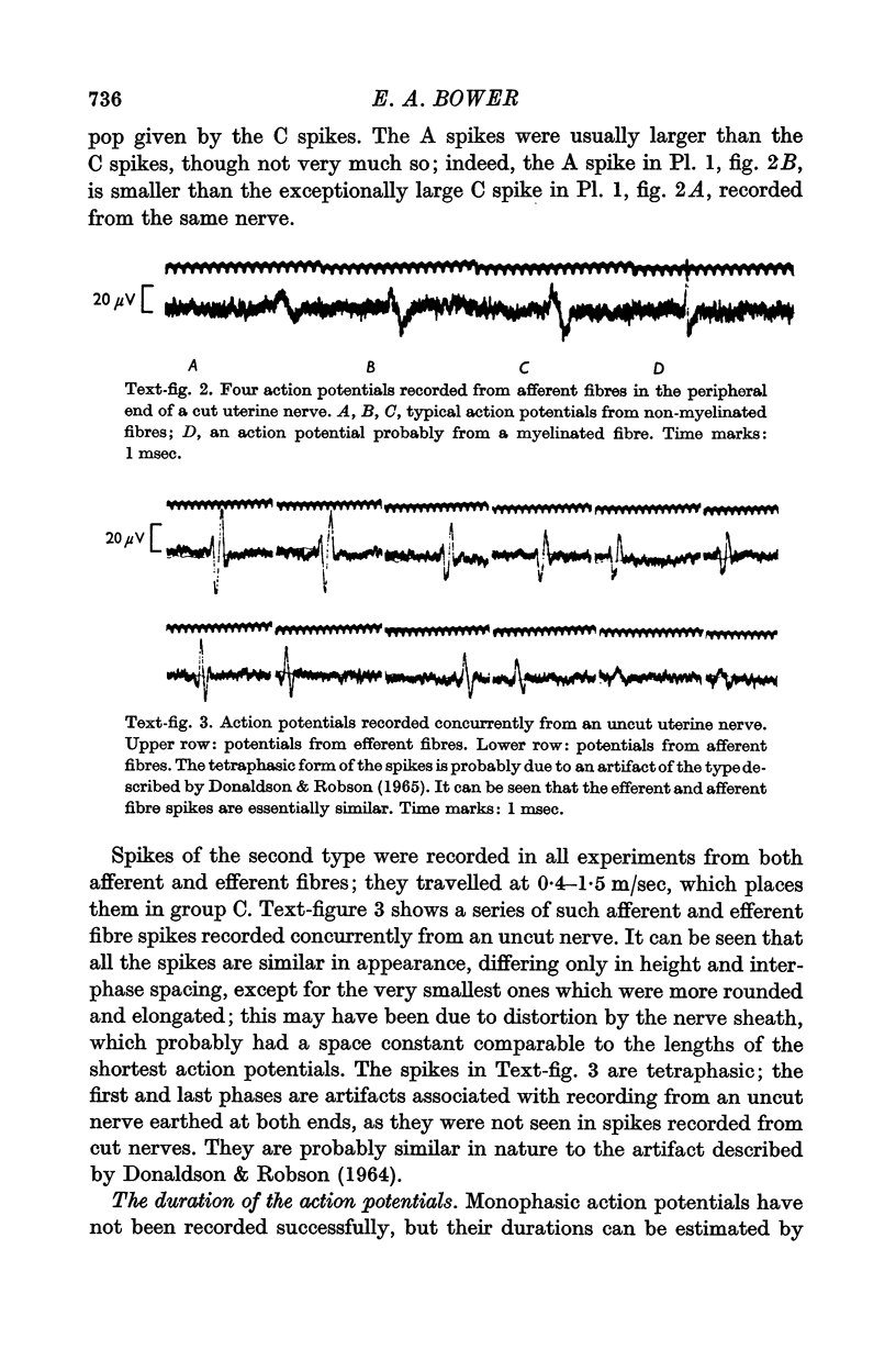
3. Two types of spontaneous action potential were observed; one travelled at about 4 m/sec and probably came from the myelinated fibres, the other travelled at 0·4-1·4 m/sec and certainly came from non-myelinated fibres.
4. The efferent fibre spikes were shown to be faster and higher than the spikes from uterine afferent fibres, but slower and smaller than spikes from broad ligament afferent fibres.
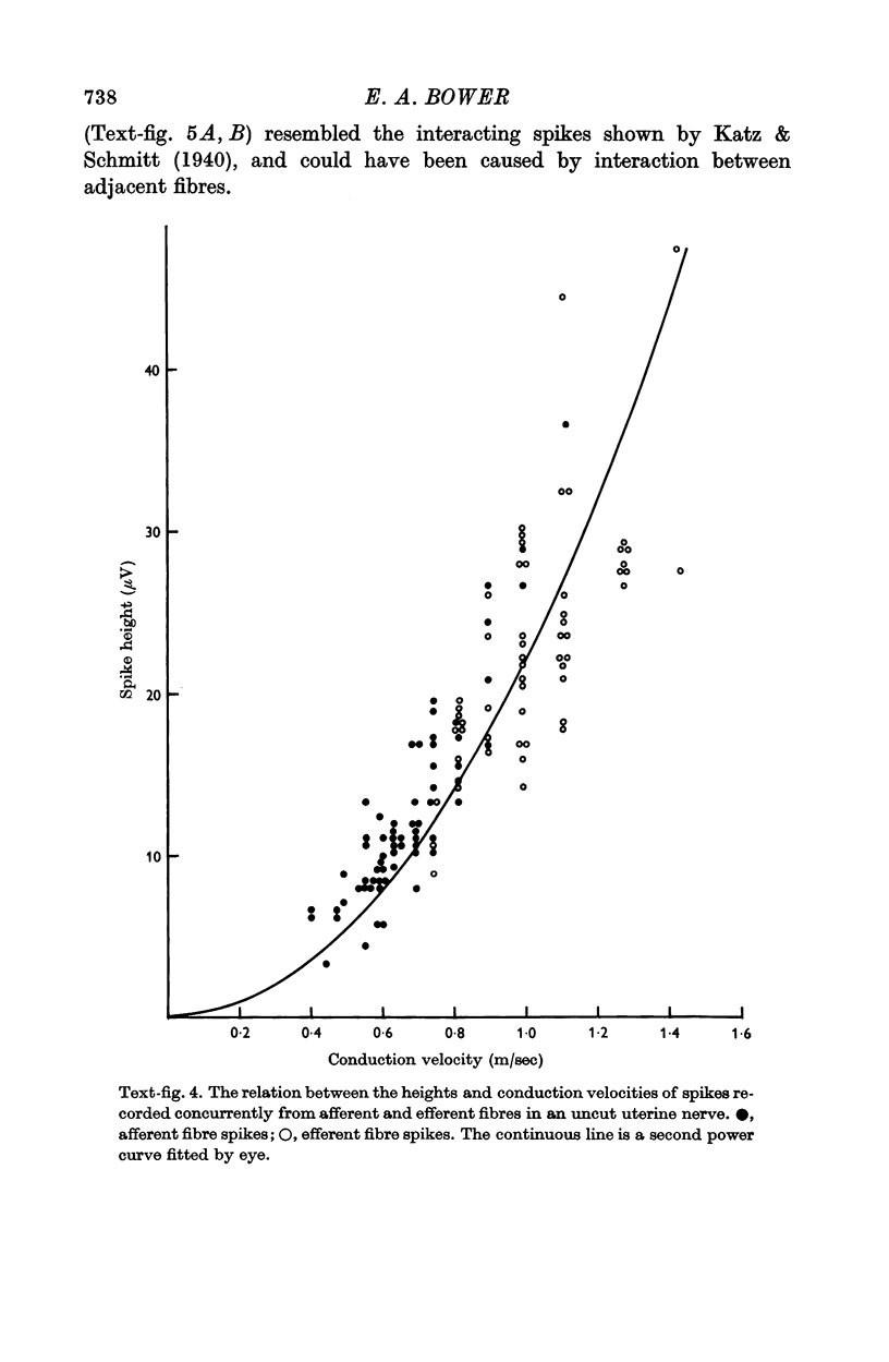
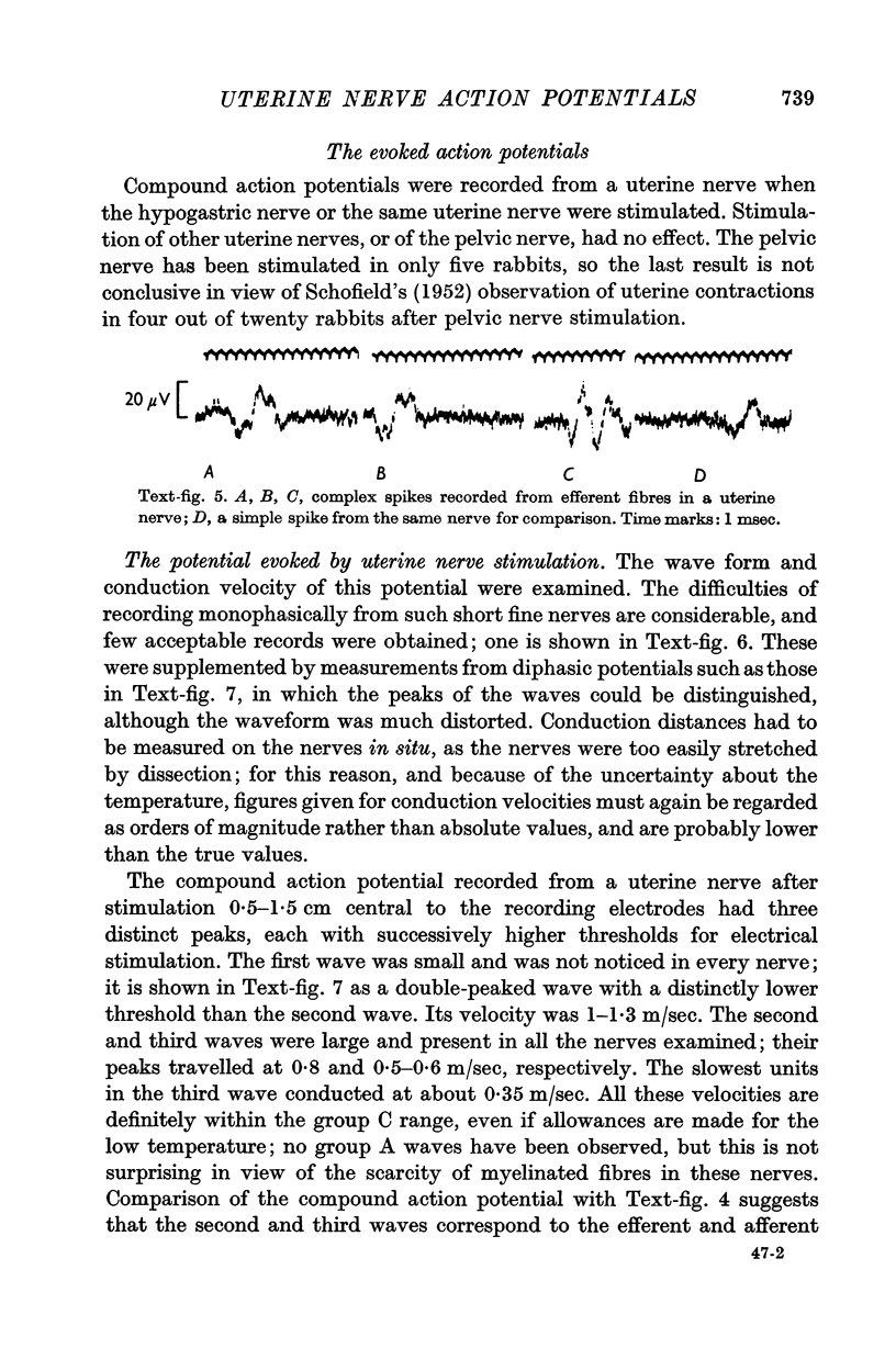
5. Apart from differences in conduction velocity and height, all spikes were basically similar, lasting about 1·5 msec. The height was related to the square of the velocity. Some more complex spikes were also observed.
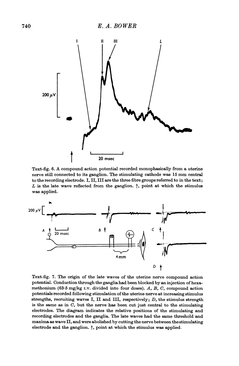
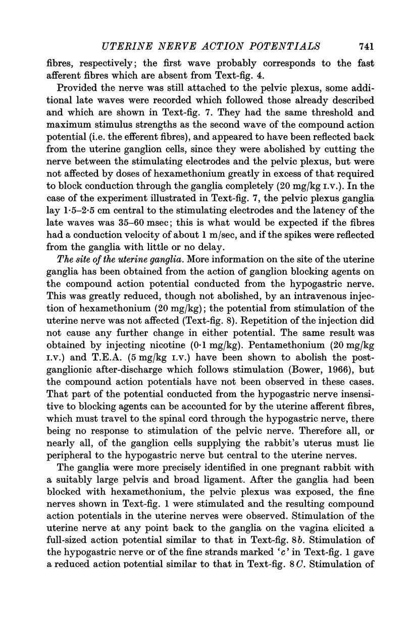
6. The compound action potential evoked by stimulation of the uterine nerve had three peaks, conducted at 1·3, 0·8 and 0·6 m/sec, respectively, and thought to correspond to the fast afferent fibres, the efferent fibres and the slow afferent fibres, respectively. There were also some late peaks due to reflexion of the antidromic action potentials from the ganglion cells.
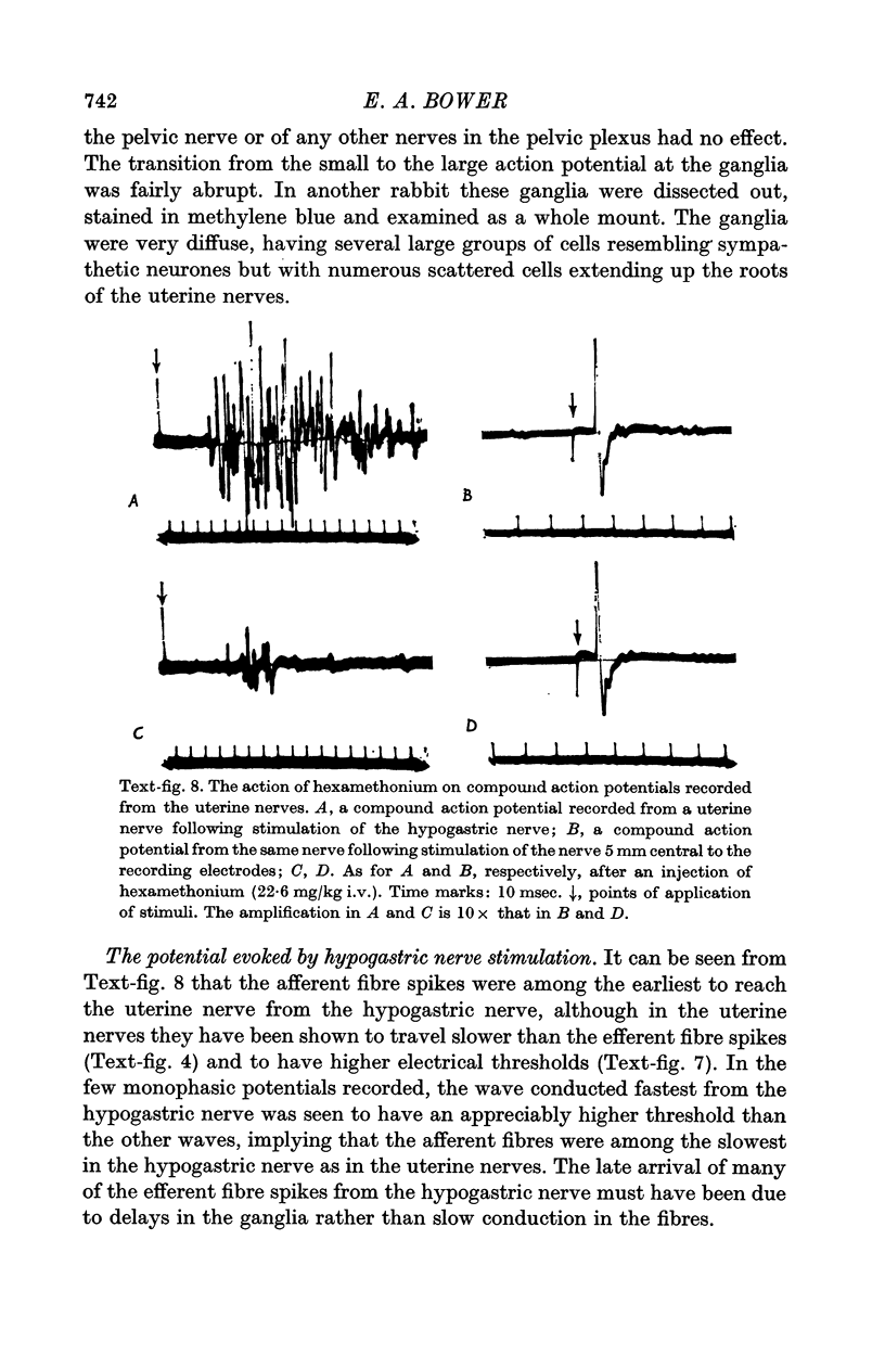
7. Stimulation of the hypogastric nerve also evoked a compound action potential in the uterine nerves. Stimulation of the pelvic nerves had no effect.
8. By means of ganglion blocking agents, the uterine ganglia were shown to lie in the pelvic plexus, peripheral to the hypogastric nerve, but central to the uterine nerves.
9. It is argued that the spontaneous action potentials came from individual fibres rather than Remak bundles, and that the recording technique used detected the activity of all but the smallest fibres.
Full text
PDF
Images in this article
Selected References
These references are in PubMed. This may not be the complete list of references from this article.
- Adrian E. D., Bronk D. W., Phillips G. Discharges in mammalian sympathetic nerves. J Physiol. 1932 Feb 8;74(2):115–133. doi: 10.1113/jphysiol.1932.sp002832. [DOI] [PMC free article] [PubMed] [Google Scholar]
- BROWN G. L., PASCOE J. W. Conduction through the inferior mesenteric ganglion of the rabbit. J Physiol. 1952 Sep;118(1):113–123. doi: 10.1113/jphysiol.1952.sp004777. [DOI] [PMC free article] [PubMed] [Google Scholar]
- Bower E. A. The activity of post-ganglionic sympathetic nerves to the uterus of the rabbit. J Physiol. 1966 Apr;183(3):748–767. doi: 10.1113/jphysiol.1966.sp007896. [DOI] [PMC free article] [PubMed] [Google Scholar]
- Bozler E. PHYSIOLOGICAL EVIDENCE FOR THE SYNCYTIAL CHARACTER OF SMOOTH MUSCLE. Science. 1937 Nov 19;86(2238):476–476. doi: 10.1126/science.86.2238.476. [DOI] [PubMed] [Google Scholar]
- Cushny A. R. On the movements of the uterus. J Physiol. 1906 Dec 29;35(1-2):1–19. doi: 10.1113/jphysiol.1906.sp001177. [DOI] [PMC free article] [PubMed] [Google Scholar]
- DONALDSON P. E., ROBSON J. G. SUPPRESSING AN ARTEFACT IN DIFFERENTIAL RECORDING FROM THIN NERVES. Med Electron Biol Eng. 1964 Jul;2:337–337. doi: 10.1007/BF02474631. [DOI] [PubMed] [Google Scholar]
- GASSER H. S. Properties of dorsal root unmedullated fibers on the two sides of the ganglion. J Gen Physiol. 1955 May 20;38(5):709–728. doi: 10.1085/jgp.38.5.709. [DOI] [PMC free article] [PubMed] [Google Scholar]
- GASSER H. S. Unmedullated fibers originating in dorsal root ganglia. J Gen Physiol. 1950 Jul 20;33(6):651–690. doi: 10.1085/jgp.33.6.651. [DOI] [PMC free article] [PubMed] [Google Scholar]
- IGGO A. The electrophysiological identification of single nerve fibres, with particular reference to the slowest-conducting vagal afferent fibres in the cat. J Physiol. 1958 Jun 18;142(1):110–126. doi: 10.1113/jphysiol.1958.sp006002. [DOI] [PMC free article] [PubMed] [Google Scholar]
- KURIYAMA H., CSAPO A. Placenta and myometrial block. Am J Obstet Gynecol. 1961 Sep;82:592–599. doi: 10.1016/0002-9378(61)90277-0. [DOI] [PubMed] [Google Scholar]
- Katz B., Schmitt O. H. Electric interaction between two adjacent nerve fibres. J Physiol. 1940 Feb 14;97(4):471–488. doi: 10.1113/jphysiol.1940.sp003823. [DOI] [PMC free article] [PubMed] [Google Scholar]
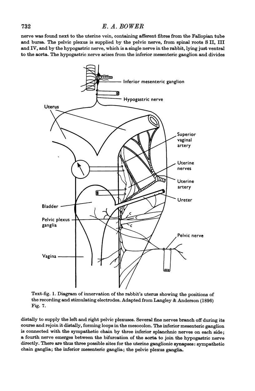
- Langley J. N., Anderson H. K. The Innervation of the Pelvic and adjoining Viscera: Part II. The Bladder. Part III. The External Generative Organs. Part IV. The Internal Generative Organs. Part V. Position of the Nerve Cells on the Course of the Efferent Nerve Fibres. J Physiol. 1895 Dec 30;19(1-2):71–139. doi: 10.1113/jphysiol.1895.sp000587. [DOI] [PMC free article] [PubMed] [Google Scholar]
- Langley J. N., Anderson H. K. The Innervation of the Pelvic and adjoining Viscera: Part VII. Anatomical Observations. J Physiol. 1896 Oct 19;20(4-5):372–406. doi: 10.1113/jphysiol.1896.sp000629. [DOI] [PMC free article] [PubMed] [Google Scholar]
- MANN M., WEST G. B. The nature of uterine and intestinal sympathin. Br J Pharmacol Chemother. 1951 Mar;6(1):79–82. doi: 10.1111/j.1476-5381.1951.tb00622.x. [DOI] [PMC free article] [PubMed] [Google Scholar]
- PALLIE W., CORNER G. W., WEDDELL G. Nerve terminations in the myometrium of the rabbit. Anat Rec. 1954 Apr;118(4):789–811. doi: 10.1002/ar.1091180407. [DOI] [PubMed] [Google Scholar]
- RUSHTON W. A. H. A theory of the effects of fibre size in medullated nerve. J Physiol. 1951 Sep;115(1):101–122. doi: 10.1113/jphysiol.1951.sp004655. [DOI] [PMC free article] [PubMed] [Google Scholar]
- SCHOFIELD B. M. The innervation of the cervix and cornu uteri in the rabbit. J Physiol. 1952 Jul;117(3):317–328. doi: 10.1113/jphysiol.1952.sp004751. [DOI] [PMC free article] [PubMed] [Google Scholar]