

Abstract
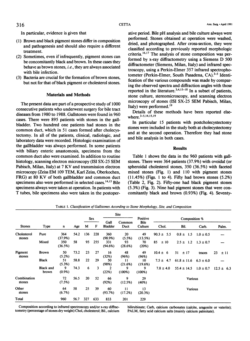
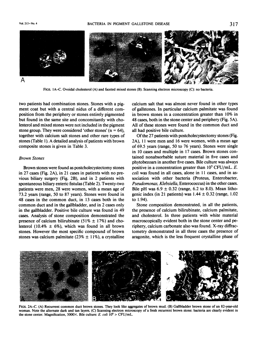
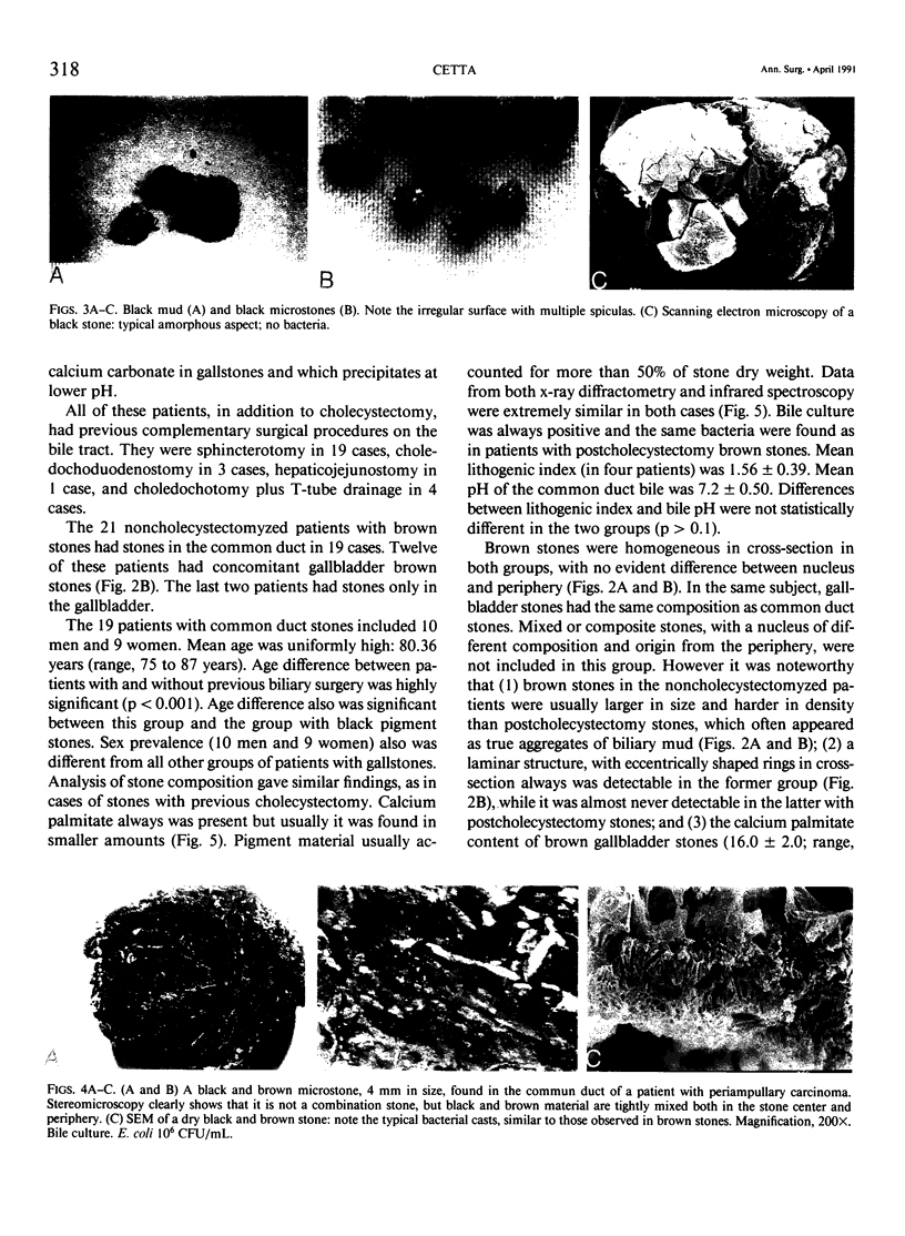
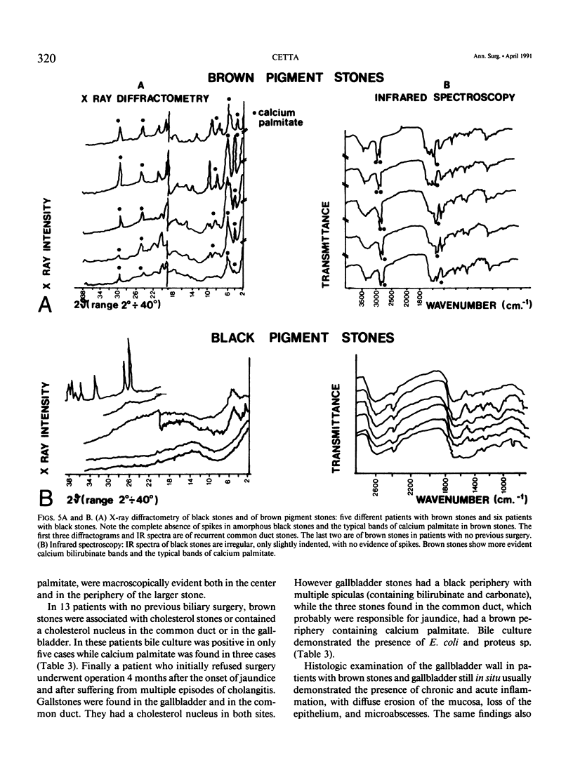
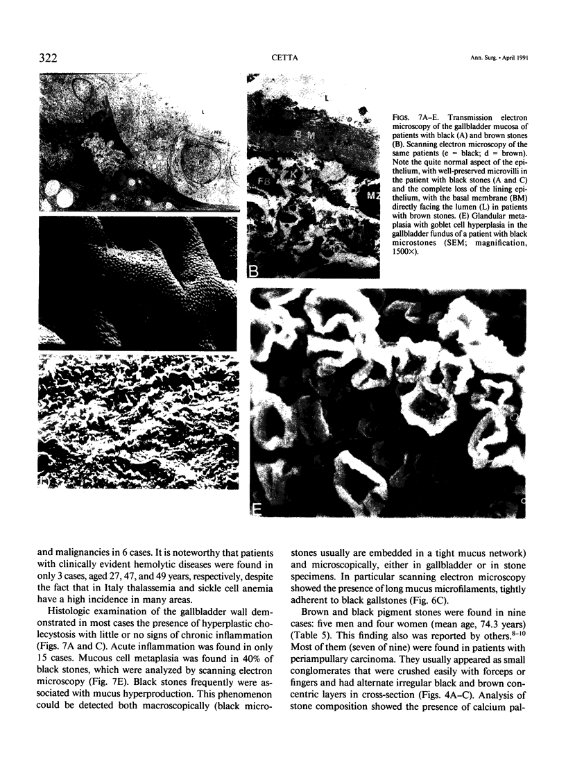
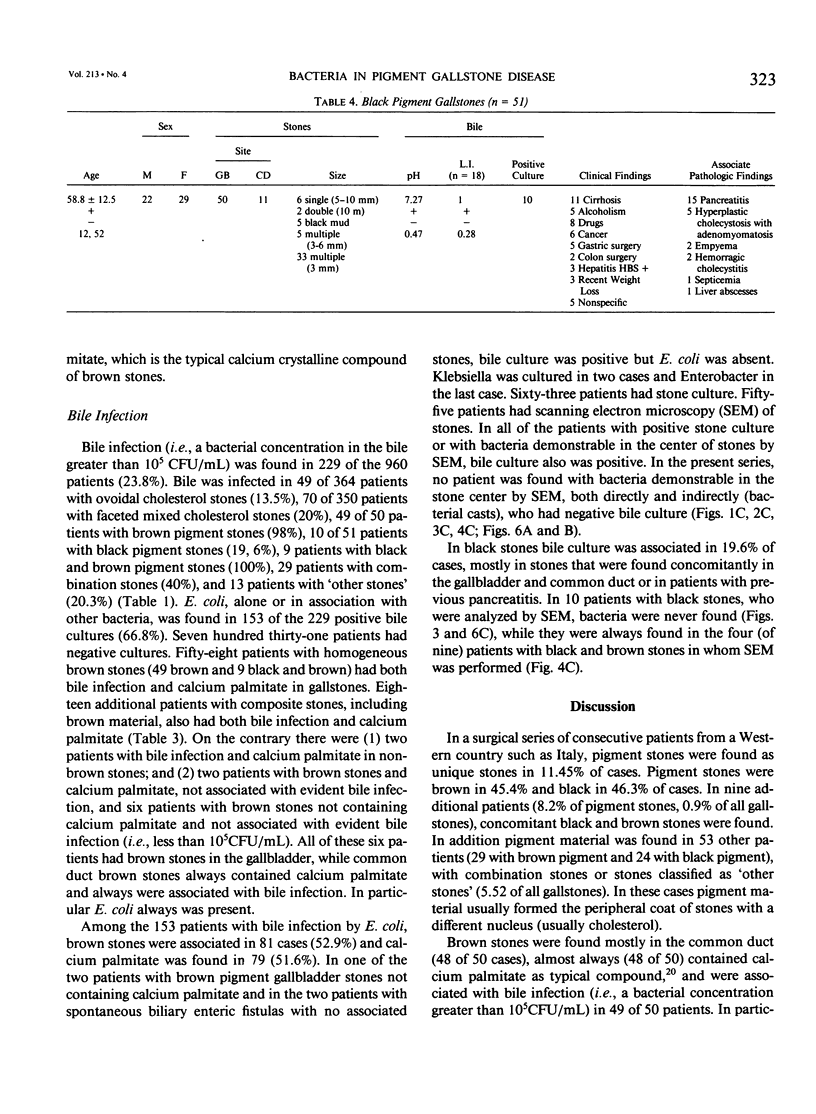
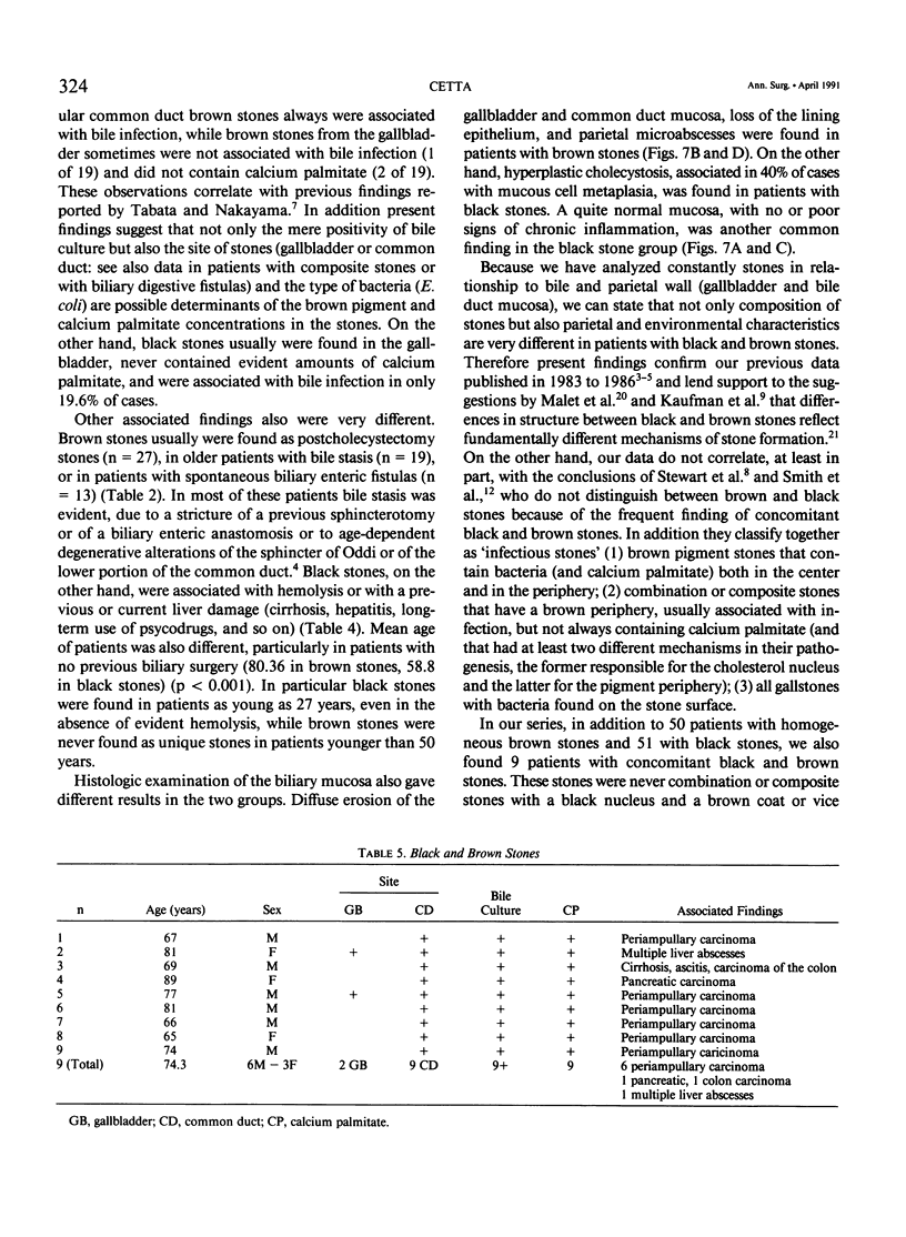
One hundred ten of nine hundred sixty consecutive patients who underwent surgery for gallstones (GS) had pigment stones (PS) (11.45%). Fifty brown PSs contained calcium bilirubinate, small amounts of cholesterol, and always calcium palmitate, were usually found in the common duct (96%), and were almost always associated with bile infection (98%) and diffuse erosion of the biliary mucosa. Fifty-one black PSs contained bilirubin polymers, calcium carbonate, and/or phosphate, seldom cholesterol, and never evident amounts of calcium palmitate, were mostly found in the gallbladder, and were associated with hemolysis or liver damage and with hyperplastic cholecystosis. Bile infection was found in 19.6% of cases, but bacteria were never found in the center of black PSs by scanning electron microscopy. Nine additional patients (8.2% of PSs, 0.9% of GSs) had concomitant black and brown PSs that were mostly found in the common duct and were always associated with bile infection. It is suggested that, even if PSs with concomitant black and brown material can be found, black and brown PSs greatly differ not only in pathogenesis but also in clinical behavior and treatment. In particular bacterial infection is important only in the pathogenesis of brown PSs while it plays no role in the initial formation of cholesterol, mixed or black GSs.
Full text
PDF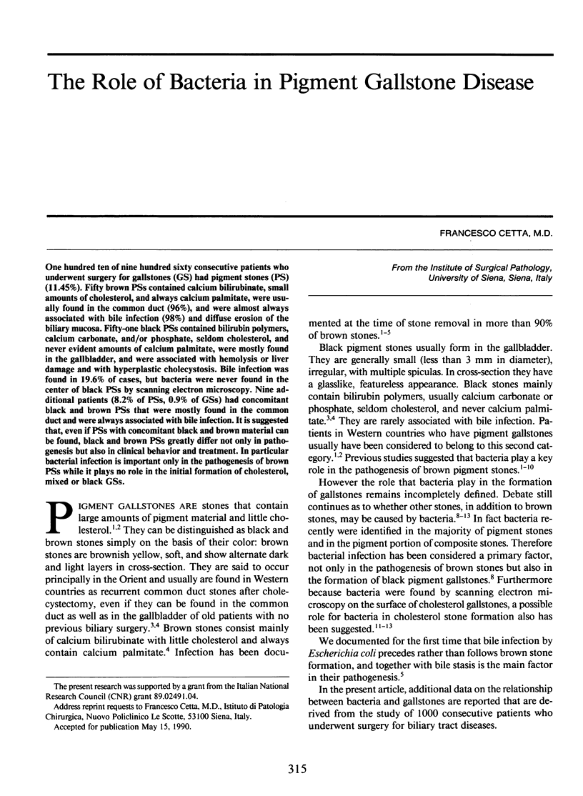

Images in this article
Selected References
These references are in PubMed. This may not be the complete list of references from this article.
- Bernhoft R. A., Pellegrini C. A., Motson R. W., Way L. W. Composition and morphologic and clinical features of common duct stones. Am J Surg. 1984 Jul;148(1):77–85. doi: 10.1016/0002-9610(84)90292-7. [DOI] [PubMed] [Google Scholar]
- Cetta F. M. Bile infection documented as initial event in the pathogenesis of brown pigment biliary stones. Hepatology. 1986 May-Jun;6(3):482–489. doi: 10.1002/hep.1840060327. [DOI] [PubMed] [Google Scholar]
- Cetta F., DeNisi A. Black and brown pigment gallstones differ in microstructure and microcomposition. Hepatology. 1986 Jan-Feb;6(1):153–154. doi: 10.1002/hep.1840060136. [DOI] [PubMed] [Google Scholar]
- Kaufman H. S., Magnuson T. H., Lillemoe K. D., Frasca P., Pitt H. A. The role of bacteria in gallbladder and common duct stone formation. Ann Surg. 1989 May;209(5):584–592. doi: 10.1097/00000658-198905000-00011. [DOI] [PMC free article] [PubMed] [Google Scholar]
- Maki T. Pathogenesis of calcium bilirubinate gallstone: role of E. coli, beta-glucuronidase and coagulation by inorganic ions, polyelectrolytes and agitation. Ann Surg. 1966 Jul;164(1):90–100. doi: 10.1097/00000658-196607000-00010. [DOI] [PMC free article] [PubMed] [Google Scholar]
- Malet P. F., Dabezies M. A., Huang G. H., Long W. B., Gadacz T. R., Soloway R. D. Quantitative infrared spectroscopy of common bile duct gallstones. Gastroenterology. 1988 May;94(5 Pt 1):1217–1221. doi: 10.1016/0016-5085(88)90015-7. [DOI] [PubMed] [Google Scholar]
- Malet P. F., Takabayashi A., Trotman B. W., Soloway R. D., Weston N. E. Black and brown pigment gallstones differ in microstructure and microcomposition. Hepatology. 1984 Mar-Apr;4(2):227–234. doi: 10.1002/hep.1840040210. [DOI] [PubMed] [Google Scholar]
- Small D. M. Cholesterol nucleation and growth in gallstone formation. N Engl J Med. 1980 Jun 5;302(23):1305–1307. doi: 10.1056/NEJM198006053022309. [DOI] [PubMed] [Google Scholar]
- Smith A. L., Stewart L., Fine R., Pellegrini C. A., Way L. W. Gallstone disease. The clinical manifestations of infectious stones. Arch Surg. 1989 May;124(5):629–633. doi: 10.1001/archsurg.1989.01410050119023. [DOI] [PubMed] [Google Scholar]
- Soloway R. D., Trotman B. W., Maddrey W. C., Nakayama F. Pigment gallstone composition in patients with hemolysis or infection/stasis. Dig Dis Sci. 1986 May;31(5):454–460. doi: 10.1007/BF01320307. [DOI] [PubMed] [Google Scholar]
- Stewart L., Smith A. L., Pellegrini C. A., Motson R. W., Way L. W. Pigment gallstones form as a composite of bacterial microcolonies and pigment solids. Ann Surg. 1987 Sep;206(3):242–250. doi: 10.1097/00000658-198709000-00002. [DOI] [PMC free article] [PubMed] [Google Scholar]
- Sutor D. J., Wooley S. E. The nature and incidence of gallstones containing calcium. Gut. 1973 Mar;14(3):215–220. doi: 10.1136/gut.14.3.215. [DOI] [PMC free article] [PubMed] [Google Scholar]
- Tabata M., Nakayama F. Bacteria and gallstones. Etiological significance. Dig Dis Sci. 1981 Mar;26(3):218–224. doi: 10.1007/BF01391633. [DOI] [PubMed] [Google Scholar]
- Trotman B. W., Soloway R. D. Pigment gallstone disease: Summary of the National Institutes of Health--international workshop. Hepatology. 1982 Nov-Dec;2(6):879–884. doi: 10.1002/hep.1840020624. [DOI] [PubMed] [Google Scholar]