Abstract
Coding sequences for the first two variable loops of the gp120 envelope glycoprotein were removed from simian immunodeficiency virus (SIV) strain 239 (SIVmac239). This deletion encompassed 100 amino acids. The resulting virus replicated poorly after transfection into immortalized T-cell lines, with peak replication occurring only after 25 to 30 days. Limited passaging of SIVmac239ΔV1V2 in cultures gave rise to a variant which had significantly improved replication kinetics but which retained the original 100-amino-acid deletion in gp120. Cloning and sequencing revealed 11 changes in the envelope, including amino acid substitutions in both gp120 (5 substitutions) and gp41(6 substitutions). Four of the five changes in gp120 are predicted to lie within and around the putative coreceptor binding domain, a region which is believed to be covered by the V1 and V2 loops in the native envelope complex. Analysis of recombinant clones surprisingly revealed that the changes in gp41 were sufficient to overcome the replication deficiency created by deletion of the V1 and V2 loops from gp120. The SIVmac239ΔV1V2 envelope displayed a significant reduction in its ability to mediate cell-cell fusion, and the infectious titer of SIVmac239ΔV1V2 was approximately four- to eightfold lower than that of parental SIVmac239. Although SIVmac239 is strongly dependent on both CD4 and a coreceptor for entry, envelope protein lacking the V1 and V2 loops was able to mediate fusion with CD4− CCR5+ cells at 60% the level observed with CD4+ CCR5+ cells. Plasma from SIVmac239-infected monkeys was at least 100 to 1,000 times more effective at neutralizing SIVmac239ΔV1V2 than SIVmac239. These results demonstrate the dispensability of the V1-V2 sequences of SIVmac239 for viral replication, a role for V1 and V2 in shielding the coreceptor binding region of the envelope, and the extreme sensitivity of a SIV lacking these sequences to antibody-mediated neutralization.
Soon after the isolation and identification of human immunodeficiency virus (HIV) type 1 (HIV-1) as the causative agent of AIDS, comparison of sequence data from different viral isolates revealed the presence of highly variable subdomains within the surface subunit (gp120) of the virus-encoded envelope protein (44, 51). The secondary structure of gp120 predicts that four of these variable sequences are largely sequestered from the rest of the protein by intrachain disulfide bonds. These four subdomains are often referred to as variable loops and are designated V1, V2, V3, and V4. Based on the assignment of disulfide bonds, the predicted folding pattern of gp120, and the crystal structure of a gp120 subunit core, it is apparent that V3 and V4 are distinct loop structures, whereas V1 and V2 share a common stem (16, 18, 24). The V1-V2 region of HIV-1 is approximately 70 amino acids long. Although the envelope genes of the simian immunodeficiency viruses (SIV) of Old World monkeys and the closely related HIV-2 are clearly distinct from that of HIV-1, their protein structures are likely to be similar (42). The region analogous to the V1 and V2 loops of HIV-1 is approximately 30 to 40 residues longer in SIV and HIV-2 and contains four additional cysteines (18). These cysteines are predicted to form two additional disulfide bonds (18).
The discovery of the variable domains in the envelope of HIV-1 quickly led to speculation that the loops may serve as an antigenic shield, screening more conserved, functionally important residues in the core of the envelope complex from immune system recognition (10). Removal of the V1 and V2 variable loops from HIV-1 strain HXB-2 (a deletion encompassing approximately 70 amino acids) gave rise to a replication-competent variant with markedly increased sensitivity to neutralization by monoclonal antibodies directed toward epitopes within the core of the envelope complex (8). Stamatatos and Cheng-Mayer reported similar results for an HIV-1 strain from which only the V2 loop had been removed (43).
It is now well established that HIV-1 entry into target cells begins with sequential binding of the envelope complex to the cellular receptor (CD4) and then to a seven-transmembrane coreceptor (typically CCR5 or CXCR4). Receptor engagement triggers the unfurling of the envelope fusion apparatus, a process which has been extensively reviewed (9, 13, 41). The gp120 protein is composed of inner and outer domains, with the CD4 binding pocket being formed by the boundary between these two domains. A binding site for CCR5 has been identified that is composed of four antiparallel β strands, which connect the distal ends of the two domains and are collectively referred to as the bridging sheet. Although the recently solved crystal structure for HIV-1 gp120 does not include coordinates for the variable loops, the positions of these domains have been inferred from the locations of the loops-stems in the structure (22) and from antibody mapping data (29, 33, 46). Specifically, it has been postulated that the V1 and V2 loops partially occlude the binding sites for the cellular receptor (CD4) and coreceptor (22, 53). Binding to CD4 induces conformational rearrangements in the envelope complex that increase the exposure of the coreceptor binding site and facilitate the interaction of the envelope complex with the coreceptor. These rearrangements presumably involve displacement of the V1 and V2 loops to unmask the coreceptor binding site.
In this report, we describe the properties of SIV strain 239 (SIVmac239) lacking V1-V2 sequences. Unlike the results of previous reports with HIV-1 (8, 43), removal of the V1-V2 structure from SIVmac239 severely impaired viral replication. Passaging of the deletion mutant in cultures gave rise to variants bearing compensatory changes; surprisingly, the changes that allowed high-level replication of the virus missing V1-V2 mapped to the gp41 transmembrane protein. The V1-V2 deletion variant of SIVmac239 characterized in this report (SIVmac239ΔV1V2) is capable of CD4-independent fusion and is extremely sensitive to antibody-mediated neutralization.
MATERIALS AND METHODS
Viral plasmids and transfections.
For manipulation of SIVmac239 DNA, plasmids containing separate 5" and 3" halves of the proviral DNA were used. The 5" clone p239SpSp5" contains the 5" long terminal repeat (LTR) and the gag, pol, and vif genes. The 3" clone pSP72-239-3" contains the viral env gene; the open reading frames for tat, rev, nef, vpx, and vpu; and the 3" LTR. To measure the replication of viruses containing the parental or altered envelope genes, 3" clones containing the relevant envelope sequences were digested with SphI, ligated to SphI-digested p239SpSp5" to generate full-length proviral DNA, and used to transfect cells by use of DEAE-dextran as a transfection reagent (30). Viral replication was measured by quantitating the appearance of the p27 gag antigen in the supernatant of transfected cells by using an antigen capture assay (Coulter Corporation, Hialeah, Fla.).
Passaging of SIVmac239ΔV1V2.
The supernatant collected at peak p27 from 221 cells transfected with SIVmac239ΔV1V2 DNA was used to infect fresh 221 cells (221 cells are a herpes virus saimiri immortalized rhesus monkey T-cell line). Supernatant was collected from infected 221 cells once a week and passaged to fresh 221 cells. Weekly samples were also frozen and kept at −80°C. To compare the infectivities of passaged derivatives, archived stocks and parental SIVmac239 stocks were thawed, normalized for p27 content, and assayed in parallel infections of fresh 221 cells.
Mutagenesis and subcloning.
The V1-V2 deletion in SIVmac239 was generated by the splice-overlap extension method (SOE) (49). For SOE, sequences 5" and 3" of the sequence targeted for deletion are amplified in separate PCRs. In each PCR, the primer adjacent to the sequence targeted for deletion contains a region that overlaps and is complementary to a primer from the other reaction. The resulting PCR products thus have a region of overlapping sequence. In the next step, the two PCR products are mixed together and fused by a second round of PCR. Primers complementary to the extreme 5" and 3" ends of the predicted fusion product are included in the final PCR to improve the yield of the fusion product. A deletion of 300 nucleotides was generated in the SIVmac239 gp120 envelope sequence by SOE. The resulting fusion product was then digested and cloned into pSP72-239-3" to replace the parental envelope sequence.
Cell-cell fusion assay.
Constructs containing parental or mutant envelope genes under the control of Tat, Rev, and the viral LTR were transfected into 293T cells by the calcium phosphate method (2). At 24 h posttransfection, the medium was removed and the cells were rinsed with phosphate-buffered saline. At 48 h posttransfection, the transfected cells were overlaid with CEMx174SIV-SEAP cells. CEMx174SIV-SEAP cells contain an integrated gene encoding secreted alkaline phosphatase (SEAP) under the control of the Tat-inducible SIV LTR. In the event of Env-mediated fusion, the cytoplasmic contents of the two cell types mix, and the Tat protein produced in the transfected 293T cells induces the expression of the SEAP indicator gene provided by the CEMx174SIV-SEAP cells. Fusion is quantitated by measuring the appearance of SEAP activity in the supernatant by using a Phospha-Light assay system (Tropix, Bedford, Mass.).
The SIVmac239 envelope expression construct was a gift from the laboratory of J. Sodroski and was described previously (25). Briefly, the expression construct contains a full-length SIVmac239 provirus with an extensive deletion of the gag gene. This construct expresses all the other viral proteins, including Tat, Rev, and Env, under the control of the viral LTR.
CD4-independent fusion assay.
The CD4-independent fusion assay was described previously (37). Briefly, effector QT6 cells were infected with vaccinia virus expressing T7 polymerase (vTF1.1) (1) and then transfected with plasmid pCDNA3 containing the designated env gene via the calcium phosphate transfection method. Target QT6 quail cells were transfected with plasmids expressing CD4, the coreceptor, and T7 luciferase. AT 24 h postinfection or posttransfection, effector cells were added to the target cells; fusion was quantitated at 8 h postmixing by lysing cells with 0.05% Triton X-100 in phosphate-buffered saline, analyzing an aliquot of lysate with an equal volume of luciferase assay reagent (Promega), and reading luminescence in a luminometer (Wallac).
Neutralization assay.
Neutralization titers of animal plasma were determined by infecting CEMx174SIV-SEAP cells; the method was described in detail previously (26). Briefly, successive dilutions of animal plasma or pooled SIV-positive plasma samples were incubated with test virus for 1 h at room temperature and then added to CEMx174SIV-SEAP cells. At 72 h postinfection, SEAP activity in culture supernatants was measured by using the Phospha-Light assay kit (Applied Biosystems). Neutralization is reported as a reduction in the percentage of SEAP activity produced by infection in the absence of plasma.
RESULTS
Construction and replication of SIVmac239ΔV1V2.
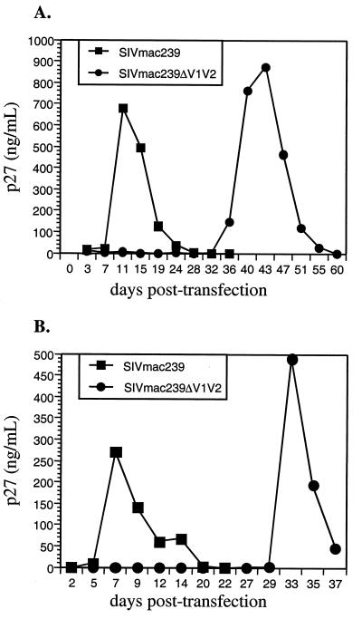
A deletion encompassing coding sequences for the first and second variable loops (V1 and V2) of gp120 (residues 110 to 209) was introduced into the SIVmac239 envelope gene (Fig. 1). The cysteines that form the disulfide-bonded stem of the V1-V2 structure were retained, and the deleted segment was replaced with a three-amino-acid (glycine-alanine-glycine) linker. The effect of this extensive deletion of the envelope sequence on viral replication was tested by transfection of proviral DNA containing the V1-V2 deletion into immortalized rhesus monkey T cells (221 cells). Peak replication of SIVmac239, as measured by the appearance of p27gag antigen in the tissue culture supernatant, typically occurred at about day 6 or 7 posttransfection. In contrast, the mutant bearing the V1-V2 deletion replicated quite poorly, with peak replication being delayed until 24 to 30 days posttransfection in repeated experiments (Fig. 2).
FIG. 1.
Variable loops V1 and V2. (A) Folding pattern of the V1 and V2 loops of HIV-1 Env protein gp120. (B) Predicted folding pattern of the V1-V2 structure of SIVmac239 (12). (C) SIVmac239ΔV1V2. The deletion mutant was created by deleting 100 amino acids (a.a.) of envelope, encompassing all of V1 and V2, and replacing the deleted residues with a Gly-Ala-Gly linker. The deletion mutant retains the stem of the V1-V2 structure. Potential sites of N-linked glycosylation attachment are indicated by asterisks.
FIG. 2.
Replication of SIVmac239ΔV1V2. Mutant DNA was used to transfect immortalized rhesus macaque T cells (221 cells), and viral replication was assayed by monitoring the expression of p27 capsid protein in the supernatant. Panels A and B show results of two independent transfections.
Virus harvested at the peak of replication of SIVmac239ΔV1V2 following transfection was used to infect fresh 221 cells; genomic DNA extracted from these cells was then used to amplify and sequence viral envelope genes. Envelope genes cloned and sequenced from three independent transfections revealed that the virus had retained the original 100-amino-acid deletion and acquired additional substitutions in the envelope. Interestingly, each clone contained changes in the ectodomain of gp41 (Table 1).
TABLE 1.
Early substitutions in SIVmac239ΔV1V2
| Transfection | Peaka (day) | Subunit | Substitution |
|---|---|---|---|
| 1 | 33 | gp120 | I437T |
| gp41 | V529G | ||
| 2 | 29 | gp120 | None |
| gp41 | F682S | ||
| 3 | 30 | gp120 | None |
| gp41 | D683G | ||
| Y690H |
Measured as the p27 concentration in transfected cell supernatants.
An indicator cell line was used to compare the infectious titer of SIVmac239 with that of SIVmac239ΔV1V2. Viral stocks were collected at peak replication from transfected cells, normalized for p27gag content, serially diluted, and used to infect CEMx174SIV-SEAP cells. CEMx174SIV-SEAP is an indicator cell line that expresses SEAP in response to viral infection. SEAP expression requires the virus to complete the early stage of infection, including entry, reverse transcription, integration of the DNA provirus, and expression of the Tat protein (26). Comparison of viral titers measured with the SEAP indicator cells (SEAP activity per nanogram of p27) revealed a four- to eightfold reduction in the infectivity of the deletion mutant virus compared to the parental virus (Fig. 3).
FIG. 3.
Infectious titer of SIVmac239ΔV1V2. Viral stocks were normalized for p27 content and then used for duplicate infections of CEMx174SIV-SEAP indicator cells, which contain a Tat-inducible gene for SEAP. SEAP activity was measured 72 h postinfection, a measure which principally reflects the first round of infection (26). Indicator cells were the same cells as those used in the neutralization assay (see Materials and Methods). Error bars show standard deviations.
SIVmac239ΔV1V2 harvested from transfected 221 cells replicated well in rhesus monkey peripheral blood mononuclear cell cultures but with delayed kinetics compared to parental SIVmac239 (data not shown).
Receptor-independent fusion.
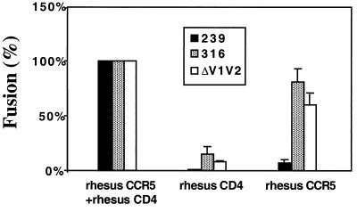
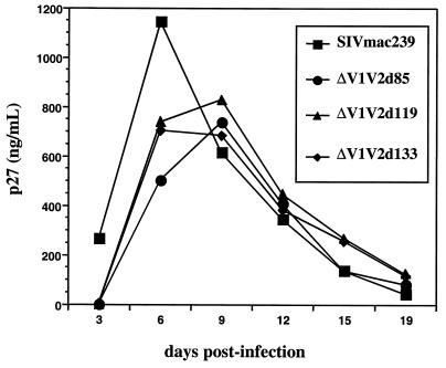
Although the recently published crystal structure for HIV-1 gp120 did not contain the variable loop domains, several lines of evidence indicate that the V1 and V2 loops partially mask the receptor and coreceptor binding sites (7, 55). Current models for the mechanism of viral entry postulate that binding to CD4 induces exposure of the coreceptor binding site. Based on this model, we hypothesized that removal of the V1 and V2 loops from SIVmac239 should increase the exposure of the CCR5 coreceptor binding site on the native envelope complex. In cell-cell fusion assays, the ability of the parental SIVmac239 envelope complex to promote membrane fusion was reduced by >95% in the absence of CD4 (CD4− CCR5−) and by 90 to 95% even when the target cells expressed high levels of the CCR5 coreceptor (CD4− CCR5+) (Fig. 4). Unlike the parental envelope complex, the deletion mutant envelope complex was able to drive fusion with CD4− CCR5+ target cells at 60% the level seen with CD4+ CCR5+ cells (Fig. 4). The Env protein of SIVmac316, a variant which can replicate in CD4− CCR5+ cell lines, was included as a positive control (27). It should be noted that the percentages reported in Fig. 4 are relative to the values for the same envelope in the context of CD4 plus CCR5. The absolute fusogenic activity of the deletion mutant virus envelope was almost 10-fold lower than that of the parental SIVmac239 envelope when tested with CD4+ CCR5+ target cells; this reduction in fusogenic activity is consistent with the markedly delayed replication of SIVmac239ΔV1V2 after transfection (Fig. 2) and its reduced infectivity (Fig. 3). Similar reductions in the levels of fusion were also observed in an independent assay (see below). Thus, the overall fusogenic activity of the envelope was significantly reduced by the removal of the V1 and V2 loops, but most of the fusogenic activity with the mutant Env was retained in the absence of CD4. Changes that compensate for the deletion of V1 and V2. To further understand the effect of the V1-V2 deletion on envelope structure and function, viral stocks generated by transfection were passaged continuously on 221 cells. Viral supernatants collected at various times during passage were normalized for p27 content and used to infect 221 cells. Virus collected between days 119 and 133 of passage had replication kinetics similar to those of parental SIVmac239 (Fig. 5). Viral envelope sequences were amplified from cells infected with the day 119 virus, cloned, and sequenced. Complete sequencing of the cloned envelope sequences revealed that the day 119 virus retained the original 100-amino-acid deletion of V1 and V2 but acquired 11 nonsynonomous changes throughout the envelope, including five substitutions in gp120 and six substitutions in the ectodomain of gp41 (Table 2). Among the six changes in gp41, there was a notable clustering of four substitutions within a 20-amino-acid stretch adjacent to the membrane-spanning segment of the protein.
FIG. 4.
The mutant envelope complex with a deletion of V1 and V2 can mediate CD4-independent fusion. Effector cells expressed parental SIVmac239 envelope (black bars), SIVmac316 envelope (grey bars), or SIVmac239ΔV1V2 envelope (white bars). Target cells expressed CD4 and coreceptor CCR5, CD4 alone, or CCR5 alone. Error bars show standard deviations. (For more details, see Materials and Methods.)
FIG. 5.
Infectivity of passaged SIVmac239ΔV1V2. SIVmac239ΔV1V2 was passaged continuously on 221 cells, and supernatants were collected weekly. Supernatants collected on days 85 (d85), 119, and 133 of passage were normalized for p27 content and used to infect fresh 221 cells.
TABLE 2.
Amino acid changes in SIVmac239ΔV1V2(d119)
| Substitution | Subunit | Domain | Function | Reference(s) |
|---|---|---|---|---|
| V32A | gp120 | N terminus | Subunit interactions | 17, 21 |
| E218K | gp120 | Bridging sheet | Coreceptor binding | 35, 36 |
| I335V | gp120 | V3 loop | Coreceptor binding | 35, 36 |
| W345R | gp120 | V3 stem | Coreceptor binding | 35, 36 |
| E454K | gp120 | Bridging sheet | Coreceptor binding | 35, 36 |
| V529A | gp41 | Fusion peptide | Membrane insertion | 4, 14, 15, 45 |
| T591A | gp41 | N peptide | Fusion (multimer) | 9, 41 |
| G679S | gp41 | Trp-rich motif | Fusion and incorporation | 38 |
| I688V | gp41 | Trp-rich motif | Fusion and incorporation | 38 |
| Y690H | gp41 | Trp-rich motif | Fusion and incorporation | 38 |
| V699A | gp41 | Membrane spanning | Membrane anchor | 50 |
To determine whether these 11 changes were sufficient to compensate for the replication defect in SIVmac239ΔV1V2, an XhoI-NheI fragment containing all 11 substitutions was used to replace the corresponding fragment in the original p239Sp3"ΔV1V2 construct. The virus containing the deletion and all 11 substitutions, SIVmac239ΔV1V2(d119), replicated significantly better following DNA transfection than the original deletion mutant (Fig. 6).
FIG. 6.
Mapping of compensatory changes in SIVmac239ΔV1V2(d119). (A) The top two lines depict the parental and mutant constructs; arrows indicate restriction fragments used to generate subclones. The third line depicts the envelope gene cloned from day 119 posttransfection, with the positions of the 11 nonsynonymous changes indicated. The last six lines depict the subclones used to map changes that compensate for the deletion of the V1 and V2 loops. The relative capacity of each variant to replicate in 221 cells is indicated to the right (++++, replication like that of the wild type; −, no detectable replication; −/+, extreme delay in replication typical of SIVΔV1V2 transfections; according to the data in Fig. 6B and other results not shown). (B) Replication of subclones of SIVmac239ΔV1V2(d119) in 221 cells. Data for variant X-C/M-N are not shown.
To determine whether some or all of the 11 changes were sufficient to compensate for the deletion of the V1 and V2 loops, subsets of the 11 changes were introduced into the original SIVmac239ΔV1V2 construct, and viral replication was measured after transfection of 221 cells (Fig. 6). Surprisingly, the subclones that replicated best were those that contained at least some of the changes in gp41, whereas the variant carrying only the five substitutions in gp120 failed to replicate at all (Fig. 6B). Thus, amino acid alterations in the ectodomain of gp41 are principally responsible for allowing high-level replication of the virus lacking the V1 and V2 loops.
Surface expression.
Western blot analysis of transiently transfected cells revealed that the envelope protein lacking the V1 and V2 loops was expressed and processed into the gp120 and gp41 subunits. To examine the expression and transport to the cell surface of the mutant envelope complexes, transfected 293T cells were examined by surface staining with an anti-gp120 monoclonal antibody followed by flow cytometry (Fig. 7A). At 48 h posttransfection, the parental SIVmac239 and SIVmac239ΔV1V2 envelope complexes were both expressed on the cell surface and at approximately similar levels (mean peak channel fluorescence values, 50.3 and 51.4, respectively). The envelope protein bearing the deletion of the V1 and V2 loops as well as the six changes in gp41 (clone C-N) was also expressed on the cell surface at a similar level (mean peak channel fluorescence value, 60.9).
FIG. 7.
(A) Surface expression of the envelope. 293T cells were transfected with Env expression constructs, and cells were harvested 48 h posttransfection, stained with monoclonal antibody KK42 and a fluorescein isothiocyanate-conjugated secondary antibody, and then sorted by flow cytometry. (B) Cell-cell fusion assay. 293T cells were transfected with envelope expression constructs as in panel A or control vectors. At 48 h posttransfection, the cells were overlaid with CEMx174SIV-SEAP indicator cells, and SEAP activity was measured at the indicated times. (See Materials and Methods for details.)
Membrane fusion.
The ability of transiently expressed envelope proteins to drive cell-cell fusion was also measured in a different assay. Briefly, 293T cells were transfected with Env expression constructs containing tat, rev, and env; at 48 h posttransfection, these cells were overlaid with CEMx174SIV-SEAP cells. In the event of Env-mediated fusion, syncitia form and SEAP expression is induced. By 24 h after mixing of the effector and target cells, the envelope protein with the deletion of the V1 and V2 loops had achieved significantly less cell-cell fusion than the parental envelope protein (Fig. 7B). Similar reductions in absolute fusogenicity were seen in the CD4-independent fusion assay described above (Fig. 4). The parental and mutant envelope proteins were expressed to comparable levels on the surfaces of transfected QT6 cells (data not shown) and 293T cells (Fig. 7A).
Interestingly, the compensatory changes in gp41 improved the fusogenic capacity of the deletion-containing envelope protein, although not to the level seen with the parental envelope protein.
Antibody-mediated neutralization of SIVmac239ΔV1V2.
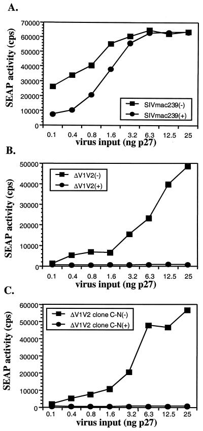
SIVmac239 is a molecular clone of a primary pathogenic SIV isolate (12, 34). In monkeys inoculated with SIVmac239, neutralizing antibody responses are slow to develop (5, 6, 11). SIVmac239 is very ineffectively neutralized by antisera, even by sera from SIVmac239-infected monkeys with high titers of antibodies that recognize Env in an enzyme-linked immunosorbent assay (26). To ascertain the influence, if any, of the V1 and V2 loops on sensitivity to antibody-mediated neutralization, SIV-positive plasma specimens from infected monkeys were tested for the ability to neutralize SIVmac239 and SIVmac239ΔV1V2 in an in vitro neutralization assay (26). Stocks of SIVmac239 and SIVmac239ΔV1V2 were harvested from transfected 221 cells and normalized for p27 content. Successive dilutions of each stock were preincubated with pooled SIV-positive plasma samples or an SIV-negative control plasma sample and then added to CEMx174SIV-SEAP indicator cells. The wild-type virus, SIVmac239, was only weakly affected by the presence of SIV-positive plasma. Moreover, the neutralizing activity of the plasma was overcome by increasing the viral input (Fig. 8A). In contrast, SIVmac239ΔV1V2 and SIVmac239ΔV1V2(clone C-N) (containing the six compensatory substitutions in gp41) were both extremely sensitive to neutralization by the SIV-positive plasma, even at the highest levels of viral input (Fig. 8B and C).
FIG. 8.
Neutralization of SIVmac239ΔV1V2 by SIV-positive rhesus monkey plasma. Increasing concentrations of stock SIVmac239 or SIVmac239ΔV1V2 were incubated with control plasma (squares) or with pooled SIV-positive plasma (circles) diluted 1:200 with RPMI medium. (A) Effect of increasing concentrations of SIVmac239 on sensitivity to SIV-positive plasma. (B) Effect of increasing concentrations of SIVmac239ΔV1V2 on sensitivity to SIV-positive plasma. (C) Effect of increasing concentrations of SIVmac239ΔV1V2(clone C-N) on sensitivity to SIV-positive plasma.
In subsequent assays, a fixed amount of each virus was incubated with serial dilutions of plasma, and the neutralization sensitivity of each virus was measured as the percent reduction in infectivity compared to that of the same virus treated with SIV-negative plasma (26). Pooled plasma samples from SIVmac239- and SIVmac251-infected animals had very little neutralizing activity against SIVmac239. However, the same plasma pool reduced the infectivity of SIVmac239ΔV1V2 by >50% at dilutions as high as 1:40,000 (Fig. 9A). SIVmac239ΔV1V2(clone C-N) exhibited sensitivity to antibody-mediated neutralization similar to that of SIVmac239ΔV1V2 (Fig. 9B).
FIG. 9.
SIVmac239ΔV1V2 is extremely sensitive to neutralization by SIV-positive plasma. CEMx174SIV-SEAP cells were infected with virus in the presence of increasing dilutions of plasma from uninfected rhesus macaques (closed symbols) or plasma from SIV-positive rhesus macaques (open symbols). (A) Neutralization of SIVmac239ΔV1V2. Background SEAP levels in this experiment are indicated by the line labeled “uninfected.” (B) Comparative neutralization of SIVmac239ΔV1V2 and SIVmac239ΔV1V2(clone C-N).
Further neutralization assays were performed with individual plasma samples collected from three SIVmac239-infected monkeys at 16 and 24 weeks postinfection. These three animals included one with a very weak overall antibody response to SIV (467-95) and two animals with moderate to strong overall antibody responses (258-95 and 259-95), as measured by an enzyme-linked immunosorbent assay, to whole, lysed virions. None of the monkey plasma samples from either time point was able to neutralize the homologous virus (Fig. 10). In contrast, plasma from all three animals had strong neutralizing activity against SIVmac239ΔV1V2 as early as 16 weeks postinfection. The potencies of the individual samples against SIVmac239ΔV1V2(d119) varied, with 50% neutralization titers as high as 1:81,920 (Fig. 10).
FIG. 10.
Neutralization of SIVmac239ΔV1V2 with various plasma samples. Plasma samples from three SIV-positive rhesus monkeys were tested for neutralizing activity against SIVmac239 (open symbols) and SIVmac239ΔV1V2 (closed symbols). (A) Plasma collected from animals at week 16 postinfection. (B) Plasma collected at week 24 postinfection.
DISCUSSION
HIV-1 isolates display a broad range of sensitivities to antibody-mediated neutralization, ranging from neutralization-sensitive, T-cell-line-adapted isolates to neutralization-resistant, primary isolates. To explain this variability, a model has been proposed in which the native HIV-1 envelope complex oscillates between open and closed states (23). According to this model, envelope spikes of T-cell-line-adapted HIV-1 isolates are predicted to have an equilibrium that favors the open state, in which the envelope core, including the receptor and coreceptor binding sites, are exposed. In contrast, difficult-to-neutralize, primary isolates are expected to favor the closed state. Structural features that are thought to contribute to the state of the envelope complex include the locations of N-linked glycans, subunit-subunit interactions, and the positioning of the variable loops (8, 20, 28, 29, 52, 54, 55). In fact, the removal of large portions of the V1-V2 structure has been reported to greatly increase the sensitivity of HIV-1 to neutralization by HIV-1-positive human sera or monoclonal antibodies directed against epitopes in the core of the gp120 protein (8, 43). We have now demonstrated a similar result with SIVmac239, a lentivirus of Old World monkeys; removal of the 100-amino-acid V1-V2 structure from SIVmac239 results in a variant with significantly increased sensitivity to plasma from SIV-infected rhesus monkeys. Deletion of the V1 and V2 loop sequences from SIVmac239 may have resulted in an envelope structure that resembles the open configuration, that is, with critical epitopes more accessible to antibody binding and therefore more readily neutralizable.
Deletion of the V1 and V2 loops may also have influenced the sensitivity of the virus to antibody-mediated neutralization through effects on envelope function. For example, if the V1 and V2 loops contribute to the structure of the coreceptor binding site itself, the 100-amino-acid deletion may create a less stable, or altered, binding site. A decrease in the efficiency of entry could prolong the window of opportunity during which antibodies could access the envelope complex or perhaps delay the process of entry in such a way as to increase the opportunity for antibodies to recognize and block the transition to a fusogenic state. This explanation is also consistent with the reduced infectivity of SIVmac239ΔV1V2 and the overall reduction in fusogenicity of the mutant envelope with a deletion of the V1 and V2 loops in cell-cell fusion assays (Fig. 3 and 4).
It has also been suggested that the density of envelope spikes on the surface of virions can influence relative sensitivity to antibody-mediated neutralization (7). If the V1-V2 deletion results in a large decrease in spike incorporation or an effective decrease in the number of functional oligomers (e.g., due to a higher level of spontaneous shedding of gp120), then the increased neutralization sensitivity of SIVmac239ΔV1V2 could reflect a reduction in the number of antibody molecules required to inactivate mutant virions. Our data do not rule out the possibility that the V1-V2 deletion has resulted in decreased incorporation of functional envelope spikes.
It has been hypothesized that high-titer antibody in HIV patients may be induced by monomeric envelope protein or viral debris (32). An alternative hypothesis for the neutralization sensitivity of SIVmac239ΔV1V2 is that the mutant virions contain a higher proportion of nonoligomeric or nonfunctional envelope proteins, and antibody bound to such molecules could neutralize infectivity through simple steric hindrance (7, 31).
Experimental infection of rhesus monkeys with SIVmac239 typically induces responses by both the cellular and the humoral branches of the immune system; nevertheless, viral loads remain high throughout the course of infection. Longitudinal studies of the antibody response in SIV-infected macaques have revealed that the maturation of neutralizing antibodies against SIVmac239 is a lengthy process, requiring months to generate antibodies with a detectable capacity to neutralize the virus present at the time of initial infection (5, 6, 11). Sera from experimentally infected animals typically exhibit weak or undetectable neutralizing activity against SIVmac239 (26). We have shown that sera collected from three animals in the early stages of experimental infection did not neutralize SIVmac239, yet these same samples clearly neutralized the SIVmac239ΔV1V2 variant, with 50% neutralization titers as high as 1:81,920. The implications of these results are as follows: (i) infection with parental SIVmac239, with the V1 and V2 loops intact, elicits antibodies which recognize viral epitopes that are largely incapable of neutralizing SIVmac239; and (ii) these antibodies can be generated during the early stages of infection. The comparable inability of high-titer antibodies to neutralize primary isolates of HIV-1 gave rise to the hypothesis that the anti-Env antibody response may be largely directed against forms of the envelope protein other than the native complex, such as shed monomeric gp120 or viral debris (33). Such antibodies are believed to be inefficient at binding to oligomeric spikes on the virion surface of primary isolates.
The V1 and V2 loops of the HIV-1 gp120 molecule are thought to lie on the surface of the envelope complex and partially mask the receptor and coreceptor binding sites in the core of the gp120 protein (54). Evidence from several studies suggests that binding of the envelope complex to CD4 moves the loops and increases the exposure of the coreceptor binding site (3, 20, 39, 40, 46-48). Furthermore, Kolchinsky et al. (19) reported that specific changes within the V1-V2 coding sequences can confer a CD4-independent phenotype on HIV-1. We observed that the SIV envelope protein with a deletion of the V1 and V2 loops was capable of mediating CD4-independent membrane fusion. Our results are consistent with a model in which the removal of the V1-V2 sequences bypasses the need for prior interactions with CD4 and allows direct interactions with a coreceptor.
We also report the novel finding that amino acid substitutions in the gp41 transmembrane subunit can compensate for the deletion of sequences from gp120. Of the six changes in gp41, four were found to cluster within a 20-amino-acid stretch of the ectodomain adjacent to the transmembrane-spanning segment (Table 2). Two additional, independent transfection experiments gave rise to SIVmac239ΔV1V2 variants with changes in this same region (Table 1). This C-terminal region of the ectodomain has been referred to as the tryptophan-rich motif and is highly conserved among lentiviruses (38). Although its exact role in viral replication has not been established, specific mutations in the region affect post-CD4-binding steps in the fusion process and the incorporation of the envelope into virions (38). Understanding how these changes compensate for the deletion of the V1 and V2 loops may also reveal novel interactions between the gp120 and gp41 subunits of the envelope complex.
Although a high-resolution structure of the SIV gp120 protein has not been reported, it is possible to map SIVgp120 residues onto the recently published structure of HIV-1 gp120 (22). The approximate structural locations of the five changes in the gp120 sequence of SIVmac239ΔV1V2(d119) are summarized in Table 2. Four of these five changes clustered at the distal end of the molecule, in or near regions thought to interact with the receptor and coreceptor. Assuming that the basic structural features of HIV-1 gp120 are conserved in the SIV protein, these changes included two changes in the bridging sheet, one change in V3, and one change in the V3 stem. Selection of changes in this region is consistent with the hypothesis that the V1-V2 domain lies close to or interacts with the coreceptor binding region of gp120. The observed substitutions alone do not rescue viral replication but may represent subtle structural or biochemical adjustments to the removal of the overlying loops. The fifth substitution in gp120 was located near the N terminus, a region previously implicated in gp120-gp41 interactions (17, 21).
Although the V1 and V2 loops of SIVmac239 differ from those of HIV-1 in size and sequence, our results demonstrate that they make similar contributions to the function and antigenicity of the envelope complex. Specifically, the V1 and V2 loops contribute to the extreme neutralization-resistant properties of SIVmac239, and removal of the loops exposes the virus to efficient antibody-mediated neutralization. The conservation of design and function between the HIV-1 and SIV envelope complexes suggests that the SIV-rhesus monkey system can be used to explore features of the virus-encoded envelope that contribute to antigenicity, immunogenicity, and pathogenesis and that may be relevant to vaccine development.
Acknowledgments
W.E.J. was supported by an Elizabeth Glaser Pediatric AIDS Foundation 2-year scholar award (grant PF-77385). This work was also supported by Public Health Service grants AI25328, AI35365, and RR00168 and by a CFAR award to the University of Massachusetts Medical School (grant AI42845). R.W.D. was supported by an Elizabeth Glaser scientist award and by NIH grant RO135383. B.A.P. was supported by Public Health Service grant F32 AI10577.
REFERENCES
- 1.Alexander, W. A., B. Moss, and T. R. Fuerst. 1992. Regulated expression of foreign genes in vaccinia virus under the control of bacteriophage T7 RNA polymerase and the Escherichia coli lac repressor. J. Virol. 66:2934-2942. [DOI] [PMC free article] [PubMed] [Google Scholar]
- 2.Ausubel, F. M., R. Brent, R. E. Kingston, D. D. Moore, J. G. Seidman, J. A. Smith, and K. Struhl (ed.). 1987. Current protocols in molecular biology. John Wiley & Sons, Inc., New York, N.Y.
- 3.Bandres, J. C., Q. F. Wang, J. O'Leary, F. Baleaux, A. Amara, J. A. Hoxie, S. Zolla-Pazner, and M. K. Gorny. 1998. Human immunodeficiency virus (HIV) envelope binds to CXCR4 independently of CD4, and binding can be enhanced by interaction with soluble CD4 or by HIV envelope deglycosylation. J. Virol. 72:2500-2504. [DOI] [PMC free article] [PubMed] [Google Scholar]
- 4.Bosch, M. L., P. L. Earl, K. Fargnoli, S. Picciafuoco, F. Giombini, F. Wong-Staal, and G. Franchini. 1989. Identification of the fusion peptide of primate immunodeficiency viruses. Science 244:694-697. [DOI] [PubMed] [Google Scholar]
- 5.Burns, D. P., C. Collignon, and R. C. Desrosiers. 1993. Simian immunodeficiency virus mutants resistant to serum neutralization arise during persistent infection of rhesus monkeys. J. Virol. 67:4104-4113. [DOI] [PMC free article] [PubMed] [Google Scholar]
- 6.Burns, D. P., and R. C. Desrosiers. 1994. Envelope sequence variation, neutralizing antibodies, and primate lentivirus persistence. Curr. Top. Microbiol. Immunol. 188:185-219. [DOI] [PubMed] [Google Scholar]
- 7.Burton, D. R., and P. W. Parren. 2000. Vaccines and the induction of functional antibodies: time to look beyond the molecules of natural infection? Nat. Med. 6:123-125. [DOI] [PubMed] [Google Scholar]
- 8.Cao, J., N. Sullivan, E. Desjardin, C. Parolin, J. Robinson, R. Wyatt, and J. Sodroski. 1997. Replication and neutralization of human immunodeficiency virus type 1 lacking the V1 and V2 variable loops of the gp120 envelope glycoprotein. J. Virol. 71:9808-9812. [DOI] [PMC free article] [PubMed] [Google Scholar]
- 9.Chan, D. C., and P. S. Kim. 1998. HIV entry and its inhibition. Cell 93:681-684. [DOI] [PubMed] [Google Scholar]
- 10.Coffin, J. M. 1986. Genetic variation in AIDS viruses. Cell 46:1-4. [DOI] [PubMed] [Google Scholar]
- 11.Cole, K. S., J. L. Rowles, B. A. Jagerski, M. Murphey-Corb, T. Unangst, J. E. Clements, J. Robinson, M. S. Wyand, R. C. Desrosiers, and R. C. Montelaro. 1997. Evolution of envelope-specific antibody responses in monkeys experimentally infected or immunized with simian immunodeficiency virus and its association with the development of protective immunity. J. Virol. 71:5069-5079. [DOI] [PMC free article] [PubMed] [Google Scholar]
- 12.Daniel, M. D., N. L. Letvin, N. W. King, M. Kannagi, P. K. Sehgal, R. D. Hunt, P. J. Kanki, M. Essex, and R. C. Desrosiers. 1985. Isolation of T-cell tropic HTLV-III-like retrovirus from macaques. Science 228:1201-1204. [DOI] [PubMed] [Google Scholar]
- 13.Doms, R. W. 2000. Beyond receptor expression: the influence of receptor conformation, density, and affinity in HIV-1 infection. Virology 276:229-237. [DOI] [PubMed] [Google Scholar]
- 14.Freed, E. O., D. J. Myers, and R. Risser. 1990. Characterization of the fusion domain of the human immunodeficiency virus type 1 envelope glycoprotein gp41. Proc. Natl. Acad. Sci. USA 87:4650-4654. [DOI] [PMC free article] [PubMed] [Google Scholar]
- 15.Gallaher, W. R. 1987. Detection of a fusion peptide sequence in the transmembrane protein of human immunodeficiency virus. Cell 50:327-328. [DOI] [PubMed] [Google Scholar]
- 16.Gregory, T., J. Hoxie, C. Watanabe, and M. Spellman. 1991. Structure and function in recombinant HIV-1 gp120 and speculation about the disulfide bonding in the gp120 homologs of HIV-2 and SIV. Adv. Exp. Med. Biol. 303:1-14. [DOI] [PubMed] [Google Scholar]
- 17.Helseth, E., U. Olshevsky, C. Furman, and J. Sodroski. 1991. Human immunodeficiency virus type 1 gp120 envelope glycoprotein regions important for association with the gp41 transmembrane glycoprotein. J. Virol. 65:2119-2123. [DOI] [PMC free article] [PubMed] [Google Scholar]
- 18.Hoxie, J. A. 1991. Hypothetical assignment of intrachain disulfide bonds for HIV-2 and SIV envelope glycoproteins. AIDS Res. Hum. Retrovir. 7:495-499. [DOI] [PubMed] [Google Scholar]
- 19.Kolchinsky, P., E. Kiprilov, P. Bartley, R. Rubinstein, and J. Sodroski. 2001. Loss of a single N-linked glycan allows CD4-independent human immunodeficiency virus type 1 infection by altering the position of the gp120 V1/V2 variable loops. J. Virol. 75:3435-3443. [DOI] [PMC free article] [PubMed] [Google Scholar]
- 20.Kolchinsky, P., E. Kiprilov, and J. Sodroski. 2001. Increased neutralization sensitivity of CD4-independent human immunodeficiency virus variants. J. Virol. 75:2041-2050. [DOI] [PMC free article] [PubMed] [Google Scholar]
- 21.Kowalski, M., J. Potz, L. Basiripour, T. Dorfman, W. C. Goh, E. Terwilliger, A. Dayton, C. Rosen, W. Haseltine, and J. Sodroski. 1987. Functional regions of the envelope glycoprotein of human immunodeficiency virus type 1. Science 237:1351-1355. [DOI] [PubMed] [Google Scholar]
- 22.Kwong, P. D., R. Wyatt, J. Robinson, R. W. Sweet, J. Sodroski, and W. A. Hendrickson. 1998. Structure of an HIV gp120 envelope glycoprotein in complex with the CD4 receptor and a neutralizing human antibody. Nature 393:648-659. [DOI] [PMC free article] [PubMed] [Google Scholar]
- 23.Labrijn, A. F., and P. W. H. I. Parren. 1999. Neutralizing epitopes of HIV-1, sect. IV, p. 18-34. In B. Korber, C. Brander, B. F. Haynes, J. P. Moore, R. Koup, B. Walker, and D. I. Watkins (ed.), HIV molecular immunology database. Los Alamos National Laboratory, Los Alamos, N.Mex.
- 24.Leonard, C. K., M. W. Spellman, L. Riddle, R. J. Harris, J. N. Thomas, and T. J. Gregory. 1990. Assignment of intrachain disulfide bonds and characterization of potential glycosylation sites of the type 1 recombinant human immunodeficiency virus envelope glycoprotein (gp120) expressed in Chinese hamster ovary cells. J. Biol. Chem. 265:10373-10382. [PubMed] [Google Scholar]
- 25.Marcon, L., and J. Sodroski. 1997. High degree of sensitivity of the simian immunodeficiency virus (SIVmac) envelope glycoprotein subunit association to amino acid changes in the glycoprotein 41 ectodomain. AIDS Res. Hum. Retrovir. 13:441-447. [DOI] [PubMed] [Google Scholar]
- 26.Means, R. E., T. Greenough, and R. C. Desrosiers. 1997. Neutralization sensitivity of cell culture-passaged simian immunodeficiency virus. J. Virol. 71:7895-7902. [DOI] [PMC free article] [PubMed] [Google Scholar]
- 27.Means, R. E., T. Matthews, J. A. Hoxie, M. H. Malim, T. Kodama, and R. C. Desrosiers. 2001. Ability of the V3 loop of simian immunodeficiency virus to serve as a target for antibody-mediated neutralization: correlation of neutralization sensitivity, growth in macrophages, and decreased dependence on CD4. J. Virol. 75:3903-3915. [DOI] [PMC free article] [PubMed] [Google Scholar]
- 28.Moore, J. P., Q. J. Sattentau, R. Wyatt, and J. Sodroski. 1994. Probing the structure of the human immunodeficiency virus surface glycoprotein gp120 with a panel of monoclonal antibodies. J. Virol. 68:469-484. [DOI] [PMC free article] [PubMed] [Google Scholar]
- 29.Moore, J. P., and J. Sodroski. 1996. Antibody cross-competition analysis of the human immunodeficiency virus type 1 gp120 exterior envelope glycoprotein. J. Virol. 70:1863-1872. [DOI] [PMC free article] [PubMed] [Google Scholar]
- 30.Naidu, Y. M., H. W. Kestler III, Y. Li, C. V. Butler, D. P. Silva, D. K. Schmidt, C. D. Troup, P. K. Sehgal, P. Sonigo, M. D. Daniel, et al. 1988. Characterization of infectious molecular clones of simian immunodeficiency virus (SIVmac) and human immunodeficiency virus type 2: persistent infection of rhesus monkeys with molecularly cloned SIVmac. J. Virol. 62:4691-4696. [DOI] [PMC free article] [PubMed] [Google Scholar]
- 31.Parren, P. W., and D. R. Burton. 2001. The antiviral activity of antibodies in vitro and in vivo. Adv. Immunol. 77:195-262. [DOI] [PMC free article] [PubMed] [Google Scholar]
- 32.Parren, P. W., D. R. Burton, and Q. J. Sattentau. 1997. HIV-1 antibody—debris or virion? Nat. Med. 3:366-367. [DOI] [PubMed] [Google Scholar]
- 33.Parren, P. W., J. P. Moore, D. R. Burton, and Q. J. Sattentau. 1999. The neutralizing antibody response to HIV-1: viral evasion and escape from humoral immunity. AIDS 13:S137-S162. [PubMed]
- 34.Regier, D. A., and R. C. Desrosiers. 1990. The complete nucleotide sequence of a pathogenic molecular clone of simian immunodeficiency virus. AIDS Res. Hum. Retrovir. 6:1221-1231. [DOI] [PubMed] [Google Scholar]
- 35.Rizzuto, C. D., and J. G. Sodroski. 1997. Contribution of virion ICAM-1 to human immunodeficiency virus infectivity and sensitivity to neutralization. J. Virol. 71:4847-4851. [DOI] [PMC free article] [PubMed] [Google Scholar]
- 36.Rizzuto, C. D., R. Wyatt, N. Hernandez-Ramos, Y. Sun, P. D. Kwong, W. A. Hendrickson, and J. Sodroski. 1998. A conserved HIV gp120 glycoprotein structure involved in chemokine receptor binding. Science 280:1949-1953. [DOI] [PubMed] [Google Scholar]
- 37.Rucker, J., B. J. Doranz, A. L. Edinger, D. Long, J. F. Berson, and R. W. Doms. 1997. Cell-cell fusion assay to study role of chemokine receptors in human immunodeficiency virus type 1 entry. Methods Enzymol. 288:118-133. [DOI] [PubMed] [Google Scholar]
- 38.Salzwedel, K., J. T. West, and E. Hunter. 1999. A conserved tryptophan-rich motif in the membrane-proximal region of the human immunodeficiency virus type 1 gp41 ectodomain is important for Env-mediated fusion and virus infectivity. J. Virol. 73:2469-2480. [DOI] [PMC free article] [PubMed] [Google Scholar]
- 39.Sattentau, Q. J., and J. P. Moore. 1991. Conformational changes induced in the human immunodeficiency virus envelope glycoprotein by soluble CD4 binding. J. Exp. Med. 174:407-415. [DOI] [PMC free article] [PubMed] [Google Scholar]
- 40.Sattentau, Q. J., J. P. Moore, F. Vignaux, F. Traincard, and P. Poignard. 1993. Conformational changes induced in the envelope glycoproteins of the human and simian immunodeficiency viruses by soluble receptor binding. J. Virol. 67:7383-7393. [DOI] [PMC free article] [PubMed] [Google Scholar]
- 41.Skehel, J. J., and D. C. Wiley. 1998. Coiled coils in both intracellular vesicle and viral membrane fusion. Cell 95:871-874. [DOI] [PubMed] [Google Scholar]
- 42.Smith, T. F., A. Srinivasan, G. Schochetman, M. Marcus, and G. Myers. 1988. The phylogenetic history of immunodeficiency viruses. Nature 333:573-575. [DOI] [PubMed] [Google Scholar]
- 43.Stamatatos, L., and C. Cheng-Mayer. 1998. An envelope modification that renders a primary, neutralization-resistant clade B human immunodeficiency virus type 1 isolate highly susceptible to neutralization by sera from other clades. J. Virol. 72:7840-7845. [DOI] [PMC free article] [PubMed] [Google Scholar]
- 44.Starcich, B. R., B. H. Hahn, G. M. Shaw, P. D. McNeely, S. Modrow, H. Wolf, E. S. Parks, W. P. Parks, S. F. Josephs, R. C. Gallo, et al. 1986. Identification and characterization of conserved and variable regions in the envelope gene of HTLV-III/LAV, the retrovirus of AIDS. Cell 45:637-648. [DOI] [PubMed] [Google Scholar]
- 45.Steffy, K. R., G. Kraus, D. J. Looney, and F. Wong-Staal. 1992. Role of the fusogenic peptide sequence in syncytium induction and infectivity of human immunodeficiency virus type 2. J. Virol. 66:4532-4535. [DOI] [PMC free article] [PubMed] [Google Scholar]
- 46.Sullivan, N., Y. Sun, Q. Sattentau, M. Thali, D. Wu, G. Denisova, J. Gershoni, J. Robinson, J. Moore, and J. Sodroski. 1998. CD4-induced conformational changes in the human immunodeficiency virus type 1 gp120 glycoprotein: consequences for virus entry and neutralization. J. Virol. 72:4694-4703. [DOI] [PMC free article] [PubMed] [Google Scholar]
- 47.Thali, M., U. Olshevsky, C. Furman, D. Gabuzda, J. Li, and J. Sodroski. 1991. Effects of changes in gp120-CD4 binding affinity on human immunodeficiency virus type 1 envelope glycoprotein function and soluble CD4 sensitivity. J. Virol. 65:5007-5012. [DOI] [PMC free article] [PubMed] [Google Scholar]
- 48.Trkola, A., T. Dragic, J. Arthos, J. M. Binley, W. C. Olson, G. P. Allaway, C. Cheng-Mayer, J. Robinson, P. J. Maddon, and J. P. Moore. 1996. CD4-dependent, antibody-sensitive interactions between HIV-1 and its co-receptor CCR-5. Nature 384:184-187. [DOI] [PubMed] [Google Scholar]
- 49.Vallejo, A. N., R. J. Pogulis, and L. R. Pease. 1995. Mutagenesis and synthesis of novel recombinant genes using PCR, p. 603-612. In C. W. Dieffenbach and G. S. Dveksler (ed.), PCR primer: a laboratory manual. Cold Spring Harbor Laboratory Press, Plainview, N.Y.
- 50.West, J. T., P. B. Johnston, S. R. Dubay, and E. Hunter. 2001. Mutations within the putative membrane-spanning domain of the siv transmembrane glycoprotein define the minimal requirements for fusion, incorporation, and infectivity. J. Virol. 75:9601-9612. [DOI] [PMC free article] [PubMed] [Google Scholar]
- 51.Willey, R. L., R. A. Rutledge, S. Dias, T. Folks, T. Theodore, C. E. Buckler, and M. A. Martin. 1986. Identification of conserved and divergent domains within the envelope gene of the acquired immunodeficiency syndrome retrovirus. Proc. Natl. Acad. Sci. USA 83:5038-5042. [DOI] [PMC free article] [PubMed] [Google Scholar]
- 52.Wyatt, R., E. Desjardin, U. Olshevsky, C. Nixon, J. Binley, V. Olshevsky, and J. Sodroski. 1997. Analysis of the interaction of the human immunodeficiency virus type 1 gp120 envelope glycoprotein with the gp41 transmembrane glycoprotein. J. Virol. 71:9722-9731. [DOI] [PMC free article] [PubMed] [Google Scholar]
- 53.Wyatt, R., P. D. Kwong, E. Desjardins, R. W. Sweet, J. Robinson, W. A. Hendrickson, and J. G. Sodroski. 1998. The antigenic structure of the HIV gp120 envelope glycoprotein. Nature 393:705-711. [DOI] [PubMed] [Google Scholar]
- 54.Wyatt, R., J. Moore, M. Accola, E. Desjardin, J. Robinson, and J. Sodroski. 1995. Involvement of the V1/V2 variable loop structure in the exposure of human immunodeficiency virus type 1 gp120 epitopes induced by receptor binding. J. Virol. 69:5723-5733. [DOI] [PMC free article] [PubMed] [Google Scholar]
- 55.Wyatt, R., and J. Sodroski. 1998. The HIV-1 envelope glycoproteins: fusogens, antigens, and immunogens. Science 280:1884-1888. [DOI] [PubMed] [Google Scholar]


