Abstract
We have described a CD4-independent variant of HXBc2, termed 8x, that binds directly to CXCR4 and mediates CD4-independent virus infection. Determinants for CD4 independence map to residues in the V3 and V4-C4 domains together with a single nucleotide deletion in the transmembrane domain which introduces a frameshift (FS) at position 706. This FS results in a truncated cytoplasmic domain of 27 amino acids. We demonstrate here that while introduction of the 8x FS mutation into heterologous R5, X4, or R5X4 Env proteins did not impart CD4 independence, it did affect the conformation of the gp120 surface subunit, exposing highly conserved domains involved in both coreceptor and CD4 binding. In addition, antigenic changes in the gp41 ectodomain were also observed, consistent with the idea that the effects of cytoplasmic domain truncation must in some way be transmitted to the external gp120 subunit. Truncation of gp41 also resulted in the marked neutralization sensitivity of all Env proteins tested to human immunodeficiency virus-positive human sera and monoclonal antibodies directed against the CD4 or coreceptor-binding sites. These results demonstrate a structural interdependence between the cytoplasmic domain of gp41 and the ectodomain of the Env protein. They also may help explain why the length of the gp41 cytoplasmic domain is retained in vivo and may provide a way to genetically trigger the exposure of neutralization determinants in heterologous Env proteins that may prove useful for vaccine development.
The human immunodeficiency virus (HIV) envelope protein is a trimeric type I integral membrane protein in which each monomer consists of a heavily glycosylated surface subunit (gp120) noncovalently associated with a transmembrane (TM) domain subunit (gp41) (reviewed in reference 62). The gp120 subunit contains highly conserved domains involved in CD4 and coreceptor binding (46). However, parts of these domains, particularly the bridging sheet, are poorly immunogenic, due in part to shielding by N-linked carbohydrate structures, the V3 loop, and the V1-V2 region (61). For its membrane fusion potential to be realized, Env must first bind CD4, which induces the exposure or formation of a highly conserved domain in gp120 that is important for coreceptor binding (33, 46, 56, 60). Binding to a coreceptor, most often the CCR5 or CXCR4 chemokine receptor (reviewed in reference 13), triggers the final conformational changes in Env that ultimately result in fusion between the viral and cellular membranes.
While the gp120 subunit mediates binding to cell surface receptors as well as attachment factors, such as DC-SIGN (reviewed in reference 4), the membrane-spanning gp41 subunit plays a critical role in the actual membrane fusion process (reviewed in references 9 and 62). The gp41 subunit contains at its N terminus a hydrophobic fusion peptide that is thought to insert into the membrane of the cell, thus linking the cellular membrane with that of the virus. A peculiar feature of gp41 is its unusually long cytoplasmic domain, typically about 150 amino acids. Truncation of the cytoplasmic domain in vitro has been associated with enhanced fusion activity but with reduced viral infectivity (19, 35, 36, 40). In vitro passage of SIVmac in certain human cell types has been shown to select for variants with truncated cytoplasmic domains, although the nature of the resulting growth advantage remains unclear (27). Interestingly, these truncated cytoplasmic domains rapidly revert in infected animals, suggesting that a long cytoplasmic domain confers an advantage to the virus in vivo (24, 27).
We have described a CD4-independent variant of HXBc2, termed 8x, which mediates CD4-independent, CXCR4-dependent infection (25, 31) as a consequence of specific mutations in the V3 and V4-C4 domains of gp120 coupled with a frameshift (FS) mutation at position 706 which results in a truncated cytoplasmic domain of 27 amino acids (18). The CD4-independent phenotype is correlated with increased exposure of the conserved coreceptor-binding site. We found that introduction of the FS into the parental HXB2c Env protein increased exposure of the coreceptor-binding site but was not sufficient to impart CD4 independence (18).
To further investigate potential functions related to the cytoplasmic domain of the HIV type 1 (HIV-1) Env protein, we placed 8x FS in several primary and laboratory-adapted R5, X4, and R5X4 HIV-1 strains. We found that truncation of the cytoplasmic domain invariably resulted in enhanced exposure of the highly conserved bridging sheet region in gp120 that is important for CCR5 binding (46). Introduction of the FS mutation also enhanced the binding of antibodies to the CD4-binding site in gp120 and to an immunodominant epitope in the ectodomain of gp41. The FS mutation had little effect on the binding of antibodies to the V1-V2 region, the V3 loop, or the C5 domain of gp120. Finally, truncation of the cytoplasmic domain in multiple Env proteins rendered them sensitive to neutralization by HIV-positive human sera. These results indicate that truncation of the cytoplasmic domain of HIV-1 Env directly affects the conformation of the ectodomain of the protein and provides a way to enhance the exposure of neutralization determinants in gp120 that may be useful for Env-based immunogens.
MATERIALS AND METHODS
Antibodies.
The following reagents were obtained through the AIDS Research and Reference Reagent Program, division of AIDS, National Institute of Allergy and Infectious Diseases, National Institutes of Health: HIV-1 gp120 monoclonal antibodies (MAbs) F105, donated by Marshall Posner (7, 41-43), 2G12, donated by Hermann Katinger (57), 48d and 17b, donated by James Robinson (34, 55), and b12 (immunoglobulin G1 [IgG1]), donated by Dennis Burton and Carlos Barbas (3, 6, 47). MAbs 1331A, 246D, 697D, and 694/98D were produced in the laboratory of S. Zolla-Pazner (2, 21, 22, 39, 63).
Plasmids and viruses.
The parental Env clones for 8x, HXB, NL4-3, UGO-24, KU-1, JRFL, ADA, and 89.6 gp160 were expressed in pSP73 as previously described (25). We introduced an FS mutation at amino acid 706 by site-directed mutagenesis with a Quickchange site-directed mutagenesis kit (Stratagene) and the conditions recommended by the manufacturer. The following primers were used: 5′-GCTGTACTTTCTATAGTGAAAAAGTTAGGCAGGGATATTCACC-3′ and 5′-GGTGAATATCCCTGCCTAACTTTTTCACTATAGAAAGTACAGC-3′ for HXB, NL4-3, KU-1, and 89.6; 5′-GCTGTGCTTTCTTTAGTAAAAAAGTTAGGCAGGGATATTCACC-3′ and 5′-GGTGAATATCCCTGCCTAACTTTTTTACTAAAGAAAGCACAGC-3′ for UGO-24; and 5′-CTGTACTTTCTATAGTGAAAAAGTTAGGCAGGGATACTCACC-3′ and 5′-GGTGAGTATCCCTGCCTAACTTTTTCACTATAGAAAGTACAG-3′ for JRFL, ADA, and ADA-CD4i (a slightly CD4-independent variant of HIV-1 ADA). Human CXCR4 and CD4 were expressed in pcDNA3 (Invitrogen), while the luciferase gene was expressed in pGEM2 (Promega) under the control of the T7 promoter. The HIV-1 ADA Env protein was made CD4 independent through the introduction of two previously described amino acid changes, R190S and S197N (30).
Cell-cell fusion assay.
The cell-cell fusion assay was described in detail previously (50). Briefly, effector quail QT6 cells were infected with recombinant vaccinia virus vTF1.1 expressing T7 polymerase (1) and transfected via Ca3(PO4)2 with Env constructs under the control of the T7 promoter. Target quail QT6 cells were transfected with CXCR4 or CCR5 and CD4 expression plasmids under the control of the cytomegalovirus promoter and the luciferase gene under the control of the T7 promoter. On the next day, effector cells were mixed with target cells and allowed to fuse for at least 7 h. Fusion was measured by quantification of luciferase in cell lysates. For neutralization experiments, serum from either HIV-1-positive individuals or seronegative controls or a given MAb was added to the effector cells at the time of mixing with the target cells. Serum and antibody were present at the same dilution during the cell-cell fusion assay, and neutralization was scored as the percent reduction in luciferase activity.
Binding assay.
The binding assay was previously described (18). Briefly, cells were infected with recombinant vaccinia virus vTF1.1 expressing T7 polymerase, transfected via Ca3(PO4)2 with a plasmid containing the Env protein of interest, and then incubated overnight at 37°C. On the next day, Env-bearing cells were washed once with phosphate-buffered saline (PBS). Cells were resuspended in binding buffer (50 mM HEPES [pH 7.4], 5 mM magnesium chloride, 1 mM calcium chloride, 0.5% bovine serum albumin) and incubated with either 1 μg of MAb/106 cells or HIV-positive human sera (1:50 dilution incubated with 106 cells) for 20 min at room temperature. Cells were washed once with PBS and resuspended in 50 μl of binding buffer. Iodinated anti-human IgG (100,000 cpm in 50 μl of binding buffer) was added to the cells, and the mixture was incubated for 1 h at room temperature. Cells were collected on Brandel-grade GF/B filters with wash buffer (same as binding buffer but with 150 mM sodium chloride and without bovine serum albumin) by using a cell harvester. Counts on filters were determined by using a Wallac Wizard 1470 automatic gamma counter. Percent binding was determined by dividing the counts on the filters by the input radioactivity. Background binding was measured with pcDNA3-transfected cells and subtracted from the sample signals obtained. The standard error of the mean was calculated from the values obtained in each experiment, i.e, three experiments done in triplicate, unless otherwise stated.
Infection assay.
Viral stocks were prepared as previously described by Ca3(PO4)2 transfection of 293T cells with plasmids expressing various Env proteins and the NL4-3 luciferase-encoding virus backbone (pNL-Luc-E−R−) (10, 11). The resulting supernatant was stored at −80°C. For infection, U87 CXCR4- and CD4-expressing cells were plated in 96-well plates, and 5 μl of pseudotyped virus was added to each well. For neutralization experiments, antibodies or serum samples were added to virus at the time of infection. Cells were lysed at 3 days postinfection by resuspension in 60 μl of 0.5% Triton X-100-PBS, and the resulting lysate was assayed for luciferase activity in a Wallac Microbeta scintillation and luminescence counter by using a luciferase assay kit from Promega. All values were within the linear range of luciferase detection.
RESULTS
Truncation of the gp41 cytoplasmic domain enhances binding of MAbs to CD4-induced epitopes in the ectodomain.
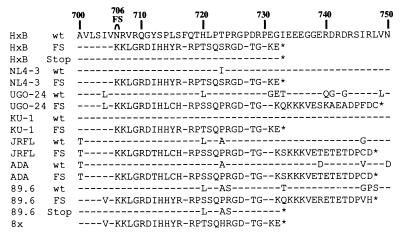
MAbs 17b and 48d bind preferentially to the HIV-1 Env protein in its CD4-bound state (34, 55). Both MAbs bind to CD4-induced epitopes in the gp120 subunit that are located largely in the bridging sheet, a highly conserved region of the Env protein that is important for binding to CCR5 and perhaps CXCR4 as well (46). Therefore, these MAbs can be used as surrogates for measuring the exposure of this region. It was previously shown that the CD4-independent 8x Env binds 17b well even in the absence of CD4, suggesting that this Env exists in a partially triggered conformation (18, 25). Sequence analysis revealed that the 8x Env had an FS mutation in gp41 that resulted in a truncated cytoplasmic domain of 27 amino acids that was necessary but not sufficient for the CD4-independent phenotype (Fig. 1). Introduction of the FS mutation into parental HXB Env also resulted in increased 17b binding (18). This observation led us to examine whether changes in the cytoplasmic domain of gp41 could influence the conformation of the ectodomain in other contexts. Therefore, we introduced the FS into R5, R5X4, and X4 HIV Env proteins (Fig. 1), transiently expressed these proteins in human 293T cells, and performed cell surface-antibody binding assays.
FIG. 1.
Impact of the FS mutation on the cytoplasmic domain sequence. The amino acid sequence of the HXB Env cytoplasmic domain (from residues 700 to 750) is shown, with the location of the FS mutation indicated at position 706. The analogous amino acid sequences of all the constructs used in this study are aligned relative to HIV-1 HXB. A dash indicates amino acid identity, and an asterisk indicates a stop. wt, wild type.
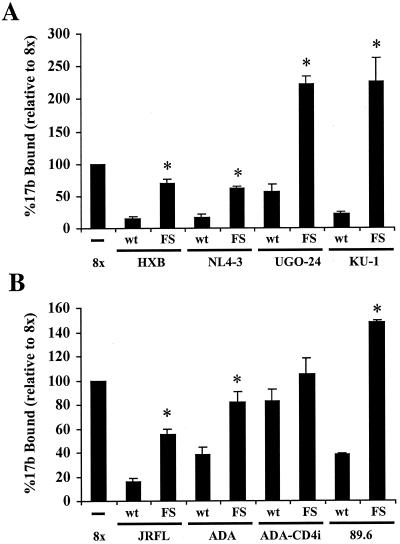
Among the four X4 Env proteins tested, all showed increased 17b binding upon introduction of the FS (Fig. 2A). Enhanced antibody binding was not due to differences in Env protein expression (see below). The FS mutation also resulted in greater 17b binding to the two R5 Env proteins tested as well as to the 89.6 Env protein, a primary R5X4 Env protein (Fig. 2B). In contrast, ADA-CD4i (29, 30) did not show a statistically significant increase in 17b binding upon truncation of the cytoplasmic domain. Thus, like the CD4-independent 8x Env protein, the ADA-CD4i Env protein exists in a partially triggered state, and the binding of CD4-induced MAb 17b was not further enhanced by the FS mutation.
FIG. 2.
Introduction of the FS mutation leads to enhanced 17b binding. 293T cells were infected with recombinant vaccinia virus vTF1.1 expressing T7 polymerase and transfected with the indicated Env expression plasmid. After incubation overnight, Env-bearing cells were washed once with PBS, resuspended in binding buffer, and incubated with 1 μg of MAb 17b/106 cells for 20 min at room temperature. Cells were washed once with PBS and resuspended in binding buffer with approximately 100,000 cpm of iodinated anti-human IgG for 1 h at room temperature. Cells were then collected, washed, and counted. The results were normalized to the amount of binding obtained with 8x Env set to 100%. Typically, between 5 and 10% of the iodinated antibody bound to the surface of Env-expressing cells. (A) X4 Env proteins. (B) R5 Env proteins and R5X4 89.6 Env. The data are compiled from three experiments each done in triplicate; standard errors of the mean are shown. Asterisks indicate statistical significance (t test; two-tailed, assuming unequal variances; P < 0.05). wt, wild type.
We also examined the impact of the FS mutation on 48d binding. Like that of 17b, the epitope of 48d is induced by binding of CD4 to gp120. Recently, its epitope was reported to overlap the CCR5-binding site in R5 Env proteins to a greater extent than that of 17b (S.-H. Xiang, J. Robinson, N. Doka, R. K. Choudhary, and J. Sodroski, Abstr. 8th Conf. Retrovir. Opportunistic Infect., abstr. 535, 2001). We found that introduction of the FS greatly increased the binding of this antibody to all X4 Env proteins tested (Fig. 3A). Likewise, all R5 Env proteins tested as well as the dualtropic 89.6 Env protein exhibited enhanced 48d binding upon introduction of the FS (Fig. 3B). In addition, while the FS mutation did not result in enhanced binding of 17b to ADA-CD4i, it did result in enhanced 48d binding (Fig. 3B). This difference may be due to the fact that ADA-CD4i is only slightly CD4 independent, mediating fusion with CD4-negative, CCR5-positive cells 15% as efficiently as with cells that are CD4 positive in our cell-cell fusion assay (data not shown). Taken together, our results indicate that shortening of the cytoplasmic domain of gp41 by introduction of the FS leads to conformational changes in gp120 that result in the exposure and/or formation of the conserved coreceptor-binding site.
FIG. 3.
Introduction of the FS mutation leads to increased 48d binding. As described in the legend to Fig. 2, Env-expressing cells were assayed for the ability to bind 48d. The results were normalized to the amount of binding obtained with 8x Env set to 100%. (A) X4 Env proteins. (B) R5 and R5X4 Env proteins. The data are compiled from three experiments each done in triplicate; standard errors of the mean are shown. Asterisks indicate statistical significance (t test; two-tailed, assuming unequal variances; P < 0.05). wt, wild type.
Length rather than amino acid sequence of the cytoplasmic domain explains enhanced 17b and 48d binding.
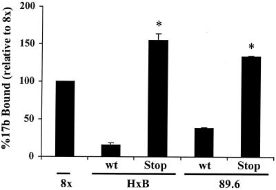
The FS mutation results in a dramatically shortened cytoplasmic domain of approximately 27 to 43 residues, depending on the Env protein (Fig. 1). To determine if enhanced 17b and 48d binding as a result of the FS mutation was due to the shortened cytoplasmic domain or the novel sequences that resulted, we introduced a stop codon at amino acid 733 in the HXB and 89.6 Env proteins (resulting in HXB-stop and 89.6-stop, respectively), thus mimicking the premature stop that occurs when the FS mutation is introduced into the 8x Env but otherwise preserving the wild-type gp41 sequence (Fig. 1). Both HXB-stop and 89.6-stop exhibited increased 17b and 48d binding (Fig. 4 and data not shown). Therefore, the length of the gp41 cytoplasmic domain rather than the artificial sequences introduced as a consequence of the FS mutation accounted for the increased exposure of the conserved coreceptor-binding site.
FIG. 4.
The length rather than a specific sequence of the cytoplasmic domain results in increased 17b binding. We engineered two Env proteins with a stop codon at position 733 for HXB (HXB-stop) or 89.6 (89.6-stop). A 17b binding assay was performed to compare the parental clones (wild type [wt]) with the stop constructs as described in the legend to Fig. 2. The results were normalized to the amount of binding obtained with 8x Env set to 100%. The data are compiled from three experiments each done in triplicate; standard errors of the mean are shown. Asterisks indicate statistical significance (t test; two-tailed, assuming unequal variances; P < 0.05).
Surface expression levels for parental and FS-containing Env proteins are similar.
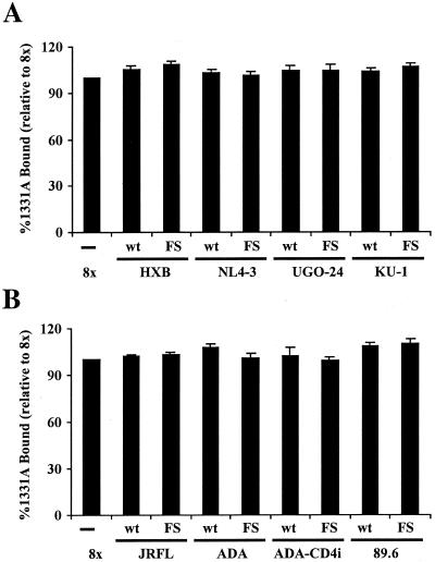
The cytoplasmic domain of the HIV-1 Env protein contains both YXXL and LL motifs that can mediate Env trafficking and internalization from the cell surface (5, 49). The FS mutation results in the loss of these sequences and could therefore result in altered cell surface expression. To examine this notion, each of the Env proteins examined in Fig. 2 and 3 were transiently expressed on 293T cells, and cell surface binding assays were carried out with HIV-positive human serum as well as MAb 1331A (Fig. 5 and data not shown). This MAb recognizes a conformation-dependent epitope in the C5 region of gp120 and binds with high affinity to a number of laboratory-adapted and primary isolates (39). Using this MAb as a probe, we detected no differences in surface expression between the various parental and FS-containing X4 (Fig. 5A), R5 (Fig. 5B), or R5X4 (Fig. 5B) Env proteins. Similar results were obtained when HIV-positive human serum was used (data not shown). Therefore, the increased 17b and 48d binding resulting from the FS mutation in multiple Env proteins cannot be accounted for by increased surface expression of the FS-containing constructs. Rather, the increased binding of 17b and 48d is due to conformational changes in gp120 which result from the introduction of a shorter cytoplasmic domain.
FIG. 5.
FS-containing envelopes have surface expression levels comparable to those of full-length envelopes. We measured surface expression levels for all envelope constructs with MAb 1331A, which binds to the C5 region of gp120, as described in the legend to Fig. 2. (A) X4 Env proteins. (B) R5 Env proteins and R5X4 89.6 Env. The results were normalized to the amount of binding obtained with 8x Env set to 100%. The data are compiled from three experiments each done in triplicate; standard errors of the mean are shown. wt, wild type.
Additional conformational changes occur in gp120 upon introduction of a truncated cytoplasmic tail into gp41.
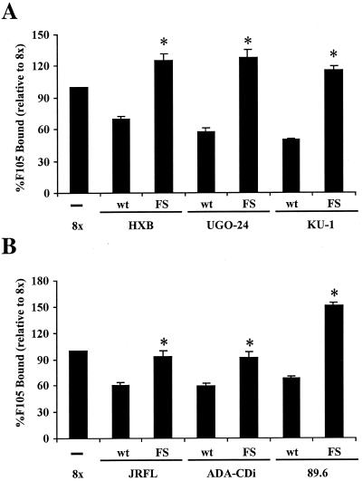
The exposure and/or formation of the coreceptor-binding site results from a significant conformational change in gp120 associated with CD4 binding (37, 45, 46). Since all of the constructs with the FS exhibited increased binding of 17b and 48d even in the absence of CD4, we tested MAbs directed against other regions of Env to further characterize the conformational changes in the ectodomain resulting from the introduction of the FS mutation into the cytoplasmic domain of gp41. Figure 6 shows that the binding of conformation-dependent MAb F105, whose epitope overlaps the CD4-binding site in gp120 (7, 8, 41-43), was enhanced by the FS mutation in all of the X4 (Fig. 6A), R5 (Fig. 6B), or R5X4 (Fig. 6B) Env proteins tested. MAb b12, which also binds to an epitope overlapping the CD4-binding site (3, 6, 47, 68), also showed enhanced binding to Env proteins containing the FS mutation (Table 1). MAb 2G12, which recognizes a conformationally sensitive, glycosylation-dependent epitope in gp120 (57), bound more efficiently to the FS-containing Env proteins, with the exception of ADA, where the FS mutation resulted in decreased binding of this antibody. In contrast, the binding of MAbs to the V3 loop or the V1-V2 region was in general enhanced only slightly or not at all by the FS mutation (Table 1). Finally, MAb 246D, which binds to the immunodominant epitope in the gp41 ectodomain (39, 63), also showed enhanced binding to Env proteins upon introduction of the FS, with the exception of ADA. In conclusion, the FS mutation results in significant structural modifications in the core of gp120 and more subtle changes in the variable loops. In addition, we also observed antigenic changes in the gp41 ectodomain, a result which might be expected given that the effects of the shortened cytoplasmic domain must in some way be transmitted to the external gp120 subunit.
FIG. 6.
Additional conformational changes result from the introduction of the FS into the cytoplasmic domain of Env. 293T cells expressing parental or FS-containing Env proteins were analyzed for the binding of F105, a conformation-dependent anti-Env MAb, as described in the legend to Fig. 2. (A) X4 Env proteins. (B) R5 Env proteins and R5X4 89.6 Env. The results were normalized to the amount of binding obtained with 8x Env set to 100%. The data are compiled from three experiments each done in triplicate; standard errors of the mean are shown. Asterisks indicate statistical significance (t test; two-tailed, assuming unequal variances; P < 0.05). wt, wild type.
TABLE 1.
Binding of antienvelope MAbs to parental and FS envelopes
| Envelopea | Mean ± SEM % binding of MAbb:
|
||||
|---|---|---|---|---|---|
| b12 (CD4bs) | 2G12 (complex) | 697D (V2) | 694/98D (V3) | 246D (gp41) | |
| 8x | 100 | 100 | 100 | 100 | 100 |
| HXB | 54 ± 8 | 53 ± 7 | 95 ± 2 | 96 ± 16 | 41 ± 11 |
| HXB/FS | 117 ± 11 | 105 ± 10 | 102 ± 5 | 108 ± 8 | 107 ± 21 |
| UGO-24 | 17 ± 6 | 42 ± 5 | 98 ± 3 | 73 ± 11 | 45 ± 15 |
| UGO-24/FS | 64 ± 5 | 100 ± 14 | 104 ± 7 | 71 ± 17 | 112 ± 13 |
| JRFL | 65 ± 5 | 57 ± 5 | 97 ± 2 | 99 ± 16 | 40 ± 13 |
| JRFL/FS | 111 ± 6 | 147 ± 12 | 104 ± 3 | 109 ± 21 | 76 ± 20 |
| ADA | 49 ± 4 | 63 ± 8 | 96 ± 3 | 90 ± 11 | 34 ± 11 |
| ADA/FS | 70 ± 4 | 44 ± 13 | 96 ± 3 | 95 ± 28 | 45 ± 17 |
| ADA-CD4i | 21 ± 10 | 54 ± 18 | 92 ± 2 | 89 ± 19 | 30 ± 7 |
| ADA-CD4i/FS | 48 ± 13 | 123 ± 35 | 90 ± 2 | 92 ± 14 | 83 ± 12 |
| 89.6 | 65 ± 3 | 66 ± 21 | 95 ± 6 | 99 ± 20 | 34 ± 10 |
| 89.6/FS | 116 ± 13 | 153 ± 33 | 123 ± 13 | 116 ± 22 | 112 ± 15 |
/FS, containing FS.
Material in parentheses indicates regions for which MAbs are specific. bs, binding site. Significantly different data are shown in bold. The data are normalized to the amount of antibody bound to the 8× Env. The data are representative of three experiments done in triplicate.
Truncation of the cytoplasmic domain leads to increased neutralization sensitivity to MAbs.
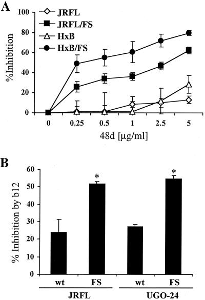
The CD4-independent HIV-1 and simian immunodeficiency virus (SIV) Env proteins that we have examined to date exhibit enhanced neutralization sensitivity to immune sera (18, 43a). Since the FS mutation appears to recapitulate some of the antigenic changes associated with CD4 independence, including enhanced exposure of the bridging sheet, we examined the consequences of the FS mutation on neutralization sensitivity using both cell-cell fusion (Fig. 7) and virus infection (Fig. 8) assays. Figure 7A shows that cell-cell fusion mediated by the FS-containing JRFL and HXB Env proteins was more efficiently inhibited by MAb 48d than was that mediated by the corresponding wild-type proteins, consistent with enhanced binding of this MAb to Env proteins bearing the FS mutation. Similar results were obtained when the FS-containing JRFL and UGO-24 Env proteins were compared to their respective parental Env proteins with 5 μg of MAb b12/ml (Fig. 7B). Additionally, Table 2 shows the fusion inhibition obtained with MAb 2G12 and the C5 MAb 1331A. Env proteins bearing the FS mutation were more sensitive to MAb 2G12 neutralization than were the full-length Env clones, with the exception of ADA. In contrast, the C5 MAb 1331A did not show any significant difference in inhibition between the parental and FS-containing constructs, consistent with the fact that it bound equally well to both full-length and truncated Env proteins (Fig. 5). We also found that the FS mutation did not render Env proteins more sensitive to inhibition by soluble CD4 over a concentration range of 0.2 to 5 μg/ml (data not shown).
FIG. 7.
FS-containing envelopes are susceptible to neutralization by MAbs 48d and b12. (A) The ability of MAb 48d to inhibit cell-cell fusion mediated by JRFL, FS-containing JRFL (JRFL/FS), HXB, or FS-containing HXB (HxB/FS) was tested at the indicated concentrations of antibody. Cells expressing the indicated Env as well as T7 polymerase were incubated with cells expressing CD4 and either CCR5 or CXCR4. The receptor-positive cells were also transfected with a plasmid containing the luciferase gene under the control of the T7 promoter. After 8 h, cells were lysed and the amount of luciferase activity, which results from cell-cell fusion, was determined. The indicated amount of MAb 48d was present throughout the 8-h time course. The data are representative of three experiments done in triplicate; standard errors of the mean are shown. (B) Two pairs of Env proteins, JRFL and FS-containing JRFL and UGO-24 and FS-containing UGO-24, were tested for their sensitivity to neutralization by MAb b12 in the cell-cell fusion assay. MAb b12 was added to the effector cells at the time of mixing with the target cells at a final concentration of 5 μg/ml. b12 was present at the same dilution during the cell-cell fusion assay, and neutralization was scored as the percent reduction in luciferase activity. The data are compiled from three experiments done in triplicate; standard errors of the mean are shown. Asterisks indicate statistical significance (P < 0.05). wt, wild type.
FIG. 8.
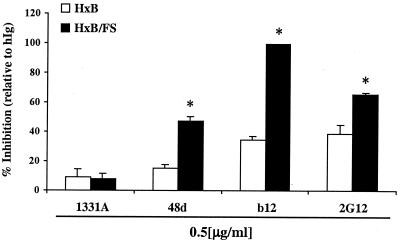
Pseudotyped virions bearing FS-containing envelopes are more sensitive to neutralization by MAbs. We compared HXB and FS-containing HXB (HxB/FS) virus pseudotypes for their sensitivity to neutralization by anti-gp120 MAbs. U87 cells stably transfected with CD4 and CXCR4 were infected as described in Materials and Methods in the presence of the indicated MAbs at a final concentration of 0.5 μg/ml. The results are combined from three independent experiments done in triplicate; standard errors of the mean are shown. Asterisks indicate statistical significance (P < 0.05). hIg, human immunoglobulins.
TABLE 2.
Fusion inhibition by antienvelope MAbs to parental and frameshift envelopes
| Envelopea | Mean ± SEM % inhibition of fusion by MAbb:
|
|
|---|---|---|
| 2G12 (complex) | 1331A (C5) | |
| 8x | 41 ± 7 | 31 ± 2 |
| HXB | 1 ± 15 | 31 ± 4 |
| HXB/FS | 52 ± 1 | 41 ± 3 |
| UGO-24 | 19 ± 10 | 29 ± 8 |
| UGO-24/FS | 42 ± 6 | 8 ± 3 |
| JRFL | 7 ± 7 | 45 ± 3 |
| JRFL/FS | 60 ± 4 | 52 ± 5 |
| ADA | 43 ± 8 | 22 ± 12 |
| ADA/FS | 36 ± 6 | 26 ± 3 |
| ADA-CD4i | 34 ± 9 | 42 ± 3 |
| ADA-CD4i/FS | 67 ± 3 | 58 ± 2 |
/FS, containing FS.
Material in parentheses indicates regions for which MAbs are specific. MAbs were used at 2.5 μg/ml. Significantly different data are shown in bold. The data are normalized to the amount of inhibition obtained with human immunoglobulins. The data are representative of at least three experiments done in triplicate.
To confirm that the enhanced neutralization sensitivity associated with the FS mutation was not an artifact of the cell-cell fusion assay, we produced HXB and FS-containing HXB pseudotyped virions and examined their susceptibility to neutralization by MAbs 1331A, 48d, b12, and 2G12 (Fig. 8). MAbs 48d, b12, and 2G12 inhibited infection by viruses bearing the FS-containing HXB Env more efficiently than HXB Env-bearing virions. As observed with the cell-cell fusion assay, no significant differences in inhibition of infection were observed with MAb 1331A.
Truncation of the cytoplasmic domain results in increased neutralization sensitivity to HIV-positive human sera.
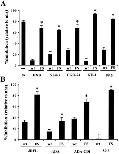
It was previously shown that the CD4-independent 8x Env is efficiently neutralized by HIV-positive human sera and that this phenotype could be related to its triggered conformation (18). We tested the ability of HIV-1-positive human sera to inhibit cell-cell fusion mediated by the panel of wild-type and FS Env proteins to determine if the FS mutation increased the sensitivity of Env proteins to neutralization. As for the MAbs to the core domain of gp120, we found that HIV-1-positive sera more easily neutralized the FS-containing constructs (Fig. 9). Similar results were obtained with viruses bearing the HXB or FS-containing HXB Env proteins (data not shown). Thus, the FS mutation in the gp41 cytoplasmic domain alters the conformation of the external gp120 subunit, enhances the binding of MAbs to regions of Env important for receptor binding, and results in a neutralization-sensitive phenotype.
FIG. 9.
Pooled HIV-positive human sera potently neutralize FS-containing envelopes. All envelope constructs were tested for neutralization with pooled HIV-1-positive human sera in the context of a fusion assay as described in the legend to Fig. 7. The percent inhibition obtained on cells expressing either CD4 and CXCR4 (A) or CD4 and CCR5 (B) is shown. The data are compiled from three experiments done in triplicate; standard errors of the mean are shown. Asterisks indicate statistical significance (t test; two-tailed, assuming unequal variances; P < 0.05). wt, wild type; nhs, normal human serum.
DISCUSSION
Primate lentiviruses have unusually long cytoplasmic tails, about 150 amino acids for HIV and SIV, compared to 20 to 40 amino acids for most other retroviruses (26). The cytoplasmic domain contains endocytosis motifs that can modulate the amount of Env expressed on the cell surface and is also palmitoylated, a characteristic which may help Env partition into lipid rafts and be incorporated into budding virions (32, 38, 48, 49, 51, 64). Several conserved structural motifs are also evident in the cytoplasmic domain of Env, including two amphipathic helices that may associate with the inner surface of viral or cellular membranes (28, 54, 58, 59). Truncation of the cytoplasmic domain of HIV-1 is typically associated with reduced incorporation of Env into virus particles and reduced infectivity, although these effects are cell type dependent (15, 20, 36, 65). The FS mutation in the 8x Env (Fig. 1) results in a cytoplasmic domain that lacks all of these structural motifs.
We found that truncation of the cytoplasmic domain did not impart a CD4-independent phenotype to any Env tested. It did, however, alter the conformation of the ectodomain in a specific, reproducible way in multiple Env backgrounds. Truncation of the cytoplasmic domain resulted in enhanced binding of MAbs directed against the highly conserved bridging sheet region in gp120 as well as to the CD4-binding site. Binding of antibodies to the V3 loop, to the V1-V2 region, and to the C5 region of gp120 either was not affected by cytoplasmic domain truncation or was affected only modestly, indicating that differences in cell surface expression levels do not account for the observed phenotype. Enhanced binding of a MAb to the immunodominant determinant in the gp41 ectodomain was also observed. Consistent with the antibody-binding results, Env proteins bearing shortened cytoplasmic domains were sensitive to neutralization by antibodies to the bridging sheet, to the CD4-binding site, and to HIV-positive human serum samples. Sensitivity to soluble CD4-mediated neutralization, however, was not affected by the FS mutation. Taken together, these results show that the truncation of the HIV-1 cytoplasmic domain that results from the FS mutation causes specific conformational changes in the core of gp120 without obviously affecting the surface expression of the protein or the accessibility of the variable loops to antibody binding.
How might alterations in the cytoplasmic domain of the HIV-1 Env glycoprotein affect ectodomain conformation? For this situation to occur, the effects of the truncation must in some way be transduced across the membrane of the virus or the cell. Since the Env protein is a homotrimer, differences in how the TM domains associate with one another could have an impact on ectodomain stability and conformation. The TM domains of the influenza virus hemagglutinin protein, for example, play an important role in hemagglutinin oligomerization and trimer stability (12, 52). Likewise, Spies and Compans have shown that truncation of the SIVmac239 Env protein alters the conformation of the TM ectodomain and enhances fusogenicity without altering surface expression levels and that oligomers formed by the truncated TM subunit exhibit enhanced stability (53). Differences in how the TM domains of HIV-1 Env interact with one another could likewise alter oligomer stability, perhaps enabling the conformational changes needed for membrane fusion to occur more easily. We found that cytoplasmic domain truncation resulted in antigenic changes in the gp41 ectodomain, a region of Env that is important for oligomerization (17). It should also be noted that the FS mutation results in the loss of both amphipathic helices in the cytoplasmic domain. While the roles that these potentially membrane-active domains play in Env structure, function, and interactions with other proteins are uncertain, their highly conserved nature suggests that they are important for one or more functions. It is also possible that the loss of palmitoylation as a consequence of the FS mutation causes Env to partition differently in the plasma membrane. If palmitoylation causes Env to preferentially partition into sphingolipid-rich lipid rafts, then local Env density and oligomer stability could be affected.
There is precedent for alterations in the cytoplasmic domain having effects on ectodomain structure and function for other viruses. The murine leukemia virus Env protein, like the HIV-1 Env protein, is an oligomer that is proteolytically cleaved by a host cell protease into surface and TM domain subunits (23). After incorporation into the virus, the TM subunit undergoes additional proteolytic cleavage, resulting in the release of a 16-amino-acid peptide, the R peptide, from the C terminus of the cytoplasmic tail (23). Cleavage of the R peptide markedly enhances the fusogenicity of the Env protein and influences the interaction of two distinct regions of the TM ectodomain (67). Truncation of the maedi-visna virus Env protein cytoplasmic domain also significantly increases its membrane fusion capacity (66).
While truncation of the cytoplasmic domain was not sufficient to confer CD4 independence, it was required for this phenotype (18). However, its effects are clearly context dependent in that truncated cytoplasmic domains are not present in the two other CD4-independent HIV-1 strains that have been described, nor is cytoplasmic domain truncation a consistent feature in CD4-independent SIV and HIV-2 strains (16, 30, 43a). Thus, there are likely many ways in which Env can be triggered in a manner that makes it CD4 independent. For an Env protein to mediate CD4-independent membrane fusion, it must successfully engage a specific coreceptor in the absence of CD4. We found that truncation of the cytoplasmic domain enabled antibodies whose determinants exhibit significant overlap with regions in Env important for coreceptor interactions to bind to Env. We do not know, however, if this phenotype necessarily translates into the ability to bind directly to a coreceptor. Furthermore, mere exposure of the coreceptor-binding site is not sufficient for a CD4-independent phenotype. Coreceptor binding must also trigger the exposure of the fusion peptide, which is thought to normally result from CD4 binding, as well as the formation of the six-helix bundle in gp41 that is important for eliciting membrane fusion (reviewed in reference 14). Thus, for an Env protein to acquire CD4 independence, it must contain mutations that enable it to bind directly to a coreceptor, as well as mutations that enable this binding event to subserve functions that are normally the result of CD4 binding. It will be important to characterize the relative contributions of the FS mutation in 8x as well as the two amino acid changes in the C4 region of gp120 to CD4 independence, as this characterization may make it possible to understand how receptor binding to gp120 ultimately results in dramatic conformational rearrangements in gp41 that lead to membrane fusion.
Finally, our results indicate that a single base change resulting in an FS mutation in the cytoplasmic domain of gp41 can render multiple Env proteins sensitive to neutralization by antibodies to the conserved region in gp120 thought to play an important role in coreceptor binding, antibodies to the CD4-binding site, and antibodies present in all HIV-positive human sera that we have tested. This scenario may provide a way to alter Env conformation, resulting in enhanced exposure of conserved neutralization epitopes in gp120. It remains to be seen whether immunization with such modified Env proteins will result in a more potent humoral immune response to primary HIV-1 isolates, but there is reason to believe that such modifications may be of benefit. Reitter et al. showed that the removal of N-linked glycosylation sites in the V1-V2 region of SIVmac239 Env resulted in an attenuated phenotype in vivo coupled with the elicitation of antibodies that could neutralize the fully glycosylated, parental Env protein (44). It was subsequently found that these modifications made these Env proteins more sensitive to neutralization by antibodies to both CCR5- and CD4-binding sites (43a). Therefore, genetic modifications of Env that alter the exposure of conserved domains that play important roles in virus entry have the potential to improve immunogenicity. The results of this study are notable in that the effects of the FS mutation were similar in multiple Env contexts and so may provide a way to alter the immunogenicity of multiple Env immunogens.
Acknowledgments
This work was supported by NIH grant R01 35383 to R.W.D., by NIH grant R01 45378 to J.A.H., and by NIH grant R01 HL59725 to S.Z.-P. This work was also supported by a Burroughs Wellcome Fund translational research award and an Elizabeth Glaser scientist award from the Pediatric AIDS Foundation to R.W.D. F.B. and S.W. were supported by a fellowship from the Swiss National Science Foundation (grants 823A-61172 and 823A-064728, respectively).
We thank James Robinson for providing us with valuable reagents.
REFERENCES
- 1.Alexander, W. A., B. Moss, and T. R. Fuerst. 1992. Regulated expression of foreign genes in vaccinia virus under the control of bacteriophage T7 RNA polymerase and the Escherichia coli lac repressor. J. Virol. 66:2934-2942. [DOI] [PMC free article] [PubMed] [Google Scholar]
- 2.Bandres, J. C., Q. F. Wang, J. O'Leary, F. Baleaux, A. Amara, J. A. Hoxie, S. Zolla-Pazner, and M. K. Gorny. 1998. Human immunodeficiency virus (HIV) envelope binds to CXCR4 independently of CD4, and binding can be enhanced by interaction with soluble CD4 or by HIV envelope deglycosylation. J. Virol. 72:2500-2504. [DOI] [PMC free article] [PubMed] [Google Scholar]
- 3.Barbas, C. F., III, J. E. Crowe, Jr., D. Cababa, T. M. Jones, S. L. Zebedee, B. R. Murphy, R. M. Chanock, and D. R. Burton. 1992. Human monoclonal Fab fragments derived from a combinatorial library bind to respiratory syncytial virus F glycoprotein and neutralize infectivity. Proc. Natl. Acad. Sci. USA 89:10164-10168. [DOI] [PMC free article] [PubMed] [Google Scholar]
- 4.Baribaud, F., S. Poehlmann, and R. W. Doms. 2001. The role of DC-SIGN and DC-SIGNR in HIV and SIV attachment, infection, and transmission. Virology 286:1-6. [DOI] [PubMed] [Google Scholar]
- 5.Boge, M., S. Wyss, J. S. Bonifacino, and M. Thali. 1998. A membrane-proximal tyrosine-based signal mediates internalization of the HIV-1 envelope glycoprotein via interaction with the AP-2 clathrin adaptor. J. Biol. Chem. 273:15773-15778. [DOI] [PubMed] [Google Scholar]
- 6.Burton, D. R., C. F. Barbas III, M. A. Persson, S. Koenig, R. M. Chanock, and R. A. Lerner. 1991. A large array of human monoclonal antibodies to type 1 human immunodeficiency virus from combinatorial libraries of asymptomatic seropositive individuals. Proc. Natl. Acad. Sci. USA 88:10134-10137. [DOI] [PMC free article] [PubMed] [Google Scholar]
- 7.Cavacini, L. A., C. L. Emes, J. Power, A. Buchbinder, S. Zolla-Pazner, and M. R. Posner. 1993. Human monoclonal antibodies to the V3 loop of HIV-1 gp120 mediate variable and distinct effects on binding and viral neutralization by a human monoclonal antibody to the CD4 binding site. J. Acquir. Immune Defic. Syndr. 6:353-358. [PubMed] [Google Scholar]
- 8.Cavacini, L. A., C. L. Emes, A. V. Wisnewski, J. Power, G. Lewis, D. Montefiori, and M. R. Posner. 1998. Functional and molecular characterization of human monoclonal antibody reactive with the immunodominant region of HIV type 1 glycoprotein 41. AIDS Res. Hum. Retrovir. 14:1271-1280. [DOI] [PubMed] [Google Scholar]
- 9.Chan, D. C., and P. S. Kim. 1998. HIV entry and its inhibition. Cell 93:681-684. [DOI] [PubMed] [Google Scholar]
- 10.Chen, B. K., K. Saksela, R. Andino, and D. Baltimore. 1994. Distinct modes of human immunodeficiency virus type 1 proviral latency revealed by superinfection of nonproductively infected cell lines with recombinant luciferase-encoding viruses. J. Virol. 68:654-660. [DOI] [PMC free article] [PubMed] [Google Scholar]
- 11.Connor, R. I., B. K. Chen, S. Choe, and N. R. Landau. 1995. Vpr is required for efficient replication of human immunodeficiency virus type-1 in mononuclear phagocytes. Virology 206:935-944. [DOI] [PubMed] [Google Scholar]
- 12.Doms, R. W., and A. Helenius. 1986. Quaternary structure of the influenza virus hemagglutinin after acid treatment. J. Virol. 60:833-839. [DOI] [PMC free article] [PubMed] [Google Scholar]
- 13.Doms, R. W., and J. P. Moore. 2000. HIV-1 membrane fusion: targets of opportunity. J. Cell Biol. 151:F9-F13. [DOI] [PMC free article] [PubMed]
- 14.Doms, R. W., and D. Trono. 2000. The plasma membrane as a combat zone in the HIV battlefield. Genes Dev. 14:2677-2688. [DOI] [PubMed] [Google Scholar]
- 15.Dubay, J. W., S. J. Roberts, B. H. Hahn, and E. Hunter. 1992. Truncation of the human immunodeficiency virus type 1 transmembrane glycoprotein cytoplasmic domain blocks virus infectivity. J. Virol. 66:6616-6625. [DOI] [PMC free article] [PubMed] [Google Scholar]
- 16.Dumonceaux, J., S. Nisole, C. Chanel, L. Quivet, A. Amara, F. Baleux, P. Briand, and U. Hazan. 1998. Spontaneous mutations in the env gene of the human immunodeficiency virus type 1 NDK isolate are associated with a CD4-independent entry phenotype. J. Virol. 72:512-519. [DOI] [PMC free article] [PubMed] [Google Scholar]
- 17.Earl, P. L., R. W. Doms, and B. Moss. 1990. Oligomeric structure of the human immunodeficiency virus type 1 envelope glycoprotein. Proc. Natl. Acad. Sci. USA 87:648-652. [DOI] [PMC free article] [PubMed] [Google Scholar]
- 18.Edwards, T. G., T. L. Hoffman, F. Baribaud, S. Wyss, C. C. LaBranche, J. Romano, J. Adkinson, M. Sharron, J. A. Hoxie, and R. W. Doms. 2001. Relationships between CD4 independence, neutralization sensitivity, and exposure of a CD4-induced epitope in a human immunodeficiency virus type 1 envelope protein. J. Virol. 75:5230-5239. [DOI] [PMC free article] [PubMed] [Google Scholar]
- 19.Freed, E. O., and M. A. Martin. 1996. Domains of the human immunodeficiency virus type 1 matrix and gp41 cytoplasmic tail required for envelope incorporation into virions. J. Virol. 70:341-351. [DOI] [PMC free article] [PubMed] [Google Scholar]
- 20.Gabuzda, D. H., A. Lever, E. Terwilliger, and J. Sodroski. 1992. Effects of deletions in the cytoplasmic domain on biological functions of human immunodeficiency virus type 1 envelope glycoproteins. J. Virol. 66:3306-3315. [DOI] [PMC free article] [PubMed] [Google Scholar]
- 21.Gorny, M. K., J. P. Moore, A. J. Conley, S. Karwowska, J. Sodroski, C. Williams, S. Burda, L. J. Boots, and S. Zolla-Pazner. 1994. Human anti-V2 monoclonal antibody that neutralizes primary but not laboratory isolates of human immunodeficiency virus type 1. J. Virol. 68:8312-8320. [DOI] [PMC free article] [PubMed] [Google Scholar]
- 22.Gorny, M. K., J. Y. Xu, S. Karwowska, A. Buchbinder, and S. Zolla-Pazner. 1993. Repertoire of neutralizing human monoclonal antibodies specific for the V3 domain of HIV-1 gp120. J. Immunol. 150:635-643. [PubMed] [Google Scholar]
- 23.Green, N., T. M. Shinnick, O. Witte, A. Ponticelli, J. G. Sutcliffe, and R. A. Lerner. 1981. Sequence-specific antibodies show that maturation of Moloney leukemia virus envelope polyprotein involves removal of a COOH-terminal peptide. Proc. Natl. Acad. Sci. USA 78:6023-6027. [DOI] [PMC free article] [PubMed] [Google Scholar]
- 24.Hirsch, V. M., P. Edmondson, M. Murphey-Corb, B. Arbeille, P. R. Johnson, and J. I. Mullins. 1989. SIV adaptation to human cells. Nature 341:573-574. [DOI] [PubMed] [Google Scholar]
- 25.Hoffman, T. L., C. LaBranche, W. Zhang, G. Canziani, J. Robinson, I. Chaiken, J. A. Hoxie, and R. W. Doms. 1999. Stable exposure of the coreceptor binding site in a CD4-independent HIV-1 envelope protein. Proc. Natl. Acad. Sci. USA 96:6359-6364. [DOI] [PMC free article] [PubMed] [Google Scholar]
- 26.Hunter, E., and R. Swanstrom. 1990. Retrovirus envelope glycoproteins. Curr. Top. Microbiol. Immunol. 157:187-253. [DOI] [PubMed] [Google Scholar]
- 27.Kodama, T., D. P. Wooley, Y. M. Naidu, H. W. Kestler III, M. D. Daniel, Y. Li, and R. C. Desrosiers. 1989. Significance of premature stop codons in env of simian immunodeficiency virus. J. Virol. 63:4709-4714. [DOI] [PMC free article] [PubMed] [Google Scholar]
- 28.Koenig, B. W., L. D. Bergelson, K. Gawrisch, J. Ward, and J. A. Ferretti. 1995. Effect of the conformation of a peptide from gp41 on binding and domain formation in model membranes. Mol. Membr. Biol. 12:77-82. [DOI] [PubMed] [Google Scholar]
- 29.Kolchinsky, P., E. Kiprilov, and J. Sodroski. 2001. Increased neutralization sensitivity of CD4-independent human immunodeficiency virus variants. J. Virol. 75:2041-2050. [DOI] [PMC free article] [PubMed] [Google Scholar]
- 30.Kolchinsky, P., T. Mirzabekov, M. Farzan, E. Kiprilov, M. Cayabyab, L. J. Mooney, H. Choe, and J. Sodroski. 1999. Adaptation of a CCR5-using, primary human immunodeficiency virus type 1 isolate for CD4-independent replication. J. Virol. 73:8120-8126. [DOI] [PMC free article] [PubMed] [Google Scholar]
- 31.LaBranche, C. C., T. L. Hoffman, J. Romano, B. S. Haggarty, T. G. Edwards, T. J. Matthews, R. W. Doms, and J. A. Hoxie. 1999. Determinants of CD4 independence for a human immunodeficiency virus type 1 variant map outside regions required for coreceptor specificity. J. Virol. 73:10310-10319. [DOI] [PMC free article] [PubMed] [Google Scholar]
- 32.LaBranche, C. C., M. M. Sauter, B. S. Haggarty, P. J. Vance, J. Romano, T. K. Hart, P. J. Bugelski, M. Marsh, and J. A. Hoxie. 1995. A single amino acid change in the cytoplasmic domain of the simian immunodeficiency virus transmembrane molecule increases envelope glycoprotein expression on infected cells. J. Virol. 69:5217-5227. [DOI] [PMC free article] [PubMed] [Google Scholar]
- 33.Lapham, C. K., J. Ouyang, B. Chandrasekhar, N. Y. Nguyen, D. S. Dimitrov, and H. Golding. 1996. Evidence for cell-surface association between Fusin and the CD4-gp120 complex in human cell lines. Science 274:602-605. [DOI] [PubMed] [Google Scholar]
- 34.Moore, J. P., H. Yoshiyama, D. D. Ho, J. E. Robinson, and J. Sodroski. 1993. Antigenic variation in gp120s from molecular clones of HIV-1 LAI. AIDS Res. Hum. Retrovir. 9:1185-1193. [DOI] [PubMed] [Google Scholar]
- 35.Murakami, T., and E. O. Freed. 2000. Genetic evidence for an interaction between human immunodeficiency virus type 1 matrix and alpha-helix 2 of the gp41 cytoplasmic tail. J. Virol. 74:3548-3554. [DOI] [PMC free article] [PubMed] [Google Scholar]
- 36.Murakami, T., and E. O. Freed. 2000. The long cytoplasmic tail of gp41 is required in a cell type-dependent manner for HIV-1 envelope glycoprotein incorporation into virions. Proc. Natl. Acad. Sci. USA 97:343-348. [DOI] [PMC free article] [PubMed] [Google Scholar]
- 37.Myszka, D. G., R. W. Sweet, P. Hensley, M. Brigham-Burke, P. D. Kwong, W. A. Hendrickson, R. Wyatt, J. Sodroski, and M. L. Doyle. 2000. Energetics of the HIV gp120-CD4 binding reaction. Proc. Natl. Acad. Sci. USA 97:9026-9031. [DOI] [PMC free article] [PubMed] [Google Scholar]
- 38.Nguyen, D. H., and J. E. K. Hildreth. 2000. Evidence for budding of human immunodeficiency virus type 1 selectively from glycolipid-enriched membrane rafts. J. Virol. 74:3264-3272. [DOI] [PMC free article] [PubMed] [Google Scholar]
- 39.Nyambi, P. N., H. A. Mbah, S. Burda, C. Williams, M. K. Gorny, A. Nadas, and S. Zolla-Pazner. 2000. Conserved and exposed epitopes on intact, native, primary human immunodeficiency virus type 1 virions of group M. J. Virol. 74:7096-7107. [DOI] [PMC free article] [PubMed] [Google Scholar]
- 40.Piller, S. C., J. W. Dubay, C. A. Derdeyn, and E. Hunter. 2000. Mutational analysis of conserved domains within the cytoplasmic tail of gp41 from human immunodeficiency virus type 1: effects on glycoprotein incorporation and infectivity. J. Virol. 74:11717-11723. [DOI] [PMC free article] [PubMed] [Google Scholar]
- 41.Posner, M. R., L. A. Cavacini, C. L. Emes, J. Power, and R. Byrn. 1993. Neutralization of HIV-1 by F105, a human monoclonal antibody to the CD4 binding site of gp120. J. Acquir. Immune Defic. Syndr. 6:7-14. [PubMed] [Google Scholar]
- 42.Posner, M. R., H. Elboim, and D. Santos. 1987. The construction and use of a human-mouse myeloma analogue suitable for the routine production of hybridomas secreting human monoclonal antibodies. Hybridoma 6:611-625. [DOI] [PubMed] [Google Scholar]
- 43.Posner, M. R., T. Hideshima, T. Cannon, M. Mukherjee, K. H. Mayer, and R. A. Byrn. 1991. An IgG human monoclonal antibody that reacts with HIV-1/GP120, inhibits virus binding to cells, and neutralizes infection. J. Immunol. 146:4325-4332. [PubMed] [Google Scholar]
- 43a.Puffer, B. A., S. Pöhlmann, A. L. Edinger, D. Carlin, M. D. Sanchez, J. Reitter, D. D. Watry, H. S. Fox, R. C. Desrosiers, and R. W. Doms. 2002. CD4 independence of simian immunodeficiency virus Envs is associated with macrophage tropism, neutralization sensitivity, and attenuated pathogenicity. J. Virol. 76:2595-2605. [DOI] [PMC free article] [PubMed] [Google Scholar]
- 44.Reitter, J. N., R. E. Means, and R. C. Desrosiers. 1998. A role for carbohydrates in immune evasion in AIDS. Nat. Med. 4:679-684. [DOI] [PubMed] [Google Scholar]
- 45.Rizzuto, C., and J. Sodroski. 2000. Fine definition of a conserved CCR5-binding region on the human immunodeficiency virus type 1 glycoprotein 120. AIDS Res. Hum. Retrovir. 16:741-749. [DOI] [PubMed] [Google Scholar]
- 46.Rizzuto, C. D., R. Wyatt, N. Hernandez-Ramos, Y. Sun, P. D. Kwong, W. A. Hendrickson, and J. Sodroski. 1998. A conserved HIV gp120 glycoprotein structure involved in chemokine receptor binding. Science 280:1949-1953. [DOI] [PubMed] [Google Scholar]
- 47.Roben, P., J. P. Moore, M. Thali, J. Sodroski, C. F. Barbas III, and D. R. Burton. 1994. Recognition properties of a panel of human recombinant Fab fragments to the CD4 binding site of gp120 that show differing abilities to neutralize human immunodeficiency virus type 1. J. Virol. 68:4821-4828. [DOI] [PMC free article] [PubMed] [Google Scholar]
- 48.Rousso, I., M. B. Mixon, B. K. Chen, and P. S. Kim. 2000. Palmitoylation of the HIV-1 envelope glycoprotein is critical for viral infectivity. Proc. Natl. Acad. Sci. USA 97:13523-13525. [DOI] [PMC free article] [PubMed] [Google Scholar]
- 49.Rowell, J. F., P. E. Stanhope, and R. F. Siliciano. 1995. Endocytosis of endogenously synthesized HIV-1 envelope protein. Mechanism and role in processing for association with class II MHC. J. Immunol. 155:473-488. [PubMed] [Google Scholar]
- 50.Rucker, J., B. J. Doranz, A. L. Edinger, D. Long, J. F. Berson, and R. W. Doms. 1997. Cell-cell fusion assay to study the role of chemokine receptors in human immunodeficiency type 1 infection. Methods Enzymol. 288:118-133. [DOI] [PubMed] [Google Scholar]
- 51.Sauter, M. M., A. Pelchen-Matthews, R. Bron, M. Marsh, C. C. LaBranche, P. J. Vance, J. Romano, B. S. Haggarty, T. K. Hart, W. M. Lee, and J. A. Hoxie. 1996. An internalization signal in the simian immunodeficiency virus transmembrane protein cytoplasmic domain modulates expression of envelope glycoproteins on the cell surface. J. Cell Biol. 132:795-811. [DOI] [PMC free article] [PubMed] [Google Scholar]
- 52.Singh, I., R. W. Doms, K. Wagner, and A. Helenius. 1990. Intracellular transport of soluble and membrane-bound glycoproteins: folding, assembly, and secretion of anchor-free influenza hemagglutinin. EMBO J. 9:631-639. [DOI] [PMC free article] [PubMed] [Google Scholar]
- 53.Spies, C. P., and R. W. Compans. 1994. Effects of cytoplasmic domain length on cell surface expression and syncytium-forming capacity of the simian immunodeficiency virus envelope glycoprotein. Virology 203:8-19. [DOI] [PubMed] [Google Scholar]
- 54.Srinivas, S. K., R. V. Srinivas, G. M. Anantharamaiah, J. P. Segrest, and R. W. Compans. 1992. Membrane interactions of synthetic peptides corresponding to amphipathic helical segments of the human immunodeficiency virus type-1 envelope glycoprotein. J. Biol. Chem. 267:7121-7127. [PubMed] [Google Scholar]
- 55.Thali, M., J. P. Moore, C. Furman, M. Charles, D. D. Ho, J. Robinson, and J. Sodroski. 1993. Characterization of conserved human immunodeficiency virus type 1 gp120 neutralization epitopes exposed upon gp120-CD4 binding. J. Virol. 67:3978-3988. [DOI] [PMC free article] [PubMed] [Google Scholar]
- 56.Trkola, A., T. Drajic, J. Arthos, J. M. Binley, W. C. Olson, G. P. Allaway, C. Cheng-Mayer, J. Robinson, P. J. Maddon, and J. P. Moore. 1996. CD4-dependent, antibody-sensitive interactions between HIV-1 and its coreceptor CCR-5. Nature 384:184-187. [DOI] [PubMed] [Google Scholar]
- 57.Trkola, A., M. Purtscher, T. Muster, C. Ballaun, A. Buchacher, N. Sullivan, K. Srinivasan, J. Sodroski, J. P. Moore, and H. Katinger. 1996. Human monoclonal antibody 2G12 defines a distinctive neutralization epitope on the gp120 glycoprotein of human immunodeficiency virus type 1. J. Virol. 70:1100-1108. [DOI] [PMC free article] [PubMed] [Google Scholar]
- 58.Trommeshauser, D., and H. J. Galla. 1998. Interaction of a basic amphipathic peptide from the carboxyterminal part of the HIV envelope protein gp41 with negatively charged lipid surfaces. Chem. Phys. Lipids 94:81-96. [DOI] [PubMed] [Google Scholar]
- 59.Venable, R. M., R. W. Pastor, B. R. Brooks, and F. W. Carson. 1989. Theoretically determined three-dimensional structures for amphipathic segments of the HIV-1 gp41 envelope protein. AIDS Res. Hum. Retrovir. 5:7-22. [DOI] [PubMed] [Google Scholar]
- 60.Wu, L., N. P. Gerard, R. Wyatt, H. Choe, C. Parolin, N. Ruffing, A. Borsetti, A. A. Cardoso, E. Desjardin, W. Newman, C. Gerard, and J. Sodroski. 1996. CD4-induced interaction of primary HIV-1 gp120 glycoproteins with the chemokine receptor CCR-5. Nature 384:179-183. [DOI] [PubMed] [Google Scholar]
- 61.Wyatt, R., P. D. Kwong, E. Desjardins, R. W. Sweet, J. Robinson, W. A. Hendrickson, and J. G. Sodroski. 1998. The antigenic structure of the HIV gp120 envelope glycoprotein. Nature 393:705-710. [DOI] [PubMed] [Google Scholar]
- 62.Wyatt, R., and J. Sodroski. 1998. The HIV-1 envelope glycoproteins: fusogens, antigens, and immunogens. Science 280:1884-1888. [DOI] [PubMed] [Google Scholar]
- 63.Xu, J. Y., M. K. Gorny, T. Palker, S. Karwowska, and S. Zolla-Pazner. 1991. Epitope mapping of two immunodominant domains of gp41, the transmembrane protein of human immunodeficiency virus type 1, using ten human monoclonal antibodies. J. Virol. 65:4832-4838. [DOI] [PMC free article] [PubMed] [Google Scholar]
- 64.Yang, C., C. P. Spies, and R. W. Compans. 1995. The human and simian immunodeficiency virus envelope glycoprotein transmembrane subunits are palmitoylated. Proc. Natl. Acad. Sci. USA 92:9871-9875. [DOI] [PMC free article] [PubMed] [Google Scholar]
- 65.Yu, X., X. Yuan, M. F. McLane, T. H. Lee, and M. Essex. 1993. Mutations in the cytoplasmic domain of human immunodeficiency virus type 1 transmembrane protein impair the incorporation of Env proteins into mature virions. J. Virol. 67:213-221. [DOI] [PMC free article] [PubMed] [Google Scholar]
- 66.Zeilfelder, U., and V. Bosch. 2001. Properties of wild-type, C-terminally truncated, and chimeric maedi-visna virus glycoprotein and putative pseudotyping of retroviral vector particles. J. Virol. 75:548-555. [DOI] [PMC free article] [PubMed] [Google Scholar]
- 67.Zhao, Y., L. Zhu, C. A. Benedict, D. Chen, W. F. Anderson, and P. M. Cannon. 1998. Functional domains in the retroviral transmembrane protein. J. Virol. 72:5392-5398. [DOI] [PMC free article] [PubMed] [Google Scholar]
- 68.Zwick, M. B., L. L. Bonnycastle, A. Menendez, M. B. Irving, C. F. Barbas III, P. W. Parren, D. R. Burton, and J. K. Scott. 2001. Identification and characterization of a peptide that specifically binds the human, broadly neutralizing anti-human immunodeficiency virus type 1 antibody b12. J. Virol. 75:6692-6699. [DOI] [PMC free article] [PubMed] [Google Scholar]


