Abstract
DNA shuffling facilitated the evolution of a human immunodeficiency virus type 1 (HIV-1) variant with enhanced replication in pig-tailed macaque peripheral blood mononuclear cells (pt mPBMC). This variant consists exclusively of HIV-1-derived sequences with the exception of simian immunodeficiency virus (SIV) nef. Sequences spanning the gag-protease-reverse transcriptase (gag-pro-RT) region from several HIV-1 isolates were shuffled and cloned into a parental HIV-1 backbone containing SIV nef. Neither this full-length parent nor any of the unshuffled HIV-1 isolates replicated appreciably or sustainably in pt mPBMC. Upon selection of the shuffled viral libraries by serial passaging in pt mPBMC, a species emerged which replicated at substantially higher levels (50 to 100 ng/ml p24) than any of the HIV-1 parents and most importantly, could be continuously passaged in pt mPBMC. The parental HIV-1 isolates, when selected similarly, became extinct. Analyses of full-length improved proviral clones indicate that multiple recombination events in the shuffled region and adaptive changes in the rest of the genome contributed synergistically to the improved phenotype. This improved variant may prove useful in establishing a pig-tailed macaque model of HIV-1 infection.
The narrow host range of human immunodeficiency virus type 1 (HIV-1) has impeded the development of a suitable animal model for the study of HIV-1 pathogenesis and for testing prophylactic and therapeutic strategies to control virus infections. Although HIV-1 infects chimpanzees, the infected animals rarely develop any AIDS-like symptoms. Furthermore, chimpanzees are endangered, expensive, and difficult to handle. Unlike the great apes, macaques are not endangered and are relatively easy to breed. Among the macaques tested, only the pig-tailed macaque (Macaca nemestrina) has been reported to support HIV-1 replication (3). However, HIV-1 replicates poorly in pig-tailed macaques (2, 15, 16), causing only small and transient declines in CD4 cells and no disease (5). In contrast, infection of macaques with simian immunodeficiency virus (SIV) often results in robust replication and AIDS-like symptoms, making this a more relevant model for HIV-1-induced disease. Its utility though, for testing anti-HIV-1 drugs and vaccines is limited by the significant genetic divergence between SIV and HIV-1 (7, 12, 13, 19).
The development of chimeric simian-human immunodeficiency viruses (SHIVs) harboring the HIV-1 envelope and regulatory genes (rev, tat, and vpu) in an SIV background has partially solved this problem (27, 29, 40, 42). These SHIVs are useful for testing HIV-1 envelope based vaccine candidates. Several pathogenic SHIVs with different HIV-1 envelopes are available (20, 22, 30, 39). Since the 5′ regions of their genomes are composed of SIV sequences, these SHIVs cannot be used for testing cytotoxic T-lymphocyte (CTL)-inducing vaccines targeted at Gag. Our ultimate goal is to address this limitation by generating a virus that replicates efficiently in macaques and comprises predominantly sequences derived from HIV-1, inclusive of all the HIV-1 structural genes.
This is a formidable task, as the restrictions to productive HIV-1 replication in macaque cells are poorly characterized. In rhesus macaque peripheral blood mononuclear cells (mPBMCs), HIV-1 replication appears to be blocked early in the viral life cycle (18, 41). These blocks may involve the release of the virion core into the cytoplasm or occur at a step immediately prior to initiation of reverse transcription. Viral entry is not the limiting step as SHIVs can infect and replicate efficiently in macaque cells. Determinants that restrict HIV-1 replication in mPBMC may reside in the sequences encompassing the 3′ half of the long terminal regional (LTR) and the gag-pol region (41, 42). Chackerian et al. (6) reported that replication of HIV-1 in macaque cells expressing human CD4 can be blocked at several steps; certain macrophage-tropic and primary HIV-1 isolates are restricted at a step subsequent to reverse transcription but prior to migration of the preintegration complex to the nucleus while other isolates are blocked prior to reverse transcription. However, expressing the appropriate human coreceptor on the surface of the rhesus macaque cells alleviates the replication blocks (6). In contrast to the complete restriction seen in rhesus macaque cells, pig-tailed macaques are semipermissive for HIV-1 replication. Some HIV-1 strains can infect T lymphocytes from this species but replicate at very low levels (15, 16). The blocks here are also poorly defined and appear to act at stages after reverse transcription (25).
The undefined nature of the replication blocks and the intrinsic complexity of the HIV-1 genome preclude the rational engineering of the virus to replicate efficiently in pig-tailed macaques. Attempts to adapt HIV-1 by serial passaging in pig-tailed macaques have not been successful (2) as replication is not sufficiently robust to favor the evolution of improved variants. Furthermore, efficient CTL responses in HIV-1-infected pig-tailed macaques limit virus replication and eventually clear the virus (23, 24).
In light of these barriers to rational and adaptive approaches, we applied DNA shuffling to kindle the process of virus evolution. In DNA shuffling, homologous sequences can be recombined in vitro by random fragmentation, followed by cycles of reassembly. This generates a pool of diverse, recombinant sequences that is then screened for novel and improved properties. The power of DNA shuffling to evolve dramatically improved phenotypes by efficiently permuting functional sequences from multiple parents has been demonstrated in many systems (8, 9, 33). We have also shown that shuffling can enhance the inherently high evolutionary potential of retroviruses. Under different selective pressures, a library of murine leukemia viruses (MLV)-containing shuffled envelopes yielded chimeras with a new tropism (44), as well as dramatically increased mechanical stabilities (36). In this study, we generated diverse libraries of HIV-1 recombinants by using DNA shuffling. By selectively passaging these libraries, we evolved a viral species that can now replicate efficiently and sustainably in pig-tailed (pt) mPBMCs.
MATERIALS AND METHODS
Cell culture.
Human and pt mPBMCs were purified from heparinized whole blood by centrifugation through Ficoll-Hypaque (Histopaque; Sigma, St. Louis, Mo.) density gradients and were either used fresh (for pt mPBMC) or cryopreserved in 90% fetal bovine serum (FBS)-10% dimethyl sulfoxide. Pt mPBMC were stimulated with complete media (RPMI 1640 supplemented with 10% FBS, 0.2 mM l-glutamine,100 U of penicillin-streptomycin/ml; all reagents from Gibco BRL, Gaithersburg, Md.) containing 1 μg of phytohemagglutinin (PHA; Sigma)/ml for 3 days and then maintained in the same medium containing 100 U of human recombinant interleukin-2 (IL-2)/ml, without PHA. To stimulate human PBMC (huPBMC), recombinant IL-2 was added together with PHA during the initial 3-day treatment.
The human T-cell line MT-4 (17) was maintained in RPMI 1640 supplemented with 10% FBS, glutamine, and antibiotics. Human 293 cells (American Type Culture Collection [ATCC]) were maintained in Dulbecco's minimal essential medium (DMEM; Gibco BRL) supplemented with 10% FBS and antibiotics.
In vitro infections.
MT-4 cells, activated huPBMC (day 5 after PHA stimulation) and pt mPBMC (day 3 after PHA stimulation) were used for in vitro virus infections. Virus inocula were normalized for reverse transcriptase (RT) activity, p24 content, or infectious titers as indicated. Infectious titers were determined by performing a limiting-dilution infection of MT-4 cells in quadruplicate and calculating the 50% tissue culture infectious dose (TCID50) (38). Supernatants from infected PBMC cultures were collected at 2- or 3-day intervals and monitored for virus-associated RT activity or for p24 levels as indicated. RT activity was measured using [32P]dTTP (>400 Ci/mmol; Amersham Pharmacia, Piscataway, N.J.), as previously described (48). RT activity was reported as counts of [32P]dTTP per minute incorporated in 10 μl (containing 1.67 μl of infected culture supernatant) of the reaction mixture. Levels of HIV-1 p24 in the supernatants were measured using an in-house p24 antigen enzyme-linked immunosorbent assay (ELISA) standardized with known amounts of p24 antigen. For the quantification of SHIV, a commercial p27 antigen ELISA (Beckman-Coulter, Miami, Fla.) was used.
Viruses.
Infectious full-length proviral clones of seven clade B (NL4-3 [1], HXB2 [37], LAI [47], JRCSF [34], YU-2 [28], AD-8 [46], and DH12 [43]), two clade D (Z2z6 [45] and ELI [4]), and the recombinant (clade A/D/I) MAL (4) provided parental sequences for shuffling. A partial clone, UG-15 (unpublished data), containing the gag and pol genes of a primary subtype D isolate was also used.
The full-length SHIV molecular clone used as a positive control in this study has been described previously (MD14YE [40]). Briefly, this SHIV consists of the DH12 env, tat, rev, and vpu genes inserted into the backbone of SIVmac239. The premature stop codon within the SIV nef sequence has been repaired, and positions 17 and 18 have been changed from RQ to YE. These changes are responsible for the high lymphocyte stimulatory activity of an SIV variant (11). MD17 is essentially a clone derived from DH12 in which the nef sequence has been replaced by the YE version of the SIVmac239 nef. In the proviral clone of MD17, the 3′ LTR is chimeric; most of the U3 region is derived from SIVmac239, and the remainder of U3 as well as the R and the U5 regions are derived from DH12. After reverse transcription, both LTRs will become chimeric. MD17 provided the infectious backbone into which the shuffled sequences were cloned.
Shuffling of viral sequences and library construction.
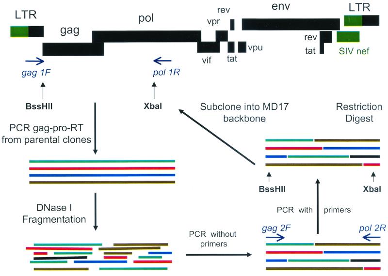
The shuffling procedure has been described previously (10) and is shown schematically (see Fig. 2). Primer sequences are denoted according to the DH12 sequence nucleotide (nt) positions. Briefly, a 3.5-kb fragment encompassing the entire gag gene, as well as the protease- and RT-coding sequences of the pol gene, was amplified from each of the 11 parental HIV-1 strains using primer gag 1F (TCT CTC GAC GCA GGA CTC GGC TTG C [nt 680 to 704]) and primer pol 1R (TCA CTA GCC ATT GCT CTC C [nt 4294 to 4276]). PCR products from the 11 parents were mixed together in equimolar amounts and subjected to random fragmentation by DNAse I (Sigma). Fragments ranging from 0.5 to 1 kb in size were eluted from an agarose gel and reassembled through cycles of denaturation, annealing, and extension in the absence of primers. Assembled fragments were then amplified using primer gag 2F (CGG CTT GCT GAA GCG CGC ACG GCA A [nt 697 to 721]) and primer pol 2R (TCT ATT CCA TCT AGA AAT AGT ACT CTC CTG ATT C [nt 4237 to 4204]), digested with BssHII and XbaI, and ligated into the MD17 backbone that had been digested with the same enzymes. Ligation mixtures were ethanol precipitated and electroporated into XL-1 Blue competent cells (Stratagene, La Jolla, Calif.). The entire transformation mixtures were plated on selective agar plates, the colonies were scraped from the plates, and plasmid DNA was isolated from the pooled bacteria without further growth in liquid culture. To assess the quality of the generated libraries, 20 sample clones from each library were analyzed for recombination frequency and viability. A rough estimate of recombination frequency was obtained through restriction fragment analysis of the gag-pro-RT region amplified from each clone. Viability was assessed by the ability of the clones to produce infectious virus. This was determined by transfecting single clones into 293 cells (FuGENE 6; Roche, Indianapolis, Ind.) followed by the infection of MT-4 cells with the transfection supernatants and monitoring for virus replication by p24 antigen assay.
FIG. 2.
Schematic overview of the construction of gag-pro-RT shuffled libraries.
Library transfection and serial passage.
Library DNA was transfected into 293 cells by the calcium phosphate precipitation method (reagents obtained from 5′→3′ Inc., Boulder, Colo.). A mixture consisting of all full-length parental HIV-1 molecular clones was transfected in parallel and served as wild-type control. As a positive control for replication in pt mPBMC, SHIV (MD14YE) was also transfected. Briefly, each 100-mm-diameter plate containing 5 × 10⁶ 293 cells was transfected with 30 μg of proviral DNA. Sixteen hours after transfection, cells were washed with PBS, and RPMI supplemented with FBS, antibiotics, and IL-2 was added. Supernatants containing virus particles were collected 48 h later and used to infect freshly stimulated pt mPBMC. To remove input virus, PBMC were washed extensively 16 h postinfection and were maintained in complete medium supplemented with IL-2 thereafter. Every 2 to 3 days, about 90% of the medium was collected for storage at −80°C and replaced with fresh medium. An aliquot was kept at −20°C for p24 ELISA or RT activity assay. Two to 3 weeks after infection, a new passage was initiated by inoculating fresh pt mPBMC with supernatants collected on the day of peak virus production. Stimulated huPBMC were infected with the pt mPBMC derived virus containing supernatants where indicated. All manipulations of infectious virus were performed under BL3 conditions.
Cloning of full-length HIV-1 and construction of chimeras.
The 1B3 virus was propagated in a short-term culture in MT-4 cells. Proviral DNA was PCR amplified from genomic DNA in two pieces. The 5′ portion of the genome (4.2 kb) was amplified using primers 5′ F (TGG AAG GGA TTT ATT ACA GTG C) and pol 2R. The 3′ portion (5.5 kb) was amplified using primers pol 2F (GAA TCA GGA AAG TAC TAT TTC TAG ATG GAA TAG A) and 3′ R (TGC TAG AGA TTT TCC ACA C [nt 9704 to 9686]). The separate PCR products were then digested at a unique XbaI restriction site (nt 4223) located in the pol gene, ligated together, and subsequently cloned into the pCR-XL-TOPO vector (Invitrogen, Carlsbad, Calif.).
gag-pro-RT chimeras were generated by exchanging the BssHII (nt 709) to XbaI (nt 4223) fragments between MD17 and 1B3 clone 1.4. Exchange of the region spanning integrase through envelope was accomplished by swapping the pieces between the XbaI site (nt 4223) and the SalI site (nt 8465). For construction of the nef-LTR chimeras, the pieces between the SalI site (nt 8465) and a BamHI site in the vector multiple cloning site were exchanged between 1B3 clone 1.4 and MD17 or between 1B3 clone 1.4 and DH12. The correct composition of each chimera was confirmed by sequence analysis. All chimeric clones were tested for viability by infection of human cells prior to use in the macaque tropism experiments.
Nucleotide sequence accession number.
The entire genome of clone 1.4 was sequenced and submitted to GenBank under accession number AF465242.
RESULTS
Parental HIV-1 strains replicate poorly on pt mPBMC.
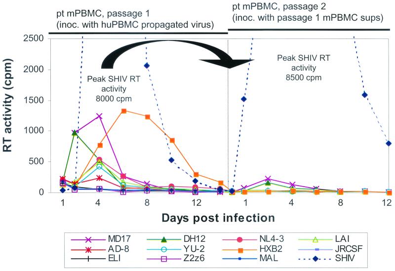
We evaluated the 10 full-length, parental HIV-1 clones for their ability to replicate on pt mPBMC. These viruses were first propagated by short-term passage on huPBMC. RT-normalized amounts of the viruses were then used to infect fresh pt mPBMC. Figure 1 shows that the HIV-1 strains replicated poorly or did not replicate at all, consistent with previous reports (3, 14, 16, 25, 35). HXB2, DH12, and its SIV nef-containing derivative MD17 produced the highest RT levels, although these were still less than 20% that of SHIV-positive control. Virus production peaked at day 4 postinfection and sharply decreased thereafter.
FIG. 1.
Replication of parental HIV-1 strains and SHIV in pt mPBMC through two passages. The maximum RT activity reached in SHIV-infected cultures was in the range of 8,000 cpm. The amount of input virus for the first pt mPBMC passage was normalized for RT activity. For infection of the second-passage pt mPBMC supernatants from passage 1 showing peak RT activity were used. Days postinfection for each passage are labeled from the start of each infection of fresh pt mPBMC.
We attempted to passage the viruses by inoculating fresh pt mPBMC with peak RT supernatants from the first passage. Only DH12 and MD17 survived the transfer and replicated slightly above background levels. A third passage resulted in no detectable viral production. No infectious virus could be rescued from this passage even by cocultivation with permissive human cells (data not shown). In contrast to the HIV-1 strains tested, SHIV showed high and sustainable replication on pt mPBMC throughout several passages. Thus, our results confirm previous studies reporting that HIV-1 replicates poorly and cannot sustain a continuous infection in pig-tailed macaque cells (16, 25).
Generating infectious HIV-1 libraries containing shuffled gag-pro-RT sequences.
Our approach consisted of shuffling a 3.5-kb region between BssHII and XbaI sites, which encompassed the entire gag, protease, and RT sequences. Determinants that restrict macaque cell tropism have been mapped to this region (41). We reasoned that shuffling different HIV-1 sequences in this region might generate favorable recombinants that would alleviate this restriction. Ten full-length clones and one partial HIV-1 clone from several clades provided the starting sequence diversity for shuffling. The homology within the shuffled region for these parent sequences ranged from 90 to 99%.
Figure 2 outlines the scheme for generating shuffled gag-pro-RT libraries. The shuffled sequences were cloned into an infectious MD17 backbone. MD17 is a derivative of the dualtropic DH12 strain (43) and, except for nef, consists entirely of HIV-1 genes. The HIV-1 nef gene has been replaced with the SIV-derived nef gene containing the YE mutations. This nef mutant induces strong proliferation of resting mPBMC in vitro and a highly pathogenic acute infection in vivo (11). The presence of the SIV YE nef alone does not confer a significant replicative advantage to MD17; like DH12, MD17 still replicated poorly in pt mPBMC and could not sustain a productive infection (Fig. 1).
We generated four shuffled HIV-1 libraries (1B3, 2B3, 2A3, and 2A6), each containing between 4 × 10⁴ and 8 × 10⁴ clones. Restriction fragment analysis using DraI and HinfI of several independent clones from each library revealed that 79 to 100% of the clones were recombinant within the shuffled region. Between 25 and 45% of the clones produced virus that productively infected human MT-4 cells (data not shown). This served as a measure of the viability of the libraries.
Emergence of an improved variant after serial passaging of shuffled HIV-1 libraries in pt mPBMC.
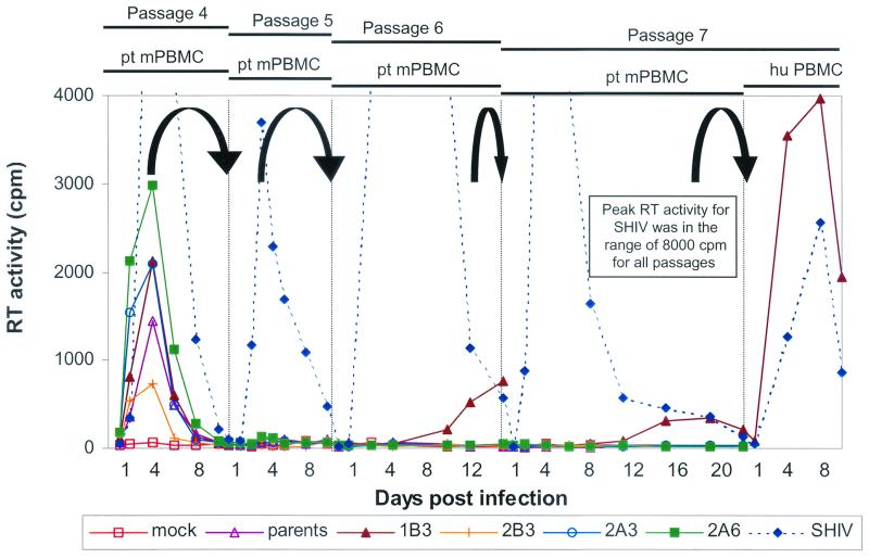
Transient transfection of the four shuffled proviral libraries and a control mixture of the parental clones into 293 cells produced the virus pools that were used to infect separate pt mPBMC cultures. All four libraries and the parental mixture replicated poorly in this first passage (data not shown). None of these viral cultures survived a second passage in fresh pt mPBMC. This result was not surprising, since clones exhibiting an augmented replication phenotype might be expected to be a minor component of each library. Also, we reasoned that additional mutations may be required to manifest incremental advantages conferred by shuffling. In a previous study, in which we applied DNA shuffling to evolve MLV for a new cell tropism (44), we selected the shuffled library for four passages before the new activity became evident. To enable the enrichment process to proceed in this previous study, the selection cultures contained a small number of semipermissive cells mixed with a majority of target cells.
We thus used a variation of this strategy here by inoculating permissive huPBMC with viral supernatants from infected pt mPBMC to rescue and amplify progeny viruses. The amplified viruses were then used to initiate the next round of infection in fresh pt mPBMC. We performed three cycles of this alternating passaging regime; all four libraries as well as the parental culture recovered and replicated to high levels when huPBMC were inoculated in the first three passages (data not shown). An infection of a fourth passage of pt mPBMC was initiated using viruses amplified in huPBMC. From this point (passage 5 and above), we increased the stringency of the selection by directly infecting fresh pt mPBMC with viral supernatants derived from the previous pt mPBMC passage, without an intervening huPBMC amplification step. Among the HIV-1 cultures, only the 1B3 and 2A6 libraries showed marginal levels (70 cpm above background of 40 cpm) of virus replication during passage 5 (Fig. 3). Another direct infection of fresh pt mPBMC was performed for passage 6 where RT levels increased only in the 1B3 culture after a 10-day delay. This pattern was repeated in passage 7.
FIG. 3.
Emergence of a viral variant in library 1B3 exhibiting improved and persistent replication pt mPBMC after extensive passaging of four gag-pro-RT shuffled libraries (1B3, 2B3, 2A3, and 2A6). The maximum RT activity reached by SHIV-infected cultures was in the range of 8,000 cpm. For infection of pt mPBMC, the supernatants from the previous passage showing peak RT activity were used. Days postinfection for each passage are labeled from the start of each infection of fresh pt mPBMC.
After four successive passages (passages 4 to 7) in pt mPBMC, we attempted to rescue viruses from the cultures using huPBMC. This was successful only with the 1B3 library and SHIV control cultures. In contrast, virus could not be recovered from the other three libraries and the parental HIV-1 cultures at passage 6 and passage 7, showing that these viral populations had not survived the stringent selection. Restriction digests of proviral gag-pro-RT sequences, amplified by PCR from the genomic DNA of 1B3 infected huPBMC, revealed a pattern distinct from any of the parental clones (data not shown). This suggests that a dominant, recombinant species had emerged from the 1B3 library.
Viral variant 1B3 shows improved pt mPBMC replication.
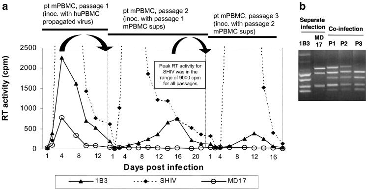
Next, we directly compared the replication kinetics of the evolved 1B3 virus to that of MD17, the parental strain that replicated best in pt mPBMC. SHIV was included as a positive control. The viruses were propagated by short-term culture in huPBMC, normalized for RT activity, and used to infect pt mPBMC. Virus production peaked on day 4 and declined thereafter (Fig. 4a). The peak RT activity of 1B3 was 3-fold higher than that of MD17 and 4.5-fold lower than that of SHIV. We performed a second pt mPBMC passage using day 4 supernatants from passage 1 pt mPBMC cultures normalized for RT activity. While no virus replication was detected in the MD17-infected cells, RT activity increased in the 1B3 culture and peaked on day 13. A third passage of all three viruses on pt mPBMC inoculated with passage 2-derived supernatants resulted in similar patterns of replication. Several attempts to rescue MD17 from passage 3-derived supernatants with permissive huPBMC failed. In contrast, infectious virus was readily recovered from passage 3 1B3-infected pt mPBMC. 1B3 could continue to replicate similarly through at least two more consecutive pt mPBMC passages (passage 5) before the experiment was terminated (data not shown). These data demonstrate that the evolved 1B3 virus has adapted to replicate better in pt mPBMC than the best HIV-1 parent, MD17. It is still not as fit as SHIV, which contains the entire gag-pol region from SIV. Further evidence of the improved fitness of 1B3 was obtained by directly competing 1B3 and MD17 in a coinfection experiment. For the first passage, pt mPBMC were coinfected with equal amounts (normalized for infectivity) of both viruses.
FIG. 4.
The 1B3 viral variant shows improved replication in pt mPBMC compared to MD17. (a) Comparison of parallel infections of pt mPBMC of the improved viral variant 1B3, the best parent MD17, and the SHIV-positive control. The maximum RT activity reached by SHIV-infected cultures was in the range of 9,000 cpm. The amount of input virus for the first pt mPBMC passage was normalized for RT activity. For the infection of subsequent pt mPBMC passages, supernatants from the previous passage showing peak RT activity were used. Days postinfection for each passage are labeled from the start of each infection of fresh pt mPBMC. (b) 1B3 outcompetes MD17 during coinfection of pt mPBMC.
Infections of fresh pt mPBMC were continued for two more passages. Virus from each passage was recovered using huPBMC. Proviral sequences amplified from these infected huPBMC were then analyzed to monitor the existing viral population at each passage. The distinctive restriction patterns of the gag-pro-RT regions of 1B3 and MD17 allowed us to ascertain the composition of the existing population. Figure 4b shows the progress of the competition. At passages 1 and 2, both viruses were present, but by passage 3, 1B3 had essentially gained complete dominance.
Molecular cloning and sequence analysis of 1B3.
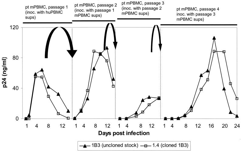
For the generation of full-length molecular clones of the 1B3 virus, the proviral genome was amplified from infected MT-4 cells in two pieces, ligated together using a unique XbaI site, and then cloned into a TA cloning vector. Of 100 clones tested, 13 generated virus that could productively infect human MT-4 cells; of these, five were able to persist through several consecutive passages in pt PBMC. In a side-by-side comparison, the replication kinetics of the improved clone 1.4 was indistinguishable from that of the uncloned 1B3 virus (Fig. 5). Similar to the uncloned 1B3 virus, clone 1.4 also outcompeted the parental MD17 in a coinfection of pt mPBMC (data not shown). Thus, clone 1.4 possessed the improved replication phenotype observed for the original evolved 1B3 virus.
FIG. 5.
The cloned 1B3 (clone 1.4) virus exhibits the same replication kinetics in pt mPBMC as the uncloned 1B3 virus stock. The amount of input virus for the first pt mPBMC passage was normalized for p24. For infection of subsequent pt mPBMC passages, supernatants from the previous passage showing peak virus production were used. Days postinfection for each passage are labeled from the start of each infection of fresh pt mPBMC.
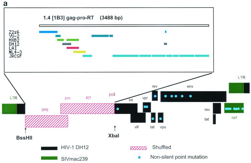
We sequenced the entire genomes of clone 1.4 (GenBank accession number listed above) and three other clones that also exhibited the improved pt mPBMC replication phenotype. These clones all shared a similar structure for the shuffled gag-pro-RT region and several similar mutations in the rest of the genome. The composition of the shuffled region and non-silent point mutations for clone 1.4 are shown diagrammatically in Fig. 6a. Analysis of the shuffled gag-pro-RT region revealed that sequences of at least seven parental HIV-1 strains recombined to generate the gag sequence of 1B3. The protease and RT coding region were most likely derived from the HIV-1 parent strain JRCSF (34).
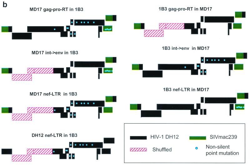
FIG. 6.
Schematic diagram of 1B3 clone 1.4 showing the recombinant structure of the shuffled gag-pro-RT region and non-silent changes in the genome (a) and the 1B3/MD17 and the 1B3/DH12 chimeras (b).
In the nonshuffled regions of the genome, we observed several amino acid changes relative to the original MD17 that were common to all the sequenced clones. These are denoted from the start of each protein. In the integrase coding region of Pol, a Ser-to-Asn change (position 730) was consistently found. Changes resulting in the substitutions of Glu to Lys at position 21 and of Gly to Glu at position 51 were observed in the vpr sequence. tat contained an Arg-to-Lys substitution at position 53, rev contained a Glu-to-Lys substitution at position 7, and in vpu, the Arg at position 49 was changed to Lys. Several similar amino acid changes were also found throughout the gp120 coding portion of env (Met to Ile at positions 150 and 468, Glu to Lys at positions 320 and 346, and Val to Ile at position 358). There were also a number of consistent changes in the SIVmac239-derived nef sequence. These changes were as follows: Asp to Asn at position 15, Arg to Lys at position 30 and 245, and Glu to Lys at positions 36, 75, and 92. Clone 1.4 and another improved clone contained an additional Glu-to-Lys mutation at position 147.
Contribution of sequence changes to improved pt mPBMC tropism.
The changes in the sequences of the 1B3-derived clones from the original MD17 parent can be attributed to shuffling in the gag-pro-RT region as well as adaptive changes in the rest of the genome. To elucidate the relative contributions of these regions to the improved phenotype, we generated six reciprocal chimeras (Fig. 6b) between clone 1.4 and the MD17 parent and assayed their ability to replicate in pt mPBMC using normalized amounts of input virus in each experiment.
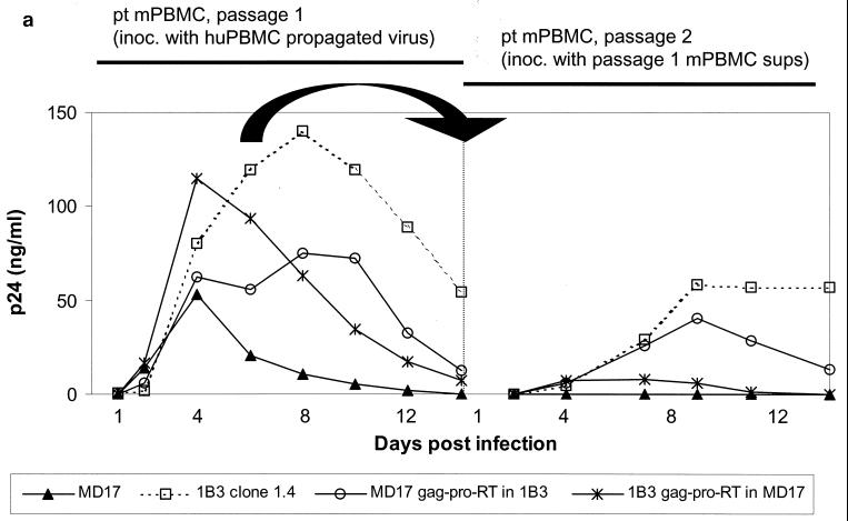
First, when the shuffled 1B3 gag-pro-RT region from clone 1.4 was transplanted into the MD17 backbone, improved replication compared to that of the parent MD17 was observed (Fig. 7a). Conversely, when the shuffled region was replaced by MD17 gag-pro-RT sequences, p24 levels decreased compared to clone 1.4. These observations suggest that the shuffled gag-pro-RT region did impart a replicative advantage, although it was by itself insufficient to confer the full, improved phenotype observed for 1B3.
FIG. 7.
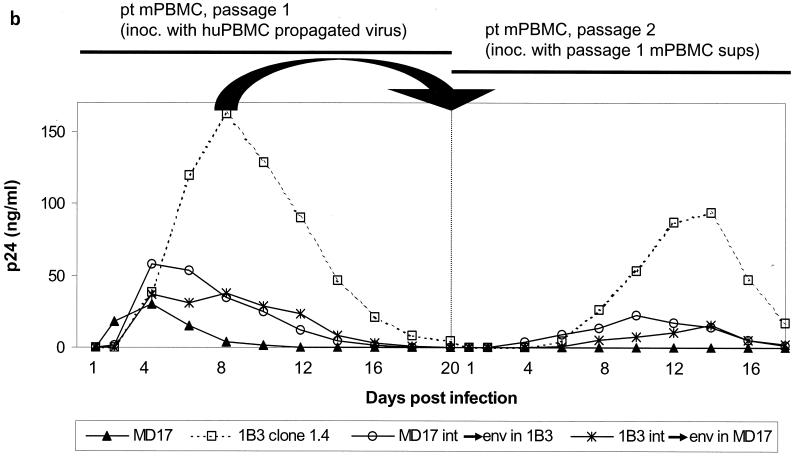
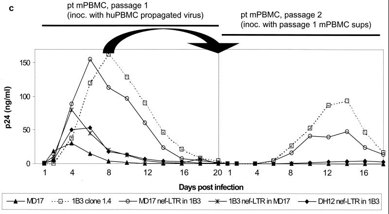
Replication in pt mPBMC for two passages of the gag-pro-RT chimeras (a), int→env chimeras (b), and nef-LTR chimeras (c) in comparison to clone 1.4 and MD17. The amount of input virus for the first pt mPBMC passage was normalized for p24. For infection of the second passage, pt mPBMC supernatants from passage 1 showing peak virus production were used. Days postinfection for each passage are labeled from the start of each infection of fresh pt mPBMC.
Next, the 4.2-kb fragment (int→env) containing integrase and envelope as well as the regulatory genes vif, vpr, vpu, tat, and rev was exchanged between 1B3 clone 1.4 and the MD17 parent. This region was not shuffled, but it had acquired several adaptive changes. Transplanting 1B3 int→env sequences into MD17 increased p24 production and persistence compared to those of MD17 (Fig. 7b). Replacing this region in clone 1.4 with MD17 sequences decreased replicative ability. Thus, adaptive changes in this nonshuffled region contributed to the augmented replication phenotype but were not sufficient to confer the full improvement observed for 1B3.
Finally, we examined whether changes in the unshuffled nef-LTR region had any effect on pt mPBMC replication (Fig 7c). The chimera containing 1B3 nef-LTR in the MD17 backbone was slightly improved in the first passage but replicated only marginally in the second passage (<1 ng/ml). The reciprocal chimera, harboring MD17 nef-LTR in clone 1.4, replicated similarly to clone 1.4 at passage 1 and at half the levels of clone 1.4 during the second passage. Collectively, the data suggest that the changes in nef-LTR also confer improvements. Replacing the SIV nef-LTR sequences in clone 1.4 with HIV-1 nef-LTR sequences resulted in a chimera (DH12 nef-LTR in 1.4) that lost the improved phenotype and that replicated similarly to MD17 (Fig. 7c). This suggested that SIV nef sequences are required for the beneficial changes in the other regions to be manifested.
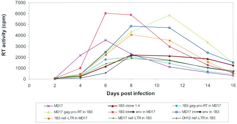
An increase in non-species-specific replicative fitness conferred by the sequence changes to 1B3 and the chimeric viruses may account for the observed improvements in pt mPBMC. To investigate this possibility, we compared the replication of these viruses in huPBMC with MD17, using RT-normalized viral supernatants to initiate the infections. All the viruses replicated robustly in huPBMC (Fig. 8). Importantly, 1B3 clone 1.4 did not exhibit any increase in viral production over MD17. Although some of the chimeras showed some small improvements (<2-fold), these were not significant as there was no correlation of their replication activities in huPBMC and pt mPBMC. Thus, we conclude that the improvements in pt mPBMC replication were not due to an overall increase in replicative fitness but to specific adaptation to pt mPBMC.
FIG. 8.
Replication of the parental MD17 virus, 1B3 clone 1.4, and the MD17-1B3 chimeric viruses in huPBMC. The amount of input virus was normalized for RT activity.
DISCUSSION
HIV-1 variants that can replicate efficiently in macaques will fill an important niche for in vivo models for AIDS (21, 26, 31, 32). Such models would enable the testing of drug candidates without many of the concerns arising from the genetic differences between SIV and HIV-1. Furthermore, vaccines based on multiple HIV-1 genes, not just on envelope sequences, can be evaluated with these models. However, the lack of information pertaining to the blocks to HIV-1 replication in macaque cells precludes structure-based, rationally designed solutions. Adaptive approaches rely on the natural ability of virus to generate diversity from which advantageous mutations can be selected and amplified. Although there have been encouraging reports of HIV-1 infection in pig-tailed macaques (3, 14-16), the level of virus replication is generally too low to favor successful adaptation to a robustly replicating strain within a reasonable time frame.
With these challenges in mind, we reasoned that supplying sufficient diversity initially would increase the probability that some variants would possess enough of a replicative advantage to spark the adaptation process. To accomplish this, DNA shuffling was employed. We shuffled the 3.5-kb fragment encompassing the gag, pro, and RT coding regions to those sequences that restrict productive HIV-1 infection of macaque cells have been mapped (41). Sequences from 11 HIV-1 isolates, gathered from different clades to increase diversity, were used. The pool of shuffled sequences was cloned into an infectious MD17 backbone, which was derived from the dualtropic, primary isolate DH12, and contains the pathogenic YE mutant SIV nef (11). Consistent with previous studies (3, 14, 16, 25, 35), MD17 and other parental HIV-1 strains replicated marginally and did not persist beyond two passages in pt mPBMC (Fig. 1). We subjected four gag-pro-RT shuffled libraries to three cycles of alternating passages in pt mPBMC and huPBMC to allow for selection, amplification, and adaptation of rare viral species that have acquired replicative advantage. The surviving populations were then stringently selected by performing four successive pt mPBMC passages. Of the four libraries, only the 1B3 library yielded a variant that survived the selection. This variant was clearly improved over all the HIV-1 parents, including MD17, although it still replicated less efficiently than SHIV (SIV genome with HIV-1 env). The 1B3 variant could replicate to high levels (>100 ng of p24/ml), and most importantly, it could be passaged continuously in fresh pt mPBMC.
Several full-length proviral clones of 1B3 that exhibited the improved replication phenotype were obtained. They all possessed a similar structure in the shuffled region, which was predicted to result from recombination between sequence fragments of at least seven of the parents (Fig. 6a). Several changes were found in the unshuffled regions of the genome, many of which were shared among the different clones. These clones likely arose from a single founder, generated by shuffling, which then acquired further adaptive mutations. Functional analyses of reciprocal chimeras between a representative improved 1B3 clone and the parental MD17 demonstrated that both the shuffled and unshuffled regions of 1B3 contributed synergistically to the augmented replication phenotype in pt mPBMC. The shuffled gag-pro-RT and int→env regions of 1B3, when transplanted individually into the original MD17 background, resulted in observable improvements in pt mPBMC replication, although replication levels were significantly lower than that for the complete 1B3 clone. Similarly, when these sequences in 1B3 were replaced by MD17 sequences, replication was adversely affected. The evolved nef-LTR of 1B3 had a smaller effect. The enhanced replication of 1B3 and the chimeric viruses were not due to an overall increase in replicative fitness since they exhibited no significant improvement in huPBMC compared to MD17.
The observation that many changes in different regions were required may reflect multiple limiting steps to HIV-1 replication in pig-tailed macaque cells. Additionally, it may reflect the requirement for different HIV-1 functions that interact with one another to be coordinately altered. This highlights the complexity involved to successfully evolve HIV-1 for pig-tailed macaque cell tropism. That only one shuffled species from the four libraries, containing an estimated 2.4 × 10⁵ total number of clones, successfully survived the selection regime and adapted suggests that improved solutions are rare and complex. Thus, natural mechanisms of retroviral recombination and mutation are unlikely to generate these improved variants easily. The control mixture of HIV-1 parents, when subjected to the same selection, became extinct (Fig. 3). Similarly, we have never achieved any observable improvements by extensive passaging of the best parent, MD17, alternately in pt mPBMC and huPBMC (data not shown). By screening larger libraries of shuffled HIV-1, other improved recombinants may be recovered.
It cannot be ruled out that unshuffled gag-pro-RT sequences from a HIV-1 parent, when transplanted into the 1B3 backbone, may also result in the fully improved replication phenotype of the complete 1B3 virus. However, in the absence of a priori information on which gag-pro-RT sequences are favorable, DNA shuffling provides an efficient means to rapidly generate and screen many permutations for the best solution. Furthermore, the fragmentation of parental sequences during the shuffling process rarely proceeds to completion. Thus, it is likely that residual, full-length parental sequences were constituents of the screened libraries. That a recombinant gag-pro-RT sequence was ultimately selected over all the other constituents in the libraries suggests that it conferred the best advantage.
In contrast to other SHIV constructs that are composed of greater than 50% SIV sequences, all the sequences in 1B3 except for SIV nef were HIV-1 derived. Although 1B3 accumulated numerous point mutations, these changes did not shift its composition towards a more SIV-like sequence. Gartner et al. (16) reported that an HIV-1 strain, CH69, derived from a chimpanzee persistently infected with HIV-1 strain IIIB showed improved viral production in pigtailed macaque cells. This variant acquired changes during its adaptation in the chimpanzee host in vivo that likely conferred replicative advantages in pigtailed macaques as well. CH69 thus underwent a different evolutionary route compared to 1B3. The activity of the evolved 1B3 virus is presently being assessed in vivo. This improved variant should be useful in establishing a macaque model of HIV-1 infection.
Acknowledgments
This work was supported by grant no. 97-01-0240 from the National Institutes of Standards and Technology's Advanced Technology Program.
We thank the AIDS Research and Reference Reagent Program and N. Landau's laboratory (Salk Institute for Biological Studies) for making available some of the parental clones used in this study. We also thank J. Ness and E. Bermudez for technical advice and M. Powers for critical reading of the manuscript.
REFERENCES
- 1.Adachi, A., H. E. Gendelman, S. Koenig, T. Folks, R. Willey, A. Rabson, and M. A. Martin. 1986. Production of acquired immunodeficiency syndrome-associated retrovirus in human and nonhuman cells transfected with an infectious molecular clone. J. Virol. 59:284-291. [DOI] [PMC free article] [PubMed] [Google Scholar]
- 2.Agy, M. B., A. Schmidt, M. J. Florey, B. J. Kennedy, G. Schaefer, M. G. Katze, L. Corey, W. R. Morton, and M. L. Bosch. 1997. Serial in vivo passage of HIV-1 infection in Macaca nemestrina. Virology 238:336-343. [DOI] [PubMed] [Google Scholar]
- 3.Agy, M. B., L. R. Frumkin, L. Corey, R. W. Coombs, S. M. Wolinsky, J. Koehler, W. R. Morton, and M. G. Katze. 1992. Infection of Macaca nemestrina by human immunodeficiency virus type-1. Science 257:103-106. [DOI] [PubMed] [Google Scholar]
- 4.Alizon, M., S. Wain-Hobson, L. Montagnier, D. Beaumont, and J. C. Gluckman. 1986. Genetic variability of the AIDS virus: nucleotide sequence analysis of two isolates from African patients. Cell 46:63-74. [DOI] [PubMed] [Google Scholar]
- 5.Bosch, M. L., A. Schmidt, M. B. Agy, L. E. Kimball, and W. R. Morton. 1997. Infection of Macaca nemestrina neonates with HIV-1 via different routes of inoculation. J. Acquir. Immune Defic. Syndr. 11:1555-1563. [DOI] [PubMed] [Google Scholar]
- 6.Chackerian, B., E. M. Long, P. A. Luciw, and J. Overbaugh. 1997. Human immunodeficiency virus type 1 coreceptors participate in postentry stages in the virus replication cycle and function in simian immunodeficiency virus infection. J. Virol. 71:3932-3939. [DOI] [PMC free article] [PubMed] [Google Scholar]
- 7.Chakrabarti, L., M. Guyader, M. Alizon, M. D. Daniel, R. C. Desrosiers, P. Tiollais, and P. Sonigo. 1987. Sequence of simian immunodeficiency virus from macaque and its relationship to other human and simian retroviruses. Nature 328:543-547. [DOI] [PubMed] [Google Scholar]
- 8.Chang, C., T. Chen, B. Cox, G. Dawes, W. P. C. Stemmer, J. Punnonen, and P. Patten. 1999. Evolution of a cytokine using DNA family shuffling. Nat. Biotechnol. 17:793-797. [DOI] [PubMed] [Google Scholar]
- 9.Crameri, A., E. Bermudez, S. Raillard, and W. P. C. Stemmer. 1998. DNA shuffling of a family of genes from diverse species accelerates directed evolution. Nature 391:288-291. [DOI] [PubMed] [Google Scholar]
- 10.Crameri, A., E. Whitehorn, E. Tate, and W. P. C. Stemmer. 1996. Improved green fluorescent protein by molecular evolution using DNA shuffling. Nat. Biotechnol. 14:315-319. [DOI] [PubMed] [Google Scholar]
- 11.Du, Z., S. M. Lang, V. G. Sasseville, A. A. Lackner, P. O. Ilyinskii, M. D. Daniel, J. U. Jung, and R. C. Derosiers. 1995. Identification of a nef allele that causes lymphocyte activation and acute disease in macaque monkeys. Cell 82:665-674. [DOI] [PubMed] [Google Scholar]
- 12.Franchini, G., and M. L. Bosch. 1989. Genetic relatedness of the human immunodeficiency viruses type 1 and 2 (HIV-1, HIV-2) and the simian immunodeficiency virus (SIV). Ann. N. Y. Acad. Sci. 554:81-87. [DOI] [PubMed] [Google Scholar]
- 13.Franchini, G., C. Gurgo, H. G. Guo, R. C. Gallo, E. Collalti, K. A. Fargnoli, L. F. Hall, F. Wong-Staal, and M. S. Reitz. 1987. Sequence of simian immunodeficiency virus and its relationship to human immunodeficiency viruses. Nature 328:539-543. [DOI] [PubMed] [Google Scholar]
- 14.Frumkin, L. R., M. B. Agy, R. W. Coombs, L. Panther, W. R. Morton, J. Koehler, M. J. Florey, J. Dragavon, A. Schmidt, M. G. Katze, and L. Corey. 1993. Acute infection of Macaca nemestrina by human immunodeficiency virus type 1. Virology 195:422-431. [DOI] [PubMed] [Google Scholar]
- 15.Gartner, S., Y. Liu, M. G. Lewis, V. Polonis, W. R. Elkins, P. M. Zack, J. Miao, E. A. Hunter, J. Greenhouse, and G. A. Eddy. 1994. HIV-1 infection in pigtailed macaques. AIDS Res. Hum. Retrovir. 10:S129-133. [PubMed] [Google Scholar]
- 16.Gartner, S., Y. Liu, V. Polonis, M. G. Lewis, W. R. Elkins, E. A. Hunter, J. Miao, K. J. Corts, and G. A. Eddy. 1994. Adaptation of HIV-1 to pigtailed macaques. J. Med. Primatol. 23:155-163. [DOI] [PubMed] [Google Scholar]
- 17.Harada, S., D. T. Purtilo, Y. Koyanagi, and N. Yamamoto. 1986. Serological differences between human T-lymphotropic virus type-I (HTLV-I) and HTLV-III/LAV. Microbiol. Immunol. 30:533-544. [DOI] [PubMed] [Google Scholar]
- 18.Himathongkham, S., and P. A. Luciw. 1996. Restriction of HIV-1 (subtype B) replication at the entry step in rhesus macaque cells. Virology 219:485-488. [DOI] [PubMed] [Google Scholar]
- 19.Hirsch, V., N. Riedel, and J. I. Mullins. 1987. The genome organization of STLV-3 is similar to that of the AIDS virus except for a truncated transmembrane protein. Cell 49:307-319. [DOI] [PubMed] [Google Scholar]
- 20.Igarashi, T., Y. Endo, G. Englund, R. Sadjadpour, T. Matano, C. Buckler, A. Buckler-White, R. Plishka, T. Theodore, R. Shibata, and M. Martin. 1999. Emergence of a highly pathogenic simian/human immunodeficiency virus in a rhesus macaque treated with anti-CD8 mAb during a primary infection with a nonpathogenic virus. Proc. Natl. Acad. Sci. USA 96:14049-14054. [DOI] [PMC free article] [PubMed] [Google Scholar]
- 21.Joag, S. V. 2000. Primate models of AIDS. Microbes Infect. 2:223-229. [DOI] [PubMed] [Google Scholar]
- 22.Joag, S. V., Z. Li, L. Foresman, E. B. Stephens, L.-J. Zhao, I. Adany, D. M. Pinson, H. M. McClure, and O. Narayan. 1996. Chimeric simian/human immunodeficiency virus that causes progressive loss of CD4+ T cells and AIDS in pig-tailed macaques. J. Virol. 70:3189-3197. [DOI] [PMC free article] [PubMed] [Google Scholar]
- 23.Kent, S. J., A. Woodward, and A. Zhao. 1997. Human immunodeficiency virus type 1 (HIV-1)-specific T cell responses correlate with control of acute HIV-1 infection in macaques. J. Infect. Dis. 176:1188-1197. [DOI] [PubMed] [Google Scholar]
- 24.Kent, S. J., L. Corey, M. B. Agy, W. R. Morton, M. J. McElrath, and P. D. Greenberg. 1995. Cytotoxic and proliferative T cell responses in HIV-1-infected Macaca nemestrina. J. Clin. Investig. 95:248-256. [DOI] [PMC free article] [PubMed] [Google Scholar]
- 25.Kimball, L. E., and M. L. Bosch. 1998. In vitro HIV-1 infection in Macaca nemestrina PBMC is blocked at a step beyond reverse transcription. J. Med. Primatol. 27:99-103. [DOI] [PubMed] [Google Scholar]
- 26.Levy, J. A. 1996. The value of primate models for studying human immunodeficiency virus pathogenesis. J. Med. Primatol. 25:163-174. [DOI] [PubMed] [Google Scholar]
- 27.Li, J., C. I. Lord, W. Haseltine, N. L. Letvin, and J. Sodroski. 1992. Infection of cynomolgus monkeys with a chimeric HIV-1/SIVmac virus that expresses the HIV-1 envelope glycoproteins. J. Acquir. Immune Defic. Syndr. 5:639-646. [PubMed] [Google Scholar]
- 28.Li, Y., J. C. Kappes, J. A. Conway, R. W. Price, G. M. Shaw, and B. H. Hahn. 1991. Molecular characterization of HIV-1 cloned directly from uncultured human brain tissue: identification of replication-competent and -defective viral genomes. J. Virol. 65:3973-3985. [DOI] [PMC free article] [PubMed] [Google Scholar]
- 29.Luciw, P. A., E. Pratt-Lowe, K. E. Shaw, J. A. Levy, and C. Cheng-Mayer. 1995. Persistent infection of rhesus macaques with T-cell-line-tropic and macrophage-tropic clones of simian/human immunodeficiency viruses (SHIV). Proc. Natl. Acad. Sci. USA 92:7490-7494. [DOI] [PMC free article] [PubMed] [Google Scholar]
- 30.Luciw, P. A., C. P. Mandell, S. Himathongkham, J. Li, T. A. Low, K. A. Schmidt, K. E. Shaw, and C. Cheng-Mayer. 1999. Fatal immunopathogenesis by SIV/HIV (SHIV) containing a variant form of the HIV-1SF33 env gene in juvenile and newborn rhesus macaques. Virology 263:112-127. [DOI] [PubMed] [Google Scholar]
- 31.Nath, B. M., K. E. Schumann, and J. D. Boyer. 2000. The chimpanzee and other non-human-primate models in HIV-1 vaccine research. Trends Microbiol. 8:426-431. [DOI] [PubMed] [Google Scholar]
- 32.Nathanson, N., V. M. Hirsch, and B. J. Mathieson. 1999. The role of nonhuman primates in the development of an AIDS vaccine. AIDS. 13(Suppl. A):S113-S120. [PubMed]
- 33.Ness, J., M. Welch, L. Giver, M. Bueno, J. Cherry, T. Borchert, W. P. C. Stemmer, and J. Minshull. 1999. DNA shuffling of subgenomic sequences of subtilisin. Nat. Biotechnol. 17:893-896. [DOI] [PubMed] [Google Scholar]
- 34.O'Brien, W. A., Y. Koyanagi, A. Namazie, J. Q. Zhao, A. Diagne, K. Idler, J. A. Zack, and I. S. Y. Chen. 1990. HIV-1 tropism for mononuclear phagocytes can be determined by regions of gp120 outside the CD4-binding domain. Nature 348:69-73. [DOI] [PubMed] [Google Scholar]
- 35.Otten, R. A., B. G. Brown, M. Simon, L. D. Lupo, B. S. Parekh, M. D. Lairmore, C. A. Schable, G. Schochetman, and M. A. Rayfield. 1994. Differential replication and pathogenic effects of HIV-1 and HIV-2 in Macaca nemestrina. AIDS 8:297-306. [DOI] [PubMed] [Google Scholar]
- 36.Powell, S. K., M. A. Kaloss, A. Pinkstaff, R. McKee, I. Burimski, M. Pensiero, E. Otto, W. P. Stemmer, and N.-W. Soong. 2000. Breeding of retroviruses by DNA shuffling for improved stability and processing yields. Nat. Biotechnol. 18:1279-1282. [DOI] [PubMed] [Google Scholar]
- 37.Ratner, L., W. Haseltine, R. Patarca, K. J. Livak, B. Starcich, S. F. Josephs, E. R. Doran, J. A. Rafalski, E. A. Whitehorn, K. Baumeister, et al. 1985. Complete nucleotide sequence of the AIDS virus, HTLV-III. Nature 313:277-284. [DOI] [PubMed] [Google Scholar]
- 38.Reed, L. J., and H. Muench. 1938. A simple method of estimating fifty per cent endpoints. Am. J. Hyg. 27:493-497. [Google Scholar]
- 39.Reimann, K. A., J. T. Li, R. Veazey, M. Malloran, I.-W. Park, G. B. Karlsson, J. Sodroski, and N. L. Letvin. 1996. A chimeric simian/human immunodeficiency virus expressing a primary patient human immunodeficiency virus type 1 isolate env causes an AIDS-like disease after in vivo passage in rhesus monkeys. J. Virol. 70:6922-6928. [DOI] [PMC free article] [PubMed] [Google Scholar]
- 40.Shibata, R., F. Maldarelli, C. Siemon, T. Matano, M. Parta, G. Miller, T. Frederickson, and M. A. Martin. 1997. Infection and pathogenicity of chimeric simian-human immunodeficiency viruses in macaques: determinants of high virus loads and CD4 killing. J. Infect. Dis. 176:362-373. [DOI] [PubMed] [Google Scholar]
- 41.Shibata, R., H. Sakai, M. Kawamura, K. Tokunaga, and A. Adachi. 1995. Early replication block of human immunodeficiency virus type 1 in monkey cells. J. Gen. Virol. 76:2723-2730. [DOI] [PubMed] [Google Scholar]
- 42.Shibata, R., M. Kawamura, H. Sakai, M. Hayami, A. Ishimoto, and A. Akio. 1991. Generation of a chimeric human and simian immunodeficiency virus infectious to monkey peripheral blood mononuclear cells. J. Virol. 65:3514-3520. [DOI] [PMC free article] [PubMed] [Google Scholar]
- 43.Shibata, R., M. D. Hoggan, C. Broscius, G. Englund, T. S. Theodore, A. Buckler-White, L. O. Arthur, Z. Israel, A. Schultz, H. C. Lane, and M. A. Martin. 1995. Isolation and characterization of a syncytium-inducing, macrophage/T-cell line-tropic human immunodeficiency virus type 1 isolate that readily infects chimpanzee cells in vitro and in vivo. J. Virol. 69:4453-4462. [DOI] [PMC free article] [PubMed] [Google Scholar]
- 44.Soong, N.-W., L. Nomura, K. Pekrun, M. Reed, L. Sheppard, G. Dawes, and W. P. C. Stemmer. 2000. Molecular breeding of viruses. Nat. Genet. 25:436-439. [DOI] [PubMed] [Google Scholar]
- 45.Srinivasan, A., R. Anand, P. Ranganathan, P. Feorino, G. Schochetman, J. Curran, V. S. Kalyanaraman, P. A. Luciw, and R. Sanchez-Pescador. 1987. Molecular characterization of human immunodeficiency virus from Zaire: nucleotide sequence analysis identifies conserved and variable domains in the envelope gene. Gene 52:71-82. [DOI] [PubMed] [Google Scholar]
- 46.Theodore, T. S., G. Englund, A. Buckler-White, C. E. Buckler, M. A. Martin, and K. W. Peden. 1996. Construction and characterization of a stable full-length macrophage-tropic HIV type 1 molecular clone that directs the production of high titers of progeny virions. AIDS Res. Hum. Retrovir. 12:191-194. [DOI] [PubMed] [Google Scholar]
- 47.Wain-Hobson, S., D. Beaumont, O. Danos, S. Cole, M. Alizon. 1985. Nucleotide sequence of the AIDS virus, LAV. Cell 40:9-17. [DOI] [PubMed] [Google Scholar]
- 48.Willey, R. L., D. H. Smith, L. A. Lasky, T. S. Theodore, P. L. Earl, B. Moss, D. J. Capon, and M. A. Martin. 1988. In vitro mutagenesis identifies a region within the envelope gene of the human immunodeficiency virus that is critical for infectivity. J. Virol. 62:139-147. [DOI] [PMC free article] [PubMed] [Google Scholar]


