Abstract
Infectious hematopoietic necrosis virus (IHNV) and viral hemorrhagic septicemia virus (VHSV) are two salmonid rhabdoviruses replicating at low temperatures (14 to 20°C). Both viruses belong to the Novirhabdovirus genus, but they are only distantly related and do not cross antigenically. By using a recently developed reverse-genetic system based on IHNV (S. Biacchesi et al., J. Virol. 74:11247-11253, 2000), we investigated the ability to exchange IHNV glycoprotein G with that of VHSV. Thus, the IHNV genome was modified so that the VHSV G gene replaced the complete IHNV G gene. A recombinant virus expressing VHSV G instead of IHNV G, rIHNV-Gvhsv, was generated and was shown to replicate as well as the wild-type rIHNV in cell culture. This study was extended by exchanging IHNV G with that of a fish vesiculovirus able to replicate at high temperatures (up to 28°C), the spring viremia of carp virus (SVCV). rIHNV-Gsvcv was successfully recovered; however, its growth was restricted to 14 to 20°C. These results show the nonspecific sequence requirement for the insertion of heterologous glycoproteins into IHNV virions and also demonstrate that an IHNV protein other than the G protein is responsible for the low-temperature restriction on growth. To determine to what extent the matrix (M) protein interacts with G, a series of chimeric pIHNV constructs in which all or part of the M gene was replaced with the VHSV counterpart was engineered and used to recover the respective recombinant viruses. Despite the very low percentage (38%) of amino acid identity between the IHNV and VHSV matrix proteins, viable chimeric IHNVs, harboring either the matrix protein or both the glycoprotein and the matrix protein from VHSV, were recovered and propagated. Altogether, these data show the extreme flexibility of IHNV to accommodate heterologous structural proteins.
Infectious hematopoietic necrosis virus (IHNV) and viral hemorrhagic septicemia virus (VHSV) are two rhabdoviruses mainly, but not exclusively, infecting trout. Like those of mammalian rhabdoviruses such as rabies virus (RV) and vesicular stomatitis virus (VSV), the IHNV and VHSV negative-strand RNA genomes encode five structural proteins: nucleoprotein (N), polymerase-associated phosphoprotein (P), matrix protein (M), glycoprotein (G), and RNA-dependent RNA polymerase (L). In contrast to those of mammalian rhabdoviruses, the IHNV and VHSV genomes contain an additional cistron, localized between the G and L genes, which encodes a small nonstructural protein of unknown function, NV (2, 21). Due to the presence of the NV gene, IHNV and VHSV have been classified as novirhabdoviruses. Mammalian rhabdoviruses are generally separated into two major genera, Lyssavirus (prototype, RV) and Vesiculovirus (prototype, VSV), based mainly on the migration pattern of the viral proteins in polyacrylamide gels. Although they are only distantly related and antigenically distinct, IHNV and VHSV have both been considered more related to Lyssavirus than to Vesiculovirus, in contrast to other fish rhabdoviruses such as spring viremia of carp virus (SVCV) (6, 31). Rhabdoviruses are enveloped viruses which possess a unique external glycoprotein G inserted into the viral membrane. This N-glycosylated class I transmembrane protein, which forms trimeric peplomers on the virion surface (13), exhibits several remarkable features common to all the rhabdoviruses: (i) G is the only protein responsible for the synthesis of neutralizing antibodies in infected animals (7, 23, 24), (ii) targeted mutations on G allow attenuation of virulence (4, 12, 19), (iii) G is the viral protein responsible for attachment to the cell membrane receptors (3), and (iv) G possesses a very short cytoplasmic tail which probably interacts with other internal proteins such as N and/or M. The putative interaction between G and other viral components is not yet very clear and has recently been studied by manipulating the genomes of negative-stranded RNA viruses by means of cloned DNA and recovering infectious viral particles (8, 32). Thus, by using a reverse-genetic approach based on VSV, it has been shown that any heterologous glycoprotein can be inserted into VSV, with no specific glycoprotein cytoplasmic tail sequence conservation (33, 34), in contrast to RV, in which, with one exception (29), insertion of glycoproteins requires conservation of the RV glycoprotein cytoplasmic tail sequence (26-28). The difference observed between RV and VSV is intriguing and could be related to the relationships between the G and M proteins or the G and N proteins. For VSV, it is now well established that a cytoplasmic tail domain (at least 9 amino acids), but one with no specific amino acid sequence, is necessary to efficiently promote viral budding. Thus, it has been hypothesized that no specific interactions between G and N or the M matrix exist but that there is a spatial topology arrangement in which the G cytoplasmic tail fits into the groove or pocket formed by M and/or N (33). In contrast, Mebatsion et al. have shown that the M protein of RV interacts with G and is probably responsible for recruiting the G protein into the virus, necessitating the conservation of specific amino acids in the G cytoplasmic tail (30).
To study whether IHNV is indeed more related to RV than to VSV regarding the requirement of the conservation of the G cytoplasmic tail domain for the insertion of foreign glycoproteins, we exchanged the IHNV G glycoprotein with that of VHSV by a recently described reverse-genetic approach (5). A recombinant virus, rIHNV-Gvhsv, was produced and was shown to grow in cell culture as well as wild-type IHNV or VHSV. This result demonstrated that cytoplasmic domain conservation is not required for efficient replacement of the IHNV glycoprotein by a heterologous glycoprotein. This observation was furthermore extended by the generation of rIHNV-Gsvcv, in which the glycoprotein from a fish vesiculovirus was inserted into IHNV particles. The putative interaction between the glycoprotein and the matrix protein was thus investigated by replacement of the matrix gene in the IHNV genome with that of VHSV. In this study we show that viable recombinant chimeric IHNVs were produced when the M- and/or G-encoding gene was replaced by that of VHSV.
MATERIALS AND METHODS
Viruses and cell cultures.
IHNV 32/87, VHSV 07/71, and SVCV “Fijan” strains were propagated in monolayer cultures of EPC cells either at 14°C (IHNV and VHSV) or at 20°C (SVCV) as previously described (10). Recombinant vaccinia virus expressing T7 RNA polymerase, vTF7-3 (11), was kindly provided by B. Moss.
Construction of pIHNV-Gvhsv and pIHNV-Gsvcv.
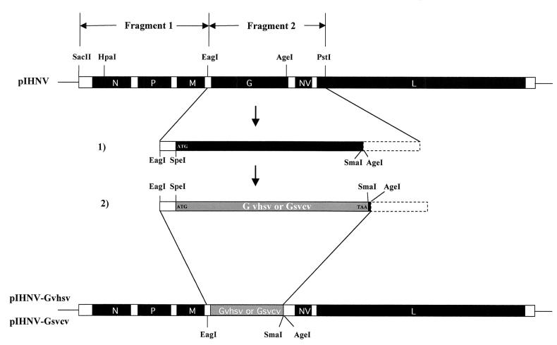
A full-length cDNA clone, pIHNV (Fig. 1), assembled from four overlapping cDNA fragments (numbered 1 to 4) and covering the complete IHNV genome (5), was used to engineer new pIHNV-derived constructs. Thus, EagI/PstI fragment 2 (pIHN2), containing the G and NV genes and the beginning of the L gene, was used to introduce, by a two-step site directed mutagenesis assay using the QuickChange Kit (Stratagene), two unique SpeI and SmaI restriction enzyme sites at the start codon and near the end of the G gene, respectively. In the first step, a SpeI restriction enzyme site was created by using primers G-SpeI-S and G-SpeI-A (Table 1), leading to pIHN2-SpeI. In the second step, a SmaI restriction enzyme site was created in pIHN2-SpeI by using primers G-SmaI-S and G-SmaI-A, leading to pIHN2-SpeI/SmaI. The VHSV or SVCV G gene was recovered by reverse transcription-PCR (RT-PCR) from the VHSV or SVCV RNA genome by using the specific primers G-SpeI-vhsv and G-SmaI-vhsv or G-NheI-svcv and G-SmaI-svcv, (the NheI restriction enzyme site is compatible with SpeI). Then the IHNV G gene was deleted from pIHN2-SpeI/SmaI by SpeI-SmaI digestion and replaced with the VHSV or SVCV G gene. Finally, an EagI-AgeI fragment was removed from the full-length pIHNV construct by restriction enzyme digestion and replaced by the modified EagI-AgeI counterpart from pIHN2-Gvhsv or pIHN2-Gsvcv, leading to pIHNV-Gvhsv or pIHNV-Gsvcv, respectively.
FIG. 1.
Construction of the pIHNV-Gvhsv and pIHNV-Gsvcv plasmids. pIHNV fragment 2 cloned into the pBluescript plasmid (5) was engineered by site-directed mutagenesis to introduce SpeI and SmaI restriction enzyme sites (step1). The mutated fragment 2 was digested with SpeI/SmaI to remove the IHNV G gene so that it could be replaced by the VHSV or SVCV G gene (step 2). Finally, the pIHNV-Gvhsv and pIHNV-Gsvcv plasmid constructs were generated by insertion into the pIHNV backbone of an EagI/AgeI fragment containing the VHSV or SVCV G gene, respectively.
TABLE 1.
Primers used in this study
| Primer | Sequencea | Restriction site |
|---|---|---|
| G-SpeI-S | GAGACCCACCAAAACTAGTGACACCACGATCAC | SpeI |
| G-SpeI-A | GTGATCGTGGTGTCACTAGTTTTGGTGGGTCTC | SpeI |
| G-SmaI-S | CCCCATGTATCACCCGGGAAACCGGTCCTAAAGG | SmaI |
| G-SmaI-A | CCTTTAGGACCGGTTTCCCGGGTGATACATGGGG | SmaI |
| G-SpeI-vhsv | GGACTAGTATGGAATGGAACACTTTTTTC | SpeI |
| G-SmaI-vhsv | TCCCCCGGGTCAGACCGTCTGACTTCTGG | SmaI |
| G-NheI-svcv | CTAGCTAGCATGTCTATCATCAGCTACATCG | NheI |
| G-SmaI-svcv | TCCCCCGGGTCAAACTAAAGACCGCATTTCG | SmaI |
| M-Mlu-ATG-S | CAGTTCAAACGAGAACGCGTCTATTTTCAAGAGAGC | MluI |
| M-Mlu-ATG-A | GCTCTCTTGAAAATAGACGCGTTCTCGTTTGAACTG | MluI |
| M-Mlu-TAG-S | GGGGAAGGAAAAATACGCGTCGACCATGCCGTC | MluI |
| M-Mlu-TAG-A | GACGGCATGGTCGACGCGTATTTTTCCTTCCCC | MluI |
| M-vhsv-S | CCGACGCGTATGGCTCTGTTCAAAAG | MluI |
| M-vhsv-A | CCGACGCGTCTACCAAGGTCGGACAG | MluI |
| M-Eco47-S | GGTTACAATACTCAGCGCTGAGGGGGAGATCAAGG | Eco47III |
| M-Eco47-A | CCTTGATCTCCCCCTCAGCGCTGAGTATTGTAACC | Eco47III |
| M-vhsv-Eco47 | GCCAGCGCTCAGGATGGTGGAGACACG | Eco47III |
Restriction enzyme sites are underlined; mutated nucleotides are boldfaced.
DNA transfection and recovery of recombinant IHNV.
Transfection experiments were carried out as described previously (5). Approximately 4 × 10⁶ EPC cells were grown in 6-well plates and infected with the recombinant vaccinia virus vTF7-3 expressing T7 RNA polymerase at a multiplicity of infection of 5 (11). After 1 h of adsorption at 37°C, cells were washed twice and transfected with a plasmid mixture containing 0.25 μg of pT7-N, 0.2 μg of pT7-P, 0.2 μg of pT7-L, and 0.12 μg of pT7-NV and with 1 μg of each of the different full-length cDNA constructs, by using the Lipofectamine reagent (Gibco-BRL) according to the supplier's instructions. The cells were incubated for 7 h at 37°C before being shifted to 14°C and incubated for 6 days. Then the cells were suspended by scratching with a rubber policeman and were subjected to two cycles of freeze-thawing. Supernatant P0 was clarified by centrifugation at 10,000 × g in a microcentrifuge and was used to inoculate fresh EPC cell monolayers in 24-well plates at 14°C.
Sucrose gradient centrifugation.
Supernatants of infected cells (60 ml) were clarified by low-speed centrifugation and then precipitated overnight at 4°C with a mixture of polyethylene glycol (PEG)-NaCl (final concentrations, 7% PEG 6000 and 2.3% NaCl). The precipitated virus was recovered by centrifugation at 20,000 rpm in an SW28 rotor for 30 min and was resuspended in 800 μl of TEN (10 mM Tris [pH 7.4], 50 mM NaCl, 1 mM EDTA). The viral suspension was layered on a 15-to-45% sucrose gradient and was centrifuged overnight at 25,000 rpm in an SW41 rotor. The virus-containing fraction was collected and diluted in TEN, and virus particles were pelleted by centrifugation at 25,000 rpm in an SW28 rotor for 90 min. The virus pellet was resuspended in 100 μl of TEN and stored at −70°C until use.
SDS-polyacrylamide gel and Western blot assay.
Proteins of aliquots of sucrose-purified virus (3 μg) were separated on a sodium dodecyl sulfate (SDS)-10% polyacrylamide gel and either stained with Coomassie blue or electrotransferred onto a ProBlott membrane (Perkin-Elmer, Applied Biosystems Division) in 3-(cyclohexylamino)-1-propanesulfonic acid (CAPS) buffer containing 10% methanol. The membrane was blocked overnight in TBSA (3% bovine serum albumin in Tris-buffered saline) and then incubated with a mixture of anti-IHNV P (generated in our laboratory [unpublished data]) and anti-VHSV G i16 (4) monoclonal antibodies (MAbs) diluted to 1:500 in TBSA containing 0.05% Tween 20. The membrane was washed and incubated with an alkaline phosphatase-conjugated anti-mouse immunoglobulin (Biosys, Compiègne, France). Bound antibodies were detected by using the Gibco-BRL nitroblue tetrazolium/5-bromo-4-chloro-3-indolylphosphate (NBT/BCIP) detection kit.
Growth curve.
EPC cells grown in 6-well plates were infected at a multiplicity of infection of 5 PFU of each virus (IHNV 32/87, VHSV 07/71, and rIHNV-Gvhsv)/cell. After 1 h of adsorption at 14°C, cells were washed to remove unadsorbed virus. Tris-buffered Glasgow modified Eagle medium (GMEM, pH 7.4) containing 2% fetal bovine serum was added. One hundred microliters of culture supernatant was removed at each time point (0, 7, 29, 50, 72, and 96 h postinfection). Infection titers of supernatants were determined on EPC cells by plaque assay.
Plaque assay.
EPC cells grown in 6-well plates were infected either with approximately 100 PFU (for plaque morphology assay) or with the supernatants at each time point postinfection (for growth curve) of each virus (IHNV 32/87, VHSV 07/71, and rIHNV-Gvhsv). After 1 h of adsorption at 14°C, cells were overlaid with agarose (0.35% in GMEM-Tris culture medium). Five days postinfection, cell monolayers were fixed with 10% formol, the agarose overlay was removed, and cells were stained with crystal violet.
Immunofluorescence assays.
Cells infected with wild-type rIHNV or rIHNV-Gvhsv (in 6-well plates) were fixed with a mixture of alcohol and acetone (1:1, vol/vol) at −20°C for 15 min. Intracellular VHSV glycoprotein and IHNV proteins were immunodetected by successive incubations with a rabbit IHNV-specific antiserum (kindly provided by P de Kinkelin, Institut National de la Recherche Agronomique [INRA], Jouy-en-Josas, France) diluted to 1:400 in phosphate-buffered saline-Tween and with a MAb against VHSV G i16 (4) diluted to 1:1,000 in phosphate-buffered saline-Tween. After 45 min at room temperature, cells were washed and incubated with a mixture containing fluorescein-conjugated anti-rabbit (Biosys) and Texas red-conjugated anti-mouse (Vector) immunoglobulins. Finally, cells were washed and examined for staining with a UV light microscope (Carl Zeiss, Inc., Thornwood, N.Y.).
Experimental fish infection and virus recovery.
Experimental fish infections were conducted as follows. Fifty virus-free juvenile rainbow trout (Oncorhynccus mykiss) were infected by static immersion with rIHNV (final titer, 5 × 10⁴ PFU/ml) for 3 h at 14°C in an aquarium filled with 3 liters of fresh water. Aquaria were then filled up to 30 liters with fresh water. Controls were fish mock infected with cell culture medium under the same conditions. The spleens, kidneys, and brain of each dead fish were homogenized in a mortar with a pestle and sea sand in 9 volumes of GMEM containing penicillin (100 IU/ml) and streptomycin (0.1 mg/ml). After centrifugation at 2,000 × g for 15 min at 4°C, the supernatant was inoculated into EPC cells.
RT-PCR.
Aliquots of supernatants from EPC cells infected with homogenized rIHNV-infected fish or with virus stocks were clarified by low-speed centrifugation. Viral RNA was then directly extracted by using the QIAamp Viral RNA purification kit (Qiagen) according to the manufacturer's recommendations. Part of the RNA genome was reverse transcribed and amplified by PCR (RT-PCR) with primers specific for the M and G genes of IHNV or VHSV. RT-PCR products were analyzed in a 1% agarose gel with or without restriction enzyme digestion and were also subjected to nucleotide sequencing.
Construction of a pIHNV series containing the chimeric M gene.
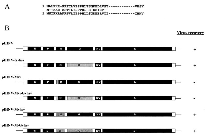
Fragment 1 (pIHN1 [Fig. 1]) containing the N, P, and M genes was used as the starting material for all the constructs. Thus, pIHN1 was modified by site-directed mutagenesis (QuickChange Kit; Stratagene) to introduce two MluI restriction enzyme sites at the start (pIHN1-Mlu) and stop (pIHN1-Mlu/Mlu) codons of the M gene by using, respectively, primer pairs M-Mlu-ATG-S-M-Mlu-ATG-A and M-Mlu-TAG-S-M-Mlu-TAG-A (Table 1). The IHNV M gene was deleted from pIHN1-Mlu/Mlu by MluI digestion and replaced by the VHSV M gene, which was recovered by RT-PCR from the VHSV RNA genome by using the specific primers M-vhs-S and M-vhs-A. The final pIHNV-Mvhsv and pIHNV-M-Gvhsv constructs (see Fig. 6B) were obtained by replacing the HpaI-EagI fragment of pIHNV or pIHNV-Gvhsv, respectively, with the modified HpaI-EagI counterpart from pIHN1-Mvhsv. For the chimeric M constructs, pIHN1-Mlu was used as a template to create, by site-directed mutagenesis, a unique Eco47III site between nucleotides 88 and 92 of the IHNV M gene by using the primer pair M-Eco47-S-M-Eco47-A, leading to the pIHN1-Mlu/Eco construct. The 5′ end of the IHNV M gene (the first 87 nucleotides) was deleted from pIHN1-Mlu/Eco by MluI-Eco47III digestion and replaced with the first 87 nucleotides of the VHSV M gene, which were amplified by RT-PCR from the VHSV RNA genome with the primer pair M-vhsv-S-M-vhsv-Eco. The final pIHNV-Mvi and pIHNV-Mvi-Gvhsv constructs (see Fig. 6B) were obtained by replacement of the HpaI-EagI fragment of pIHNV or pIHNV-Gvhsv, respectively,with the modified HpaI-EagI counterpart from pIHN1-Mvi.
FIG. 6.
Diagram of the chimeric M and/or G pIHNV constructs. (A) Alignment of the first 29 amino acid sequences of IHNV and VHSV. +, conserved amino acid residues. (B) The complete IHNV M or G gene or parts of these genes (solid) were replaced by the respective VHSV counterparts (shaded). Recovery (+) or no recovery (−) of recombinant virus is indicated on the right.
Nucleotide sequencing.
All the sequences of the plasmid constructs used in this study and those of RT-PCR products were checked by nucleotide sequencing reactions carried out on an ABI 373A DNA automatic sequencer using the DyeDeoxy Terminator-Prism Kit (Applied Biosystems) and specific primers.
RESULTS
Recovery of recombinant IHNV expressing the VHSV glycoprotein.
The complete gene encoding the IHNV G glycoprotein was removed from the pIHNV construct following the introduction of two unique restriction enzyme sites at the start codon and near the end of the gene. As described previously (5), the pIHNV construct, encoding an antigenomic T7-driven IHNV RNA, was assembled from four fragments covering the full-length IHNV genome. Thus, fragment 2 (Fig. 1), which contains the G and NV genes and the beginning of the L gene, was used as a template to create, by site-directed mutagenesis, two new unique SpeI and SmaI restriction enzyme sites at the beginning and the end of the G gene, respectively. Then the IHNV G gene was replaced with the VHSV G gene in the mutated fragment 2, following excision and insertion using the SpeI and SmaI restriction enzyme sites. Finally, an EagI/AgeI fragment was excised from pIHNV fragment 2 and inserted back into the pIHNV backbone, leading to the final pIHNV-Gvhsv plasmid construct. By using the established conditions (5), pIHNV-Gvhsv was cotransfected with pT7-N, pT7-P, pT7-NV, and pT7-L into vTF7-3-infected EPC cells and incubated for 6 days at 14°C. By passage of the supernatant on fresh EPC cells, a typical virus-induced cytopathic effect was observed. The recombinant virus, rIHNV-Gvhsv, was amplified by several cell passages and used for further studies.
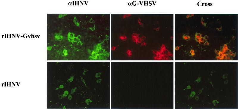
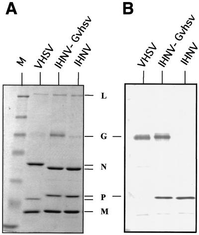
Expression of the VHSV G glycoprotein together with the IHNV viral proteins in rIHNV-Gvhsv-infected EPC cells was demonstrated by an indirect immunofluorescence assay using double labeling against the VHSV G and IHNV proteins (Fig. 2). The presence of VHSV glycoprotein G in the IHNV virions was investigated following analysis of sucrose gradient-purified virus. Figure 3A shows the protein patterns of the purified viruses separated on a Coomassie blue-stained SDS-polyacrylamide gel; the migration of G is indistinguishable between IHNV and VHSV, in contrast to the N and P proteins. A demonstration that VHSV G was incorporated into the IHNV virions was provided by Western blot analysis (Fig. 3B) using MAbs directed against the IHNV P protein and VHSV glycoprotein G. As shown in Fig. 3B, rIHNV-Gvhsv was recognized by both MAbs, whereas IHNV and VHSV were exclusively recognized by the anti-IHNV P MAb and the anti-VHSV G MAb, respectively.
FIG. 2.
Expression of VHSV glycoprotein in rIHNV-Gvhsv-infected cells. EPC cells were infected with wild-type rIHNV or rIHNV-Gvhsv as indicated on the left. Cells were alcohol-acetone fixed and incubated first with a rabbit anti-IHNV polyclonal antiserum (αIHNV) and then with a mouse anti-VHSV G MAb (αG-VHSV). Expression was revealed with a mixture containing Texas red-conjugated goat anti-mouse immunoglobulin G (for αG-VHSV) and fluorescein isothiocyanate-conjugated goat anti-rabbit immunoglobulin G (for αIHNV). Overlapping of double labeling is shown in the right panels (Cross).
FIG. 3.
VHSV G is incorporated into IHNV virions. (A) Sucrose gradient-purified virus proteins were separated on an SDS-10% polyacrylamide gel and visualized following Coomassie blue staining. (B) The gel was electrotransferred onto a polyvinylidene difluoride membrane and incubated with a mixture of MAbs against VHSV G and IHNV P. Immunodetected viral antigens were visualized with an alkaline phosphatase-conjugated goat anti-mouse immunoglobulin G. M, standard molecular weight marker. Viral proteins: L, RNA polymerase; G, glycoprotein; N, nucleoprotein; P, phosphoprotein; M, matrix protein.
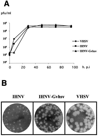
The characteristics of rIHNV-Gvhsv in cell culture were studied in comparison to those of IHNV and VHSV. Thus, comparison of the growth curves of IHNV, rIHNV-Gvhsv, and VHSV when EPC cells were infected at the same multiplicity of infection of 5 (Fig. 4A) showed that viral production reached a plateau at 30 h postinfection, with a viral yield of roughly 4 × 10⁸ PFU/ml for all three viruses. For rIHNV-Gvhsv, virus production started with a small delay (7 h postinfection) compared to IHNV or VHSV. The plaque morphologies produced by rIHNV-Gvhsv, IHNV, and VHSV were compared following a plaque assay (Fig. 4B). The plaques induced by rIHNV-Gvhsv were of a size intermediate between the small and large plaques induced by IHNV and VHSV, respectively.
FIG. 4.
Growth curves (A) and plaque morphologies (B) of IHNV, VHSV, and rIHNV-Gvhsv. (A) EPC cells were infected at a multiplicity of infection of 5 with either VHSV 07/71 (♦), IHNV 32/87 (▪), or rIHNV-Gvhsv (▴). Supernatants (100 μl) were taken at 0, 7, 29, 50, 72, and 96 h postinfection (h.p.i.) and titered. (B) EPC cells were infected with 50 PFU of each virus and cultured under an agarose-containing medium. At 5 days postinfection, cell monolayers were fixed and stained.
rIHNV-Gvhsv is pathogenic for rainbow trout.
The observation that rIHNV-Gvhsv replicates well in cell culture prompted us to ensure that rIHNV-Gvhsv was also capable of infecting, and replicating in, fish. Thus, juvenile rainbow trouts were experimentally infected by the bath immersion route (see Materials and Methods) with 5 × 10⁴ PFU of rIHNV-Gvhsv or wild type IHNV/ml. Deaths occurred, and dead fish were analyzed in more detail. Thus, kidneys, spleens, and brains were taken separately and homogenized to inoculate EPC cells. A few days later, a typical virus-induced cytopathic effect appeared. The presence of the respective IHNV or rIHNV-Gvhsv virus in infected cells was confirmed by an indirect immunofluorescence assay using an anti-IHNV P or anti-VHSV G MAb, respectively (data not shown). In all cases, with one exception, the virus was recovered in cell culture from the kidney and spleen homogenates, indicating that fish died from the usual virus-induced disease. The only exception was a dead fish infected with rIHNV-Gvhsv for which the virus was recovered exclusively from the brain. To examine whether that rIHNV-Gvhsv had a particular phenotype which could explain the nervous system tropism, cDNA from the G gene was amplified by RT-PCR from total RNA extracted from the infected brain. The expected 1,600-nucleotide RT-PCR product (data not shown) was gel purified and subjected to direct nucleotide sequencing. No mutations were detected in the G gene.
The SVCV glycoprotein can replace IHNV G.
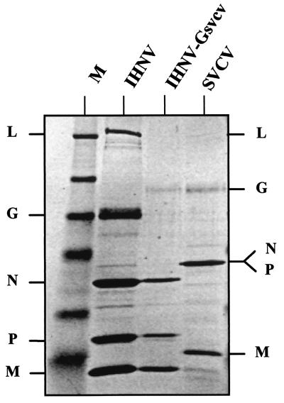
The observation that VHSV G can be efficiently incorporated into IHNV virions instead of IHNV G confirmed the previous observations made with VSV (33, 34), indicating that the conservation of the G cytoplasmic tail sequence is not critical for the formation of an infectious viral particle containing heterologous glycoproteins. To extend this observation, a construct in which the IHNV G gene was removed and replaced by a G gene from a fish vesiculovirus (SVCV) was engineered (Fig. 1). By cotransfection into EPC cells of pIHNV-Gsvcv together with expression vectors pT7-N, pT7-P, and pT7-L, a recombinant virus, IHNV-Gsvcv, was recovered. rIHNV-Gvhsv replicated well in cell culture and reached a titer of 2 × 10⁷ PFU/ml, which was approximately 10-fold lower than those obtained with wild-type IHNV and SVCV (>2 × 10⁸ PFU/ml). Due to the lack of availability of specific anti-SVCV G antibodies, it was not possible to directly demonstrate, by a Western blot assay, the presence of SVCV G on purified rIHNV-Gsvcv. However, comparison of the migration pattern of the viral proteins of rIHNV-Gsvcv on a Coomassie blue-stained polyacrylamide gel with those of wild type-IHNV and SVCV (Fig. 5) evidenced the insertion of SVCV G into IHNV virions. In contrast to IHNV and VHSV, which cannot grow at temperatures higher than 20°C, SVCV replicates well up to 28°C. However, attempts to grow rIHNV-Gsvcv at a temperature higher than 20°C as for wild-type SVCV failed, suggesting that IHNV proteins other than the glycoprotein are responsible for the “low temperature” growth phenotype of IHNV.
FIG. 5.
SVCV glycoprotein insertion into IHNV virions. Sucrose gradient purified virus proteins were separated on an SDS-10% polyacrylamide gel and visualized following Coomassie blue staining.
The IHNV matrix protein together with the G glycoprotein can be replaced by their VHSV counterparts.
The possible interaction between the matrix protein and glycoprotein G in IHNV virions was investigated by generation of chimeric viruses. Alignment of the VHSV and IHNV M amino acid sequences revealed high conservation of the first 29 amino acids (Fig. 6A), whereas overall identity was only 38%. Thus, we hypothesized that this short sequence conservation may represent the interacting domain between the M and G proteins. To verify this, the pIHNV construct was modified so that the M gene encodes a chimeric matrix protein in which the first 29 amino acids from IHNV have been replaced by the VHSV counterpart (Fig. 6B, pIHNV-Mvi). Despite several attempts, no recombinant virus could be recovered when the pIHNV-Mvi construct was cotransfected with the pT7-N, pT7-P, and pT7-L expression vectors. Similarly, when pIHNV-Mvi was further modified to replace the IHNV G gene with the VHSV G gene, yielding pIHNV-Mvi-Gvhsv (Fig. 6B), no virus could be recovered. However, when the complete IHNV M gene was replaced with that of VHSV (pIHNV-Mvhsv), a recombinant virus, rIHNV-Mvhsv, was generated. The recovery efficiency for rIHNV-Mvhsv was very low (see below) (Table 2).
TABLE 2.
Recovery efficiencies of the various recombinant IHNVs
| Virus | Titer (PFU/ml)a |
|---|---|
| rIHNV | 104 |
| rIHNV-Gvhsv | 105 |
| rIHNV-Gsvcv | 5 × 10⁴ |
| rIHNV-Mvhsv | 50 |
| rIHNV-M-Gvhsv | 50 |
Measured on the first passage (P1) of the transfection supernatant.
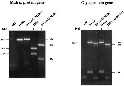
When both the complete M and G genes from IHNV were replaced by their VHSV counterparts (pIHNV-M-Gvhsv), a recombinant virus, rIHNV-M-Gvhsv, was also successfully recovered. The generation of rIHNV-M-Gvhsv was demonstrated by an RT-PCR assay using the genomic RNA extracted from the supernatant of rIHNV-M-Gvhsv-infected cells after four consecutive passages. As shown in Fig. 7, PCR products of the expected sizes and restriction enzyme digestion patterns were generated and clearly differentiated the recombinant rIHNV-M-Gvhsv from the wild-type IHNV.
FIG. 7.
Successful replacement of both the IHNV M and G genes by their VHSV counterparts. After four passages, viral genomic RNA extracted from supernatants of cells infected with either wild-type IHNV or rIHNV-M-Gvhsv was amplified by RT-PCR with specific M or G primers derived from IHNV or VHSV sequences. PCR products were analyzed on a 1% agarose gel with (+) or without (−) restriction enzyme digestion. A control without reverse transcriptase (−RT) was added to ensure that no contaminating plasmids were present in the rIHNV-M-Gvhsv-infected cell supernatant. The sizes of the fragments (in nucleotides) are given on the left for the IHNV M and G genes and on the right for the VHSV M and G genes.
One common feature of rIHNV-Mvhsv and rIHNV-M-Gvhsv is their very slow growth compared to that of the other recombinant viruses (rIHNV, rIHNV-Gvhsv, and rIHNV-Gsvcv), indicating that viral assembly is severely impaired. Indeed, the very low recovery efficiency for these two viruses (Table 2) does not fully explain why, after several passages, cytopathic effect in cells infected with rIHNV-Mvhsv or rIHNV-M-Gvhsv appears only at 6 days postinfection, whereas it takes only 2 days for the other recombinant IHNVs.
DISCUSSION
IHNV and VHSV both belong to the Novirhabdovirus genus, although they are only distantly related; for example, the amino acid identity between the two glycoproteins is less than 40%. The migration pattern of the viral polypeptides in SDS-polyacrylamide gel electrophoresis is roughly similar to that of RV, the Lyssavirus prototype. Previous studies on RV show that heterologous glycoproteins can be inserted into viral particles when the G cytoplasmic tail domain is conserved (26, 27, 28), in contrast to VSV, the Vesiculovirus prototype, for which the process appears to be sequence independent (33, 34).
These differences between RV and VSV have led to contradictory conclusions for introducing the G protein into the virus. For RV, it appears that the G cytoplasmic tail contains a signal sufficient and necessary to direct the efficient incorporation not only of G (28), but also of chimeric proteins with foreign transmembrane domains and ectodomains, into the envelopes of budding virions (26, 27). Moreover, it has been shown that the M protein, which is located between the ribonucleoprotein (RNP) complex and the membrane, interacts with the G protein (30). The M protein is thus responsible for recruiting the G protein into the virion. However, one exception has been described for RV: the functional replacement of an RV glycoprotein by a heterologous lyssavirus glycoprotein from the Eth-16 virus (Mokola virus) (29). Despite the sequence heterogeneity between the two cytoplasmic domains, this heterologous glycoprotein can functionally replace RV G in this rescue experiment, but approximately 30-fold fewer infectious particles were generated after expression of the heterologous Eth-16 G than after expression of the homologous G protein. As suggested by Mebatsion et al., a critical motif might be located in the carboxyl-terminal region of the G tail within the Lyssavirus genus (26).
In contrast to RV, VSV appears to use a nonspecific way of incorporating the VSV G protein (33) or heterologous transmembrane proteins (18, 20, 34) into the envelope. For example, substitution of the CD4 transmembrane and cytoplasmic domains for those from VSV G did not increase the level of CD4 incorporation into the VSV particle (34, 35); conversely, CD4 and CXCR4 proteins were efficiently incorporated into RV only when the autologous RV G tail was present (27). Moreover, it has been shown that 9 amino acids of the normal VSV G tail were sufficient to drive nearly normal budding efficiency but that the G protein with only 1 cytoplasmic amino acid presented only 10% of normal budding (33). However, as with RV, the human immunodeficiency virus type 1 (HIV-1) envelope protein was incorporated into VSV virions only when its cytoplasmic tail was replaced with the VSV G cytoplasmic tail (17) or deleted except for 3 amino acids (16) These observations suggest that a negative signal is present within the membrane-proximal 10 amino acids of the Env tail, sequestering the Env protein at sites distinct from VSV budding sites (16). These studies all suggest that the VSV G cytoplasmic domain is important in budding but that specific sequences are not required in that domain. So, as the direct interaction of VSV M and VSV G has not yet been unequivocally demonstrated (25) and if all the M protein is inside the RNP, as suggested by Barge et al. (1), important differences between the assembly mechanisms of VSV and RV appear. Since the localization of the VSV M protein is controversial (for a review, see reference 22), the differences between G protein incorporation by VSV and RV may be explained by a less stringent and weaker interaction between M and G in VSV virions.
The present study describes the recovery of a recombinant virus, rIHNV-Gvhsv, in which the IHNV G gene has been replaced with that of VHSV, another antigenically distinct novirhabdovirus. The data presented here demonstrate that rIHNV-Gvhsv buds efficiently and incorporates the VHSV G glycoprotein. rIHNV-Gvhsv, which grows as well as wild-type IHNV and VHSV in cell culture (reaching a titer of 4 × 10⁸ PFU/ml), is also pathogenic in fish; it was shown to grow even better than the wild-type recombinant IHNV in cell culture and was even more virulent in vivo (data not shown). The plaques induced by rIHNV-Gvhsv are intermediate in size between the small and large plaques induced by IHNV and VHSV, respectively. The recovery of rIHNV-Gvhsv demonstrates that for IHNV there is no cytoplasmic domain conservation requirement for replacement of the IHNV glycoprotein.
The absence of any requirement of the autologous IHNV G tail can be explained by the conservation of a critical motif in the G tail within the same genus, as suggested by Mebatsion et al. (29) for the functional replacement of RV G by that of the Mokola virus. A similar result was observed for the parainfluenza viruses. The hemagglutinin-neuraminidase (HN) and fusion (F) glycoprotein genes of parainfluenza virus type 3 (PIV3) can be efficiently replaced with their counterparts from PIV1, which also belongs to the Respirovirus genus (36). In contrast, when the PIV2 HN and F proteins were used to replace those of PIV3, a viable recombinant virus was recovered only when the PIV3 cytoplasmic domains were retained (37). Thus, exchange of full-length glycoprotein open reading frames was not achieved between different genera, i.e., between Respirovirus (PIV3) and Rubulavirus (PIV2). To study whether IHNV exhibits similar behavior, we have replaced IHNV G with that of SVCV, which belongs to the Vesiculovirus genus. A recombinant virus, rIHNV-Gsvcv, was recovered, demonstrating that a glycoprotein originating from another viral genus can be efficiently incorporated into IHNV virions. rIHNV-Gsvcv grows fairly well in cell culture. In addition, we demonstrate that IHNV proteins other than the glycoprotein are responsible for the temperature growth restriction. Indeed, in contrast to IHNV, which cannot grow at a temperature above 20°C, SVCV replicates well up to 28°C; however, rIHNV-Gsvcv does not replicate at temperatures higher than 20°C.
Thus, these data show that the IHNV glycoprotein can be efficiently replaced with that of either of two unrelated fish rhabdoviruses, VHSV (Novirhabdovirus) and SVCV (Vesiculovirus). The observation that this process does not require specific IHNV G-derived sequences fits well with the results from previous studies on VSV (18, 20, 34) but is not in good accordance with the results obtained with RV (26, 27), although IHNV is more related to lyssaviruses such as RV than to vesiculoviruses such as VSV.
The possible interaction between the matrix M protein and glycoprotein G, which has been demonstrated clearly for RV (30) but not unequivocally for VSV (25), was investigated for IHNV by the generation of chimeric viruses. Alignment of the VHSV and IHNV M amino acid sequences revealed high conservation of the first 29 amino acids (Fig. 6A). It has previously been shown that the N-terminal region of VSV M contains one of the two membrane-binding sites (38), and for VSV and RV, a highly conserved PPxY motif is present in this region. That motif, which interacts with WW domains of specific cellular proteins (14), can substitute for the L domain of the Rous sarcoma virus (RSV) Gag protein to promote the release of chimeric particles (9) and plays an important role in the release of VSV particles from the cell surface (15). While the M proteins of many rhabdoviruses maintain the PPxY motif at their amino termini, it should be noted that the M proteins from IHNV and VHSV possess a PPxH motif for which interaction with either WW domains or a WW-like domain has not yet been demonstrated. For all these reasons (high conservation and membrane location), it could be hypothesized that this short sequence may represent the interacting domain between the M and G proteins. Thus, to study this, the sequence encoding the first 29 amino acids from the IHNV M protein was replaced with the VHSV counterpart in the full-length IHNV pIHNV constructs. However, no chimeric M recombinant virus could be recovered when the construct was transfected into the cells. Thus, despite the high amino acid conservation in the N-terminal region between IHNV and VHSV, replacement of the first 29 amino acids of IHNV M with those of VHSV probably induces a misfolding of the IHNV M protein, preventing the M-G interaction. In contrast, when the complete IHNV M gene was replaced by the VHSV counterpart, a recombinant virus, rIHNV-Mvhsv, was generated, demonstrating that the VHSV matrix protein can functionally replace the IHNV matrix protein. Furthermore, when both M and G full-length open reading frames from IHNV were replaced with those of VHSV, a recombinant virus, rIHNV-M-Gvhsv, was successfully recovered. Therefore, VHSV glycoprotein G can accommodate its own and heterologous matrix M proteins as well for efficient budding of the chimeric recombinant IHNV. The same observation is true for IHNV. The recovery of rIHNV-M-Gvhsv and rIHNV-Mvhsv reinforces the hypothesis of a misfolding of the chimeric VHSV/IHNV M protein, which could explain the lack of recovery of recombinant IHNV particles.
Taken together, these data show the extreme flexibility of IHNV with regard to the functional replacement of two major structural proteins, M and G, with those of a distantly related fish rhabdovirus.
Acknowledgments
This work was carried out with financial support from the Commission of the European Communities, Agriculture and Fisheries (FAIR) RDT program, CT98-4398.
We thank Maria-Isabel Thoulouze (INRA) for helpful discussions, Thanh Lan Lai and Aurélie Schwartz for expert technical assistance, and Wendy Brand-Williams (INRA) for careful reading of the manuscript. Michel Dorson and the fish facility staff (INRA) are acknowledged for the animal experiments.
REFERENCES
- 1.Barge, A., Y. Gaudin, P. Coulon, and R. W. Ruigrok. 1993. Vesicular stomatitis virus M protein may be inside the ribonucleocapsid coil. J. Virol. 67:7246-7253. [DOI] [PMC free article] [PubMed] [Google Scholar]
- 2.Basurco, B., and A. Benmansour. 1995. Distant strains of the fish rhabdovirus VHSV maintain a sixth functional cistron which codes for a nonstructural protein of unknown function. Virology 212:741-745. [DOI] [PubMed] [Google Scholar]
- 3.Bearzotti, M., B. Delmas, A. Lamoureux, A. M. Loustau, S. Chilmonczyk, and M. Bremont. 1999. Fish rhabdovirus cell entry is mediated by fibronectin. J. Virol. 73:7703-7709. [DOI] [PMC free article] [PubMed] [Google Scholar]
- 4.Bearzotti, M., A. F. Monnier, P. Vende, J. Grosclaude, P. de Kinkelin, and A. Benmansour. 1995. The glycoprotein of viral hemorrhagic septicemia virus (VHSV): antigenicity and role in virulence. Vet. Res. 26:413-422. [PubMed] [Google Scholar]
- 5.Biacchesi, S., M. I. Thoulouze, M. Bearzotti, Y. X. Yu, and M. Bremont. 2000. Recovery of NV knockout infectious hematopoietic necrosis virus expressing foreign genes. J. Virol. 74:11247-11253. [DOI] [PMC free article] [PubMed] [Google Scholar]
- 6.Bjorklund, H. V., K. H. Higman, and G. Kurath. 1996. The glycoprotein genes and gene junctions of the fish rhabdoviruses spring viremia of carp virus and hirame rhabdovirus: analysis of relationships with other rhabdoviruses. Virus Res. 42:65-80. [DOI] [PubMed] [Google Scholar]
- 7.Boudinot, P., M. Blanco, P. de Kinkelin, and A. Benmansour. 1998. Combined DNA immunization with the glycoprotein gene of viral hemorrhagic septicemia virus and infectious hematopoietic necrosis virus induces double-specific protective immunity and nonspecific response in rainbow trout. Virology 249:297-306. [DOI] [PubMed] [Google Scholar]
- 8.Conzelmann, K. K. 1998. Nonsegmented negative-strand RNA viruses: genetics and manipulation of viral genomes. Annu. Rev. Genet. 32:123-162. [DOI] [PubMed] [Google Scholar]
- 9.Craven, R. C., R. N. Harty, J. Paragas, P. Palese, and J. W. Wills. 1999. Late domain function identified in the vesicular stomatitis virus M protein by use of rhabdovirus-retrovirus chimeras. J. Virol. 73:3359-3365. [DOI] [PMC free article] [PubMed] [Google Scholar]
- 10.Fijan, N., M. Sulimanovic, M. Béarzotti, D. Muzinic, L. O. Zwillenberg, S. Chilmonczyk, J. F. Vautherot, and P. de Kinkelin. 1983. Some properties of the Epithelioma papulosum cyprini EPC cell line from carp Cyprinus carpio. Ann. Inst. Pasteur/Virol. 134E:207-220.
- 11.Fuerst, T. R., E. G. Niles, F. W. Studier, and B. Moss. 1986. Eukaryotic transient-expression system based on recombinant vaccinia virus that synthesizes bacteriophage T7 RNA polymerase. Proc. Natl. Acad. Sci. USA 83:8122-8126. [DOI] [PMC free article] [PubMed] [Google Scholar]
- 12.Gaudin, Y., P. de Kinkelin, and A. Benmansour. 1999. Mutations in the glycoprotein of viral haemorrhagic septicaemia virus that affect virulence for fish and the pH threshold for membrane fusion. J. Gen. Virol. 80:1221-1229. [DOI] [PubMed] [Google Scholar]
- 13.Gaudin, Y., R. W. Ruigrok, C. Tuffereau, M. Knossow, and A. Flamand. 1992. Rabies virus glycoprotein is a trimer. Virology 187:627-632. [DOI] [PMC free article] [PubMed] [Google Scholar]
- 14.Harty, R. N., J. Paragas, M. Sudol, and P. Palese. 1999. A proline-rich motif within the matrix protein of vesicular stomatitis virus and rabies virus interacts with WW domains of cellular proteins: implications for viral budding. J. Virol. 73:2921-2929. [DOI] [PMC free article] [PubMed] [Google Scholar]
- 15.Jayakar, H. R., K. G. Murti, and M. A. Whitt. 2000. Mutations in the PPPY motif of vesicular stomatitis virus matrix protein reduce virus budding by inhibiting a late step in virion release. J. Virol. 74:9818-9827. [DOI] [PMC free article] [PubMed] [Google Scholar]
- 16.Johnson, J. E., W. Rodgers, and J. K. Rose. 1998. A plasma membrane localization signal in the HIV-1 envelope cytoplasmic domain prevents localization at sites of vesicular stomatitis virus budding and incorporation into VSV virions. Virology 251:244-252. [DOI] [PubMed] [Google Scholar]
- 17.Johnson, J. E., M. J. Schnell, L. Buonocore, and J. K. Rose. 1997. Specific targeting to CD4+ cells of recombinant vesicular stomatitis viruses encoding human immunodeficiency virus envelope proteins. J. Virol. 71:5060-5068. [DOI] [PMC free article] [PubMed] [Google Scholar]
- 18.Kahn, J. S., M. J. Schnell, L. Buonocore, and J. K. Rose. 1999. Recombinant vesicular stomatitis virus expressing respiratory syncytial virus (RSV) glycoproteins: RSV fusion protein can mediate infection and cell fusion. Virology 254:81-91. [DOI] [PubMed] [Google Scholar]
- 19.Kim, C. H., J. R. Winton, and J. C. Leong. 1994. Neutralization-resistant variants of infectious hematopoietic necrosis virus have altered virulence and tissue tropism. J. Virol. 68:8447-8453. [DOI] [PMC free article] [PubMed] [Google Scholar]
- 20.Kretzschmar, E., L. Buonocore, M. J. Schnell, and J. K. Rose. 1997. High-efficiency incorporation of functional influenza virus glycoproteins into recombinant vesicular stomatitis viruses. J. Virol. 71:5982-5989. [DOI] [PMC free article] [PubMed] [Google Scholar]
- 21.Kurath, G., and J. C. Leong. 1985. Characterization of infectious hematopoietic necrosis virus mRNA species reveals a nonvirion rhabdovirus protein. J. Virol. 53:462-468. [DOI] [PMC free article] [PubMed] [Google Scholar]
- 22.Lenard, J. 1996. Negative-strand virus M and retrovirus MA proteins: all in a family? Virology 216:289-298. [DOI] [PubMed] [Google Scholar]
- 23.Lorenzen, N., P. M. Cupit, K. Einer-Jensen, E. Lorenzen, P. Ahrens, C. J. Secombes, and C. Cunningham. 2000. Immunoprophylaxis in fish by injection of mouse antibody genes. Nat. Biotechnol. 18:1177-1180. [DOI] [PMC free article] [PubMed] [Google Scholar]
- 24.Lorenzen, N., N. J. Olesen, and P. E. Jorgensen. 1990. Neutralization of Egtved virus pathogenicity to cell cultures and fish by monoclonal antibodies to the viral G protein. J. Gen. Virol. 71:561-567. [DOI] [PubMed] [Google Scholar]
- 25.Lyles, D. S., M. McKenzie, and J. W. Parce. 1992. Subunit interactions of vesicular stomatitis virus envelope glycoprotein stabilized by binding to viral matrix protein. J. Virol. 66:349-358. [DOI] [PMC free article] [PubMed] [Google Scholar]
- 26.Mebatsion, T., and K. K. Conzelmann. 1996. Specific infection of CD4+ target cells by recombinant rabies virus pseudotypes carrying the HIV-1 envelope spike protein. Proc. Natl. Acad. Sci. USA 93:11366-11370. [DOI] [PMC free article] [PubMed] [Google Scholar]
- 27.Mebatsion, T., S. Finke, F. Weiland, and K. K. Conzelmann. 1997. A CXCR4/CD4 pseudotype rhabdovirus that selectively infects HIV-1 envelope protein-expressing cells. Cell 90:841-847. [DOI] [PubMed] [Google Scholar]
- 28.Mebatsion, T., M. Konig, and K. K. Conzelmann. 1996. Budding of rabies virus particles in the absence of the spike glycoprotein. Cell 84:941-951. [DOI] [PubMed] [Google Scholar]
- 29.Mebatsion, T., M. J. Schnell, and K. K. Conzelmann. 1995. Mokola virus glycoprotein and chimeric proteins can replace rabies virus glycoprotein in the rescue of infectious defective rabies virus particles. J. Virol. 69:1444-1451. [DOI] [PMC free article] [PubMed] [Google Scholar]
- 30.Mebatsion, T., F. Weiland, and K. K. Conzelmann. 1999. Matrix protein of rabies virus is responsible for the assembly and budding of bullet-shaped particles and interacts with the transmembrane spike glycoprotein G. J. Virol. 73:242-250. [DOI] [PMC free article] [PubMed] [Google Scholar]
- 31.Morzunov, S. P., J. R. Winton, and S. T. Nichol. 1995. The complete genome structure and phylogenetic relationship of infectious hematopoietic necrosis virus. Virus Res. 38:175-192. [DOI] [PubMed] [Google Scholar]
- 32.Pekosz, A., B. He, and R. A. Lamb. 1999. Reverse genetics of negative-strand RNA viruses: closing the circle. Proc. Natl. Acad. Sci. USA 96:8804-8806. [DOI] [PMC free article] [PubMed] [Google Scholar]
- 33.Schnell, M. J., L. Buonocore, E. Boritz, H. P. Ghosh, R. Chernish, and J. K. Rose. 1998. Requirement for a non-specific glycoprotein cytoplasmic domain sequence to drive efficient budding of vesicular stomatitis virus. EMBO J. 17:1289-1296. [DOI] [PMC free article] [PubMed] [Google Scholar]
- 34.Schnell, M. J., L. Buonocore, E. Kretzschmar, E. Johnson, and J. K. Rose. 1996. Foreign glycoproteins expressed from recombinant vesicular stomatitis viruses are incorporated efficiently into virus particles. Proc. Natl. Acad. Sci. USA 93:11359-11365. [DOI] [PMC free article] [PubMed] [Google Scholar]
- 35.Schnell, M. J., J. E. Johnson, L. Buonocore, and J. K. Rose. 1997. Construction of a novel virus that targets HIV-1-infected cells and controls. HIV-1 infection. Cell 90:849-857. [DOI] [PubMed] [Google Scholar]
- 36.Tao, T., A. P. Durbin, S. S. Whitehead, F. Davoodi, P. L. Collins, and B. R. Murphy. 1998. Recovery of a fully viable chimeric human parainfluenza virus (PIV) type 3 in which the hemagglutinin-neuraminidase and fusion glycoproteins have been replaced by those of PIV type 1. J. Virol. 72:2955-2961. [DOI] [PMC free article] [PubMed] [Google Scholar]
- 37.Tao, T., M. H. Skiadopoulos, F. Davoodi, J. M. Riggs, P. L. Collins, and B. R. Murphy. 2000. Replacement of the ectodomains of the hemagglutinin-neuraminidase and fusion glycoproteins of recombinant parainfluenza virus type 3 (PIV3) with their counterparts from PIV2 yields attenuated PIV2 vaccine candidates. J. Virol. 74:6448-6458. [DOI] [PMC free article] [PubMed] [Google Scholar]
- 38.Ye, Z., W. Sun, K. Suryanarayana, P. Justice, D. Robinson, and R. R. Wagner. 1994. Membrane-binding domains and cytopathogenesis of the matrix protein of vesicular stomatitis virus. J. Virol. 68:7386-7396. [DOI] [PMC free article] [PubMed] [Google Scholar]


