

Abstract
We previously demonstrated that formation of complexes between the DNA binding domains of the Hepatocyte Nuclear Factor 6 (HNF6; HNF-6 or OC-1) and Forkhead Box a2 (Foxa2; HNF3β) proteins stimulated Foxa2 transcriptional activity. Here, we used HepG2 cell cotransfection assays to demonstrate that HNF6 transcriptional activity was stimulated by CCAAT/enhancer-binding protein α (C/EBPα), but not by the related C/EBPβ or C/EBPδ proteins. Formation of the C/EBPα-HNF6 protein complex required the HNF6 Cut-domain and the C/EBPα activation domain 1 (AD1)/AD2 sequences. This C/EBPα-HNF6 transcriptional synergy required both the N-terminal HNF6 poly-Histidine and STP Box sequences as well as the C/EBPα AD1/AD2 sequences, the latter of which are known to recruit the CREB binding protein (CBP) transcriptional coactivator. Consistent with these findings, adenovirus E1A mediated inhibition of p300/CBP histone acetyltransferase activity abrogated C/EBPα-HNF6 transcriptional synergy in cotransfection assays. Co-immunoprecipitation assays with liver protein extracts demonstrate an association between the HNF6 and C/EBPα transcription factors and the CBP coactivator protein in vivo. Furthermore, Chromatin Immunoprecipitation assays with hepatoma cells demonstrated that increased levels of both C/EBPα and HNF6 proteins were required to stimulate association of these transcription factors and the CBP coactivator protein with the endogenous mouse Foxa2 promoter region. Taken together, our results suggest that formation of the C/EBPα-HNF6 protein complex stimulates recruitment of the CBP coactivator protein for expression of Foxa2, a transcription factor critical for regulating expression of hepatic gluconeogenic genes during fasting.
Keywords: Basic leucine zipper, Cut-Homeodomain, Hepatocyte Nuclear Factor, CREB binding protein, One Cut transcription factor, mouse liver
Abbreviations: HNF6, Hepatocyte Nuclear Factor 6; OC-1, Onecut-1 or HNF-6; Foxa2, Forkhead Box a2; CREB, cAMP binding protein; CBP, CREB binding protein; TTR, Transthyretin; CMV, Cytomegalovirus virus immediate early promoter; C/EBPα, CCAAT/Enhancer Binding Protein α; AD1, activation domain 1; AD2, activation domain 2; STP box, Serine Threonine and Proline Box; PH, poly histidine; GST, Glutathione-S-Transferase; IVT, in vitro transcription and translation; GFP, Green Fluorescent Protein; NMR, Nuclear Magnetic Resonance; pfu, plaque forming units; ifu, infectious particle units; HA, influenza hemagglutinin epitope tag; bp, base pair; AdHNF6, adenovirus expressing HNF6 cDNA; CMVTetO, 7 copies of the Tetracycline Operator sequence linked to the minimal CMV promoter; AdCMV-TA, adenovirus expressing Tetracycline Transcriptional Activator; AdC/EBPα, adenovirus with inducible expression of HA-C/EBPα (CMV-TetO HA-C/EBPα plus AdCMVTA); AdGFP, adenovirus expressing GFP; Co-IP, co-immunoprecipitation; ChIP, Chromatin Immunoprecipitation
INTRODUCTION
Transfection studies in hepatoma cell lines demonstrated that synergistic transcriptional activation of hepatocyte-specific genes requires simultaneous binding of multiple Hepatocyte Nuclear Factor (HNF) to their promoter/enhancer regions (1). However, deciphering the mechanisms by which these HNF transcription factors functionally interact to elicit transcriptional synergy remains to be completed. The CCAAT/Enhancer binding protein α (C/EBPα) transcription factor regulates expression of hepatocyte-specific genes, whose protein products are involved in glucose, lipid and circulatory homeostasis (2–9). The C/EBPα and C/EBPβ transcription factors are co-expressed in hepatocytes and utilize a C-terminal basic region leucine zipper (bZIP) bipartite motif to bind to DNA as either homodimers or heterodimers (10). Structure-function studies demonstrate that the C/EBPα protein stimulates transcription through the conserved C/EBPα Activation Domain 1 sequences (AD1; amino acids 51 to 136), which recruits CREB binding protein (CBP)/p300 transcriptional coactivators (11).
Hepatocyte Nuclear Factor 6 (HNF6) protein regulates transcription of hepatocytespecific genes critical for adult liver function and for development of the hepatic biliary tree (1, 12, 13). The HNF6 protein belongs to the ONECUT (OC) family of transcription factors, which utilize a C-terminal DNA binding motif consisting of a single Cut-domain and Homeodomain (14–18). Formation of HNF6 and Forkhead Box a2 (Foxa2) protein complexes stimulated Foxa2 transcriptional activity by recruiting the CBP coactivator protein through the HNF6 Cut-Homeodomain sequences (19). Nuclear Magnetic Resonance (NMR) structural determination of the HNF6 protein-DNA complex demonstrates that the HNF6 Cut-Homeodomain sequences that are necessary for recruitment of the CBP coactivator are inaccessible when this HNF6 motif is bound to its DNA recognition sequence (20). These NMR structural studies predicts that DNA recognition by the HNF6 Cut-Homeodomain sequences precludes recruitment of the CBP coactivator, suggesting that HNF6 stimulates transcription through its N-terminal Serine, Threonine and Proline (STP) Box domain (16). Furthermore, we recently showed that the HNF6 Cut domain Lysine 339 residue is acetylated by the CBP coactivator protein and that this modification is essential for HNF6 protein stability (21).
In our current study, we examine whether HNF6 associates with another liver transcription factor(s) to further stimulate HNF6 activity. We showed that formation of the C/EBPα and HNF6 complex stimulated HNF6-dependent transcription through recruitment of the CBP coactivator protein and defined sequences required for this protein association. Co-immunoprecipitation (Co-IP) experiments with liver protein extracts and Chromatin Immunoprecipitation (ChIP) assays with hepatoma cells have allowed us to demonstrate a functional association between the HNF6 and C/EBPα transcription factors to stimulate recruitment of the CBP coactivator promoter to the endogenous Foxa2 promoter region.
MATERIALS AND METHODS
Expression and reporter plasmids and cotransfection assays.
The CMV promoter expression vectors containing the HNF6 cDNA alone or tagged with Green Fluorescent Protein (GFP) or V5 tagged and the C/EBPα, C/EBPβ or C/EBPδ expression vectors were described previously (17, 21, 22). The SV40 promoter pECE expression vectors containing mutant HNF6 ΔSTP Box (Δ98–123) or HNF6 ΔPolyhistidine (ΔPH) cDNAs were a generous gift from Frédéric Lemaigre (Brussels, Belgium) and were described previously (16). The CMV pCDNA3.1 expression vectors containing the HA-tagged C/EBPα or HA-C/EBPα deletion mutants lacking either the transcriptional activation domain 2 (AD2: amino acids Δ116 to 254) or AD1 sequences (119–360) and the C/EBPα K300E DNA binding mutant were described previously (23, 24). The pGEX4-T-1 plasmid was used to express the Glutathione-S-Transferase (GST)-C/EBPα fusion proteins consisting of either AD1 (1 to 119), both AD1 and AD2 (1 to 226 or 1 to 286), the bZIP DNA binding domain (280 to 336) or the basic domain (313 to 336) was described previously (23). To generate the GST-C/EBPα AD2 (92–271) fusion protein, we blunted the BssHII fragment from the human C/EBPα cDNA into the SmaI site of the pGEX4T-1 expression plasmid. The pGEM1 HNF6 expression plasmids were used to synthesize radioactively labeled full-length or truncated HNF6 proteins and were described previously (19). The HNF6 dependent reporter gene (6X HNF6 TATA Luciferase plasmid) was described previously (17, 19).
Human hepatoma HepG2 cells, mouse hepatoma Hepa1-6 cells (ATCC #CRL-1830) or human osteosarcoma U20S cells were maintained in monolayer cultures and grown as previously described (18, 25) and cotransfected using Fugene 6 reagent (Roche) according to the manufacturer’s protocol. HepG2 or U2OS cells were transfected with 1.6 μg of the 6X HNF6 TATA Luciferase plasmid and 200 ng of CMV C/EBPα (WT or mutant) or CMV C/EBPβ expression plasmid with or without 200 ng of the CMV HNF6 expression vector. We also performed cotransfection assays with 1.6 μg of the 6X HNF6 TATA Luciferase reporter plasmid and 200 ng of CMV HNF6 and CMV C/EBPα with or without 200 ng of the CMV adenovirus E1A expression vector.
In vitro GST-C/EBPα pull-down assay, Western and co-immunoprecipitation with transfected HepG2 nuclear extracts.
Production and isolation of Glutathione-S-Transferase (GST) fusion proteins from BL21 E. coli was performed as described previously (26). Binding buffer and wash buffer for the in vitro GST pull-down assay and co-immunoprecipitation assays with nuclear extracts were performed as described previously (19, 22). The immunoprecipitates were eluted by the addition of SDS sample buffer containing 1% β-mercaptoethanol and boiled for five minutes. Eluted proteins were resolved by SDS-PAGE and transferred to nitrocellulose membranes for Western blot analysis with either monoclonal antibody specific to GFP antibody (Clontech, for GFP-HNF6), HA epitope tag (Invitrogen, for HA-C/EBPα), V5 epitope tag (Invitrogen, for V5-HNF6) or C/EBPβ {Santa Cruz Biotechnology; sc-150 (C-19)} and detected as described previously (19, 21, 27). For co-immunoprecipitation assays, 750 μg of total protein extract from quiescent mouse liver (0h) or regenerating mouse liver was immunoprecipitated with HNF6 (27) or rabbit serum (Vector Laboratories) and then subjected to Western analysis with rabbit anti-C/EBPα antibody (Santa Cruz Biotechnology; sc-61), C/EBPβ antibody {Santa Cruz Biotechnology; sc-150 (C-19)} or rabbit anti-CBP C-terminal antibody (Upstate; Catalog #06-294; Lot # 13094). Peroxidase conjugated ImmunoPure Recombinant Protein A/G (Pierce, Rockford, IL, product # 32490) was used to recognize Antibody-antigen bands and detected with Enhanced Chemiluminescence Plus (ECL-plus, Amersham Pharmacia Biotech, Piscataway, NJ) followed by autoradiography.
Construction of adenovirus with inducible C/EBPα expression and adenovirus purification.
The Ad-Tetracycline Responsive Element (TRE)-HA-C/EBPα adenoviral construct was produced using the Adeno-X™ Tet-Off™ Expression System by inserting the HA-tagged human C/EBPα cDNA (1–360) into the TRE shuttle vector following the manufacturer’s protocol (BD Biosciences, Palo Alto, CA). The adenovirus vector used 7 copies of the Tetracycline Operator sequence linked to the minimal CMV promoter (CMV-TetO) to drive expression of the HA-C/EBPα protein. Induced expression was accomplished by coinfection with a second adenovirus containing the CMV promoter driving expression of the Tetracycline Transcriptional Activator (AdCMV-TA), which is able to transcriptionally activate in the absence of Doxycycline (Tet-off system). Expression of HA-C/EBPα from this inducible adenovirus delivery system was detected by Western blot analysis with either the C/EBPα antibody or HA-tag antibody (Upstate Catalog #07-221). Recombinant adenoviruses were used to infect HEK 293A cells and cell lysates were harvested at 72 hours post infection (PI). Generation and infection with the adenovirus containing the CMV-HNF6 cDNA expression cassette (AdHnf6) was described previously (28) and adenovirus CMV Green Fluorescent protein (GFP) was purchased from BD Biosciences (Palo Alto, CA). The large-scale production of recombinant Adenovirus particles was performed by infection of QBO-293 cells and subsequent purification following the protocol of Quantum Biotechnologies (Montreal, Canada). The number of infectious particle units (ifu) was determined using the Adeno-X Rapid Titer Kit (BD Biosciences, Palo Alto, CA) with protocols provided by the manufacturer.
Infection of hepatoma Hepa1-6 cells with recombinant adenoviruses and Chromatin Immunoprecipitation (ChIP) Assays.
Mouse hepatoma Hepa1-6 cells (1 X 10⁷ cells per 150-mm dish) were infected at a multiplicity of infection (MOI) of 10 ifu per cell with AdHNF6 or AdC/EBPα (AdCMV-TetO-HA-C/EBPα and AdCMV-TA) either separately or together or with AdGFP control virus alone, as described previously (19, 29). We also used the Nucleofector II apparatus and buffers recommended by manufacturer (Amaxa, Gaithersburg, MD) to transfect Hepa1-6 cells with CMV expression vectors containing either mouse HNF6 cDNA or rat C/EBPβ cDNA separately or together or with the CMV-GFP expression plasmid alone. The Nucleofector II system transfected approximately 60% of the Hepa1-6 cells as determined by electroporation of CMV GFP expression vector and counting the number of cells positive for GFP fluorescence (data not shown). At 24 hours after infection or electroporation, the cells were processed for Chromatin Immunoprecipitation (ChIP) assays using published methods (30, 31). The following antibodies were used for ChIP assays, as described previously (30): Hnf6 rabbit antibody (27), CBP rabbit antibody {Santa Cruz Biotechnology; sc-369 (A-22)}, C/EBPα or C/EBPβ rabbit antibody (Santa Cruz Biotechnology; see above) or control rabbit serum (Vector Laboratories). Crosslinks were reversed on all chromatin samples by RNase A and Proteinase K digestion and then DNA was purified using PCR purification columns per manufacturer’s instructions (Qiagen, Valencia, CA) as described previously (30). The total input sample was diluted 1:10 and 2.5 μl was used for PCR (10% total input). To amplify the mouse Foxa2 promoter in ChIP assays we used the −166 bp sense primer (ctcctgaagtcatcccacaagg) and −67 bp anti-sense primer (ggtgcccaaagcatttcgtaac) with 54°C annealing temperature. The following reaction mixture was used for all PCR samples: 1X of IQ SybrGreen Supermix (Biorad, Carlsbad, CA), 100 nM of each primer, and 2.5μl of each purified ChIP extract in a 25μl total volume. Reactions were amplified and analyzed in triplicate using a MyiQ Single Color Real- Time PCR Detection System (Biorad, Carlsbad, CA). Normalization was carried out using the ΔΔCT method as described previously (30, 31). The levels of either AdGFP-infected or CMV GFP transfected control samples were set at one. The experimental ChIP binding levels were expressed as a fold induction with respect to these control samples (relative promoter binding) ± SD.
ChIP assays with wild type mouse liver.
Liver tissue was isolated from three separate wild type mice, washed in 1 X Sterile PBS, and weight of mouse livers were recorded. The liver tissue was diced with razor blade, homogenized in a 1X Sterile PBS solution for 2 seconds with a PowerGen 125 Polytron (Fisher Scientific), and then fixed in 10 ml 1% Formaldehyde (diluted in 1 X sterile PBS) for 15 min at room temperature with rotation. Glycine was added to 0.125 M and suspension was incubated with rotation for an additional 5 min at room temperature to neutralize the Formaldehyde. Fixed liver tissue was then pelleted 1500–2500 rpm at 4°C for 10 min. The cell pellet was homogenized for 1 minute in a Medimachine (Becton-Dickinson, Franklin Lakes, NJ) by re-suspending tissue pellet in 1 ml cold 1 X PBS containing protease inhibitor cocktail (Roche) and transferring to a 50 μm medicon using an 18 gauge needle and syringe. Nuclei were recovered through the port in medicon using syringe and needle and the homogenate was re-applied to the medicon two more times to purify liver nuclei. The liver nuclei were then pelleted and frozen at −80°C overnight. At this point, the chromatin was sonicated and the ChIP Assay was performed and analyzed as described above for cultured cells.
RESULTS
HNF6 transcriptional synergy requires the C/EBPα AD1/AD2 sequences and is abrogated by E1A mediated inhibition of CBP activity.
In our current study, we examine whether HNF6 associates with another liver transcription factor to stimulate HNF6 activity. We cotransfected human hepatoma HepG2 cells with the HNF6 dependent reporter plasmid (6X HNF6 TATA Luciferase) and the CMV HNF6 expression plasmid with or without expression vectors containing either HNF4α, Fetoprotein Transcription Factor (FTF), C/EBPα, C/EBPβ or C/EBPδ cDNAs. These cotransfection experiments revealed that only the C/EBPα expression vector stimulated HNF6 transcriptional activity (data not shown). Cotransfection assays with C/EBPα and HNF6 expression vectors and 6X HNF6 TATA Luciferase plasmid demonstrated that C/EBPα stimulated HNF6-dependent transcription in either human Hepatoma HepG2 or Osteosarcoma U2OS cells (Fig. 1A–B). Furthermore, a DNA binding deficient C/EBPα (K300E) mutant was able to stimulate HNF6-dependent transcription in cotransfection assays (Fig. 1A), suggesting that this transcriptional activation was independent of C/EBPα DNA binding activity. This HNF6 transcriptional activation was specific to the C/EBPα isoform because neither C/EBPβ nor C/EBPδ expression vectors provided significant stimulation of HNF6 transcriptional activity in HepG2 or U2OS cotransfection assays (Fig. 1A–B and data not shown).
Fig. 1. C/EBPα synergistically stimulates HNF6 transcriptional activity in cotransfection assays.
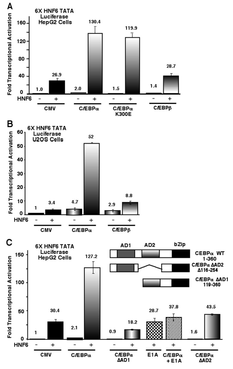
(A–B) HNF6-dependent transcription is stimulated by C/EBPα, but not C/EBPβ, in cotransfection assays with either HepG2 or U2OS cells. Human hepatoma HepG2 cells (A) or osteosarcoma U2OS cells (B) were cotransfected with 6X HNF6 TATA Luciferase reporter construct and CMV expression constructs containing no insert (CMV), C/EBPα, DNA binding deficient C/EBPα mutant protein (C/EBPα K300E) or C/EBPβ with or without the CMV HNF6 expression vector. The CMV Renilla-Luciferase reporter construct was included in all of the transfections and used as a normalization control. Protein extracts were prepared from transfected HepG2 or U2OS cells at 24 hours following DNA transfection and the Dual-Luciferase Assay System was used to measure Luciferase enzyme activity as described previously (19). (C) C/EBPα-HNF6 transcriptional synergy requires the C/EBPα AD1 and AD2 sequences and is inhibited by adenovirus E1A. Schematically shown is the location of the C/EBPα transcriptional activation domain 1 (AD1) and AD2 sequence and the Basic Leucine Zipper (bZIP) DNA binding domain. HNF6 cotransfection assays were performed with deletion of the C/EBPα AD1 (Δ1 to 118 amino acids) or the AD2 (Δ116 to 254 amino acids) sequences. HepG2 cells were also cotransfected with the 6X HNF6 TATA-Luciferase construct together with the CMV HNF6, CMV C/EBPα and CMV adenovirus E1A (an inhibitor of p300/CBP histone acetyltransferase activity) plasmids or with HNF6 and E1A expression vectors. All of the transfection results are presented as mean fold induction of promoter activity ± SD from two separate experiments in triplicate. Transcriptional activity of the CMV empty is set at 1.0.
Cotransfection studies with expression vectors containing C/EBPα deletion mutants demonstrated that both Activation Domain 1 (AD1) and AD2 sequences were essential for C/EBPα-HNF6 transcriptional synergy (Fig. 1C; C/EBPα ΔAD1 or ΔAD2). Because the AD1 sequences were shown to mediate transcriptional activation by the p300/CBP coactivators (11), we performed cotransfection assays with the CMV vector expressing the adenovirus E1A protein, which inhibits histone acetyltransferase activity of the p300/CBP coactivator proteins (32). Cotransfection assays showed that the E1A protein suppressed C/EBPα’s ability to stimulate HNF6 transcriptional activity (Fig. 1C), a finding that supports the hypothesis that C/EBPα stimulates HNF6 transcriptional activity by recruiting the CBP coactivator proteins. In contrast, cotransfection assays with the E1A and HNF6 expression vectors and the 6X HNF6 TATA Luciferase plasmid was unable to inhibit the basal transcriptional activity of HNF6, a finding consistent with previous studies (19). Taken together, these structure-function studies indicated that both the C/EBPα AD1 and AD2 sequences are essential for C/EBPα-HNF6 transcriptional synergy.
The HNF6 poly-Histidine and STP box sequences are essential for C/EBPα transcriptional synergy in cotransfection assays.
We examine whether deletion of either the N-terminal HNF6 transcriptional activation STP box domain (HNF6ΔSTP; Δ98–123) or the poly Histidine (HNF6ΔPH; Δ122–139) sequences were necessary for transcriptional synergy with the C/EBPα protein (Fig. 2A). We performed HepG2 cotransfection studies with the 6X HNF6 TATA Luciferase plasmid and expression vectors containing WT HNF6, HNF6ΔSTP or HNF6ΔPH mutant proteins with or without the CMV C/EBPα expression construct. Consistent with published studies without C/EBPα (16), the HNF6ΔPH mutant displayed higher transcriptional activity than the WT HNF6 protein, while the HNF6ΔSTP mutant exhibited a 30% reduction in transcriptional activity (Fig. 2B). However, both the HNF6ΔSTP and HNF6ΔPH mutant proteins showed a reduction in HNF6-dependent transcriptional activity when combined with the CMV C/EBPα expression vector (Fig. 2B). These results demonstrated that retention of both the HNF6 N-terminal STP box and PH sequences was required for HNF6-C/EBPα transcriptional synergy.
Fig. 2. N-terminal activation domain of HNF6 is required for the transcriptional synergy with C/EBPα.
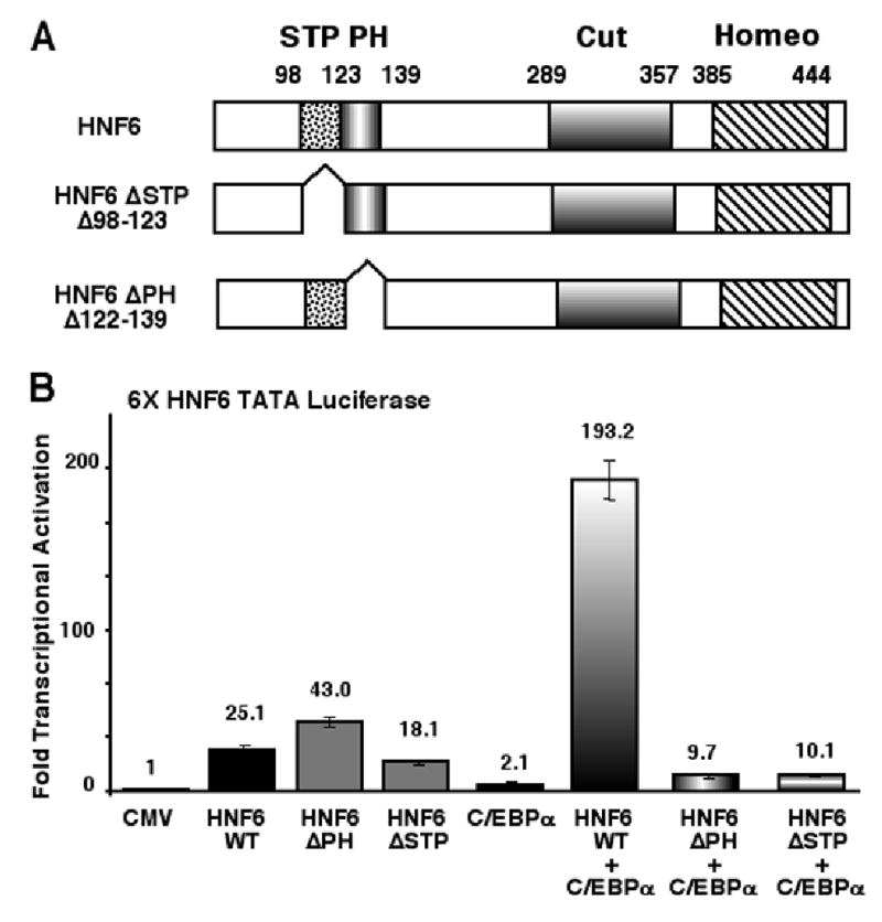
(A) Schematic representation of HNF6 wild type (WT) protein with N-terminal transcriptional activation Serine/Threonine/Proline (STP) box domain and the poly-Histidine (PH) region and the C-terminal Cut-Homeodomain DNA binding motif (16). Also shown in the diagram are the HNF6 deletion mutant proteins: HNF6 ΔSTP (Δ98–123) or HNF6 ΔPH (Δ122–139). (B) The HNF6 STP Box and PH sequences are required for C/EBPα-HNF6 transcriptional synergy. HepG2 cells were cotransfected with the 6X HNF6 TATA Luciferase reporter and expression vectors containing either the HNF6 WT, HNF6 ΔPH or HNF6 ΔSTP box with or without the CMV C/EBPα expression vector. Transfections and analysis of transcriptional activity using the dual Luciferase assay were described in Fig. 1 legend.
HNF6 associates with the C/EBPα protein: HNF6 Cut-domain and C/EBPα AD 1/2 sequences are required for the interaction.
To define sequences required for association between HNF6 and C/EBPα proteins, we performed pull-down experiments with Glutathione-S-Transferase (GST) C/EBPα fusion proteins and radioactively labeled in vitro transcription and translation (IVT) HNF6 proteins as diagrammed in Figures 3A and 3B (see Figure 3 Legends). The HNF6 expression plasmids was used to synthesize 35S Methionine labeled HNF6 proteins by IVT (Fig. 3B, right panel) and then these labeled HNF6 proteins were utilized for binding reactions with various GST-C/EBPα fusion proteins (Fig. 3A) immobilized on Glutathione-Sepharose beads as described previously (19). These GST-C/EBPα pull-down assays demonstrated that labeled HNF6 proteins that retained the Cut-domain sequences (Fig. 3C, WT HNF6, Cut-Homeo and Cut) bound to the GST-C/EBPα AD1/AD2 fusion proteins (Fig. 3C, GST-C/EBPα proteins 2 and 3), and that deletion of the HNF6 Cut-Homeodomain sequences abrogated this binding (Fig. 3C, ΔCut-Homeo). None of the labeled HNF6 proteins interacted with GST-C/EBPα fusion proteins containing only the AD1, the bZIP or basic domain sequences (Fig. 3A and C, proteins 1, 4 and 5). Furthermore, labeled WT HNF6 protein bound weakly to the GST-C/EBPα AD2 fusion protein (Fig. 3D, protein 6), suggesting that both the C/EBPα AD1 and AD2 sequences were required for efficient HNF6 binding. Taken together, these studies demonstrated that formation of the C/EBPα -HNF6 complex required the HNF6 Cut domain and the AD1/AD2 C/EBPα sequences.
Fig. 3. Formation of the HNF6 and C/EBPα protein complex requires the HNF6 Cut domain sequence and C/EBPα Activation domain 2 sequence.
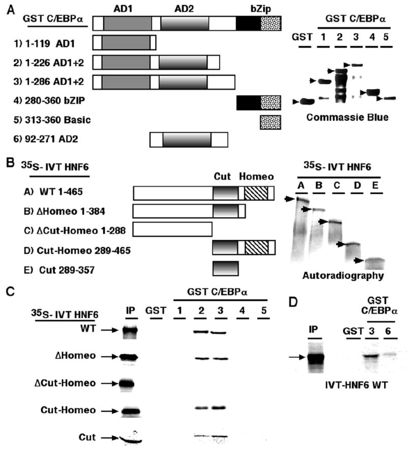
(A) Schematic representation of the various Glutathione-S-Transferase (GST)-C/EBPα fusion proteins used for the in vitro GST-pull down assays. Diagram depicting the GST-C/EBPα fusion proteins containing either transcriptional activation domain 1 (AD1, 1 to 119; construct 1), both AD1 and AD2 (1 to 226 or 1 to 286; construct 2 and 3), the basic Leucine Zipper (bZIP) DNA binding domain (280 to 336; Construct 4) or the basic domain (313 to 336; construct 5) or AD2 (92–271; construct 6). Right panel shows the expression levels of these GST-C/EBPα fusion proteins as visualized by Commassie blue staining following SDS-polyacrylamide gel electrophoresis (PAGE). (B) Diagram of HNF6 expression plasmids containing either the full-length HNF6 protein (WT), or N-terminal HNF6 protein that deleted either the Homeodomain (ΔHomeo) or the Cut- Homeodomain (ΔCut-Homeo) from the C-terminus, the Cut-Homeodomain alone (Cut-Homeo), or the Cut-domain alone (Cut). These HNF6 deletion plasmids were used to synthesize radioactively labeled HNF6 proteins using in vitro transcription and translation (IVT) with 35S Methionine and used for the in vitro GST-C/EBPα pull-down assays. Right panel shows the expression of these 35S-labelled HNF6 deletion proteins as visualized by autoradiography following SDS-PAGE. (C–D) The in vitro GST pull-down assays demonstrate that complex formation between the HNF6 and C/EBPα transcription factors requires the HNF6 Cut domain sequences and the C/EBPα AD1 and AD2 sequences (amino acids 1 to 226). GST-C/EBPα fusion proteins (3 μg preabsorbed to 20ul of glutathione-Sepharose) were incubated with IVT 35S labeled full-length HNF6 or deletion mutants, then washed, and boiling sample in SDS loading buffer eluted bound proteins. Labeled bound HNF6 proteins are resolved by SDS-PAGE and visualized by autoradiography.
The C/EBPα protein co-immunoprecipitates with HNF6 in transfected HepG2 nuclear extracts.
The C/EBPα protein is a potent inhibitor of cell proliferation (23, 33), and hepatoma cell lines therefore do not express C/EBPα at high levels. In order to perform HNF6-C/EBPα co-immunoprecipitation (Co-IP) assays, we prepared nuclear extracts from HepG2 cells cotransfected with plasmids expressing either the GFP-tagged HNF6 protein (19) or the HAtagged C/EBPα protein (23). These transfected HepG2 nuclear extracts were subjected to immunoprecipitation with an HA-tag antibody followed by Western blot analysis using a monoclonal GFP antibody (Fig. 4A). These Co-IP studies demonstrated that the GFP-HNF6 protein was able to associate with the HA-tagged C/EBPα protein (Fig. 4A, right panel). Co-IP assays with nuclear extracts prepared from HepG2 cells transfected with V5-tagged HNF6 (21) and C/EBPβ (22) expression vectors demonstrated that HNF6 protein failed to associate with the C/EBPβ protein (Fig. 4B, right panel). Western blot analysis of these transfected HepG2 nuclear extracts with the GFP, HA, V5 or C/EBPβ antibody showed protein expression from the transfected vectors (Fig. 4A and B, left panel 10% input). These Co-IP results demonstrate that the HNF6 transcription factor associates with only the C/EBPα protein.
Fig. 4. C/EBPα protein interacts with HNF6 protein in vivo.
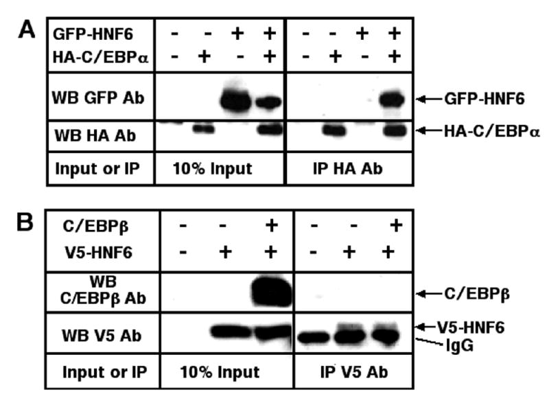
(A) Coimmunoprecipitation assays showed that the HNF6 and C/EBPα proteins form a complex in vivo. Nuclear protein extracts were prepared from HepG2 cells transfected with the Green Fluorescent Protein (GFP)-HNF6 and influenza hemagglutinin (HA)-epitope tagged C/EBPα cDNA expression vectors. These nuclear extracts were subjected to immunoprecipitation with an HA monoclonal antibody followed by Western blot analysis using a monoclonal GFP or HA antibody (right panel). The transfected GFP-HNF6 or HA-tagged C/EBPα proteins were expressed as determined by Western blot analysis of the transfected HepG2 nuclear extracts with either the GFP or HA monoclonal antibody (left panel 10% input). (B) Coimmunoprecipitation assays showed that the HNF6 and C/EBPβ proteins fail to associate in vivo. Nuclear protein extracts were prepared from HepG2 cells transfected with the V5-epitope tagged HNF6 protein and rat C/EBPβ cDNA expression vectors. These nuclear extracts were subjected to immunoprecipitation with an V5 monoclonal antibody followed by Western blot analysis using antibodies specific to either V5- epiptope or C/EBPβ (right panel). The transfected V5-HNF6 or rat C/EBPβ proteins were expressed as determined by Western blot analysis of the transfected HepG2 nuclear extracts with antibodies specific to either V5-epiptope or C/EBPβ (left panel 10% input). Note that the antibody band migrates slightly faster than the V5-HNF6 protein in the control lanes.
Co-immunoprecipitation assays with mouse liver extracts demonstrate a functional association between the HNF6 and C/EBPα transcription factors and the CBP coactivator.
In order to verify that HNF6 and C/EBPα transcription factors formed a complex with the CBP coactivator protein in vivo, we performed Co-IP assays with protein extracts from quiescent mouse liver (0h) or regenerating mouse liver isolated at 40 hours (40h) following partial hepatectomy (PH), the latter of which represents the peak in hepatocyte DNA replication (34). Quiescent or regenerating mouse liver extracts were Co-IP with either the HNF6 or control rabbit serum and then IP proteins were subjected to Western blot analysis with either the C/EBPα or C/EBPβ antibody. This Co-IP experiment demonstrated that HNF6 and C/EBPα proteins formed a stable complex in extracts from both quiescent and regenerating liver, but HNF6 failed to interact with the related C/EBPβ protein (Fig. 5A). Furthermore, Western blot analysis demonstrated that the CBP coactivator protein was Co-IP with C/EBPα in liver extracts using the HNF6 antibody (Fig. 5A). None of these transcription factors was immunoprecipitated by the control IgG antibody (Fig. 5A). Taken together, these Co-IP studies with liver protein extracts demonstrated an association between the HNF6 and C/EBPα transcription factors and the CBP coactivator protein in vivo.
Fig. 5. Co-immunoprecipitation assays with mouse liver extracts demonstrate functional association between the HNF6 and C/EBPα transcription factors and the CBP coactivator.
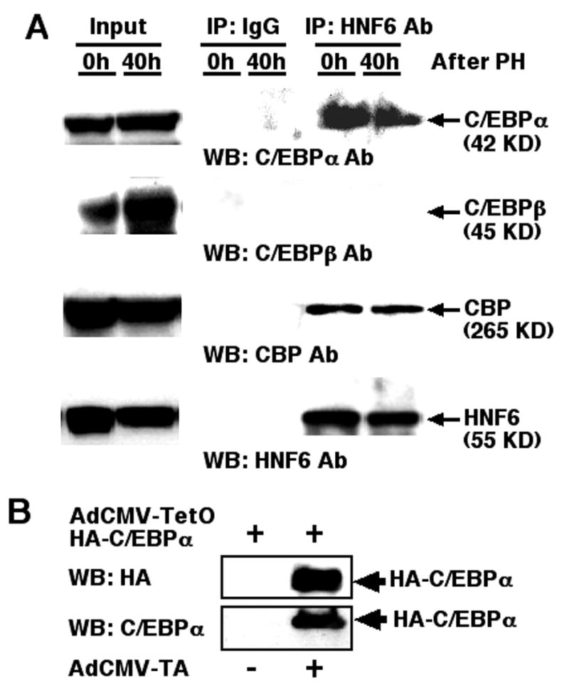
(A) Co-IP assays with liver extracts and HNF6 antibody shows association between HNF6 and the CBP coactivator protein. We performed co-immunoprecipitation (Co-IP) assays with protein extracts from quiescent mouse liver (0h) or regenerating mouse liver isolated at 40 hours (40h) following partial hepatectomy (PH). Quiescent or regenerating mouse liver extracts were Co-IP with either the HNF6 or rabbit serum control antibody and then IP proteins were subjected to Western blot analysis with either C/EBPα, C/EBPβ, CBP or HNF6 antibody as described in Materials and Methods. (B) Adenovirus vectors mediating inducible expression of HA-tagged human C/EBPα protein. Coinfection of HepG2 cells with both AdCMV-TetO-HA-C/EBPα and AdCMV-TA induces expression of the HA-C/EBPα as visualized by Western blot analysis with either HA-epitope or C/EBPα antibody.
ChIP assays demonstrated that increased expression of HNF6 and C/EBPα stimulates their binding and CBP recruitment to the endogenous mouse Foxa2 promoter.
In order to increase expression of C/EBPα in hepatoma cells, we generated an adenovirus vector that conditionally expressed the HA-tagged human C/EBPα protein under the control of the Tetracycline inducible promoter (CMV-TetO). Expression of HA-C/EBPα protein is induced by coinfection with a separate adenovirus containing the CMV promoter driving expression of the Tetracycline Transcriptional Activator (AdCMV-TA), which is active in the absence of Doxycycline (Tet-off system). Coinfection of hepatoma cells with both AdCMV-TetO-HAC/EBPα and AdCMV-TA (AdC/EBPα) is sufficient to induce expression of the HA tagged C/EBPα protein as evidenced by Western blot analysis with antibody specific to either the HAtag or the C/EBPα protein (Fig. 5B). The proximal mouse Foxa2 promoter region contains a high affinity binding site for the HNF6 (−138 and −126 bp) and C/EBP (−84 to −74 bp) proteins as described previously (18, 22). To determine whether increased expression of both C/EBPα and HNF6 protein enhances association of these transcription factors and the CBP coactivator protein with the endogenous mouse Foxa2 promoter region, we used Chromatin Immunoprecipitation (ChIP) assays as described previously (30, 31).
We prepared cross-linked chromatin from mouse hepatoma Hepa1-6 cells at 24 hours after infection with AdHNF6 or AdC/EBPα alone or combined and then sonicated chromosomal DNA into fragments between 500 to 1000 nucleotides in length. We also prepared cross linked and sonicated chromatin from Hepa1-6 cells that were efficiently transfected with CMV-HNF6 or CMV-C/EBPβ expression vector alone or combined by electroporation as described in Materials and Methods. This chromatin was then immunoprecipitated (IP) with antibodies specific to HNF6, CBP, C/EBPα, C/EBPβ or rabbit serum (control) and the IP genomic DNA was analyzed for the amount of mouse Foxa2 promoter DNA using quantative Real-Time PCR with primers specific to the mouse Foxa2 promoter region. We also performed ChIP assays with Hepa1-6 cells either infected with AdGFP or electroporated with CMV-GFP expression plasmid as normalization controls.
These hepatoma cell ChIP assays demonstrated that increased expression of both HNF6 and C/EBPα proteins stimulated association of either HNF6 (Fig. 6A) or C/EBPα (Fig. 6B) protein to the endogenous Foxa2 promoter region compared to hepatoma cells infected separately with AdHNF6 or AdC/EBPα (Fig. 6A–B). Interestingly, ChIP assays revealed that increased levels of both HNF6 and C/EBPα stimulated recruitment of the CBP coactivator protein to the endogenous Foxa2 promoter region compared to control hepatoma cells infected singly with AdHNF6 or AdC/EBPα (Fig. 6C). Although we were unable to demonstrate that HNF6 and C/EBPβ proteins interact using Co-IP assays with transfected cells or liver protein extract, cotransfection of both HNF6 and C/EBPβ expression vectors stimulated HNF6 binding to the endogenous Foxa2 promoter region (Fig. 6D), suggesting that these factors may interact in the context of the chromatin associated Foxa2 promoter region. However, cotransfection of both HNF6 and C/EBPβ expression vectors was unable to efficiently recruit either C/EBPβ (Fig. 6E) or the CBP coactivator protein (Fig. 6F) to the endogenous Foxa2 promoter region. ChIP assays with cross linked and sonicated mouse liver tissue showed that HNF6, C/EBPα and C/EBPβ bound at similar levels to the endogenous mouse Foxa2 promoter region (Fig. 6G). Interestingly, we observed reduced levels of CBP coactivator recruitment to the mouse liver Foxa2 promoter compared to that observed in Hepa1-6 cells with increased levels of HNF6 and C/EBPα proteins (compare Fig. 6C and 6G). These results are consistent with the inability of the HNF6-C/EBPβ complex to efficiently recruit the CBP coactivator protein to the endogenous Foxa2 promoter region. These studies support our hypothesis that formation of the C/EBPα-HNF6 complex stimulated binding of these transcription factors and recruitment of the CBP coactivator protein to the chromatin associated Foxa2 promoter region.
Fig. 6. ChIP assays demonstrated that increased expression of HNF6 and C/EBPα stimulates their binding and CBP recruitment to the endogenous mouse Foxa2 promoter.
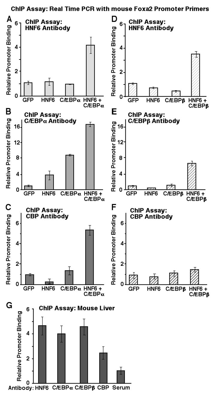
Mouse hepatoma Hepa1-6 cells were infected with AdHNF6 or AdC/EBPα either separately or together or with AdGFP control virus. We also used the Nucleofector II apparatus and buffers recommended by manufacturer (Amaxa, Gaithersburg, MD) to electroporate Hepa1-6 cells with CMV expression vectors containing either mouse HNF6 cDNA or rat C/EBP cDNA separately or together or with the CMV-GFP expression plasmid alone. At 24 hours after infection or electroporation, the cells were processed for Chromatin Immunoprecipitation (ChIP) assay as described in Materials and Methods. The cross-linked and sonicated chromatin was immunoprecipitated (IP) with blocked Protein A-Sepharose CL4B and antibodies specific to Hnf6 (A, D), C/EBPα (B), C/EBPβ (E) CBP (C, F), or IgG (control), washed, and protein was digested and removed from chromosomal DNA as described in Materials and Methods. The IP genomic DNA was analyzed for the amount of mouse Fox2 promoter region using a MyiQ Single Color Real-Time PCR Detection System and primers designed for the proximal mouse Foxa2 promoter region (−166 to −67 base pair; in triplicate). Normalization was carried out using the ΔΔCT method as described previously (19, 29). These relative values were then normalized to either the AdGFP-infected or CMV-GFP electroporated samples and shown graphically as relative promoter binding ± standard deviation (SD). (G) ChIP assays for Foxa2 binding factors with mouse liver. Wild type liver from three distinct mice were cross linked and sonicated and then immunoprecipitated with antibodies specific for HNF6, C/EBPα, C/EBPβ, CBP or rabbit serum and then analyzed for the mouse Foxa2 promoter region by real time PCR (performed in triplicate) as described in materials and methods. These relative values were averaged from three distinct mouse livers, normalized to rabbit serum value (value set at 1) and shown graphically as relative promoter binding ± SD.
DISCUSSION
Our previous cotransfection studies demonstrated that formation of complexes between the HNF6 and Foxa2 DNA binding domains stimulated Foxa2 transcriptional activity through recruitment of the CBP coactivator protein by the HNF6 Cut-Homeodomain (19). In our current study, we used cotransfection assays in human hepatoma HepG2 cells to demonstrate that HNF6 transcriptional activity was stimulated by C/EBPα, but not by the related C/EBPβ or C/EBPδ proteins. Formation of the C/EBPα-HNF6 protein complex required the HNF6 Cut DNA binding-domain sequences and the C/EBPα AD1/AD2 transcriptional activation domain sequences (Fig. 7A). Structure-function cotransfection assays demonstrated that stimulation of HNF6 transcriptional activity required the C/EBPα AD1/AD2 sequences, which were shown to mediate transcriptional activation by the p300/CBP coactivators (Fig. 7A–B). Consistent with these studies, cotransfection of the adenovirus E1A protein, which inhibits the activity of the p300/CBP histone acetyltransferase, abrogated the C/EBPα-HNF6 transcriptional synergy. Coimmunoprecipitation (Co-IP) experiments with liver protein extracts and Chromatin Immunoprecipitation (ChIP) assays with hepatoma cells have allowed us to demonstrate a functional association between the HNF6 and C/EBPα transcription factors to stimulate recruitment of the CBP coactivator promoter to the endogenous Foxa2 promoter region. The p300/CBP coactivators are known to stimulate transcription by acetylating positively charged Lysine residues on histone proteins, causing their dissociation from DNA regulatory regions, and by interacting with the basal transcriptional machinery (32).
Fig. 7. Model for C/EBPα-HNF6 transcriptional synergy on an HNF6-dependent promoter.
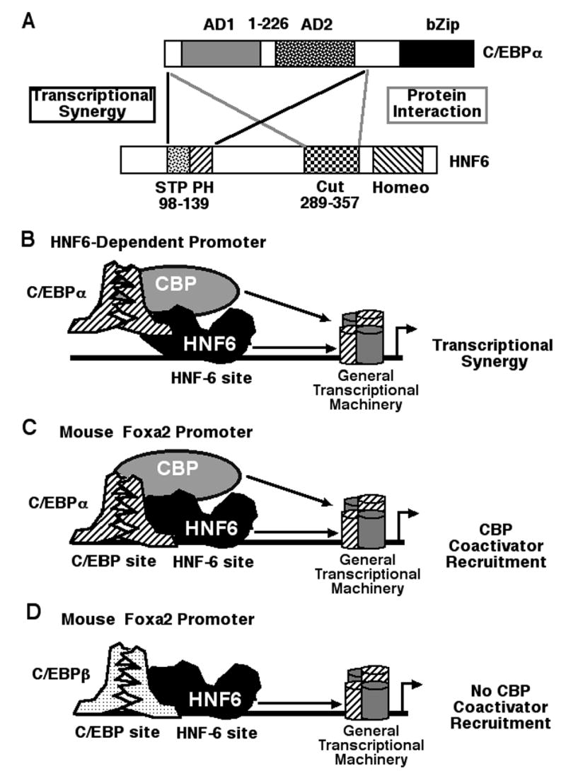
(A) Schematically depicted are the C/EBPα and HNF6 sequences required for protein interaction and transcriptional synergy. Structure-function studies demonstrated that formation of the C/EBPα-HNF6 complex required the HNF6 Cut-domain and C/EBPα activation domain 1 (AD1) and AD2 sequences (gray lines). C/EBPα-HNF6 transcriptional synergy required the HNF6 Serine, Threonine and Proline (STP) box and poly histidine (PH) sequences and the C/EBPα AD1/AD2 sequences (black lines). (B) On a HNF6 dependent promoter, formation of the C/EBPα and HNF6 protein complex synergistically stimulated HNF6-dependent transcription, which is likely mediated by C/EBPα AD1 recruitment of the CBP coactivator protein. (C) On a promoter containing both C/EBP and HNF6 sites (Foxa2 promoter) C/EBPα and HNF6 associate on the endogenous promoter region and recruit the CBP coactivator protein. (D) On a promoter containing both C/EBP and HNF6 sites (Foxa2 promoter) C/EBPβ and HNF6 associate on the endogenous promoter region and this complex is unable to recruit the CBP coactivator protein.
We also used ChIP assays with mouse hepatoma Hepa1-6 cells to demonstrate that increased levels of both the C/EBPα and HNF6 proteins were required to stimulate binding of these transcription factors and recruitment of the CBP coactivator protein to the endogenous mouse Foxa2 promoter region (Fig. 7C). Although elevated levels of both C/EBPβ and HNF6 proteins increased binding of these transcription factors to the endogenous Foxa2 promoter, they were unable to stimulate recruitment of the CBP coactivator to this endogenous promoter region as determined by ChIP assay (Fig. 7D). ChIP assays with cross linked and sonicated mouse liver tissue showed that HNF6, C/EBPα and C/EBPβ bound at similar levels to the endogenous mouse Foxa2 promoter region. Interestingly, we observed reduced levels of CBP coactivator recruitment to the mouse liver Foxa2 promoter when C/EBPα and C/EBPβ proteins are recruited equally well to the Foxa2 promoter region. One possible explanation for this result is that C/EBPβ and C/EBPα proteins are forming heterodimers to bind to the endogenous FoxA2 promoter region and HNF6 protein forms a complex with C/EBPα protein of this heterodimer complex. Recent studies using mouse livers that are conditionally deleted in the Foxa2 gene demonstrated that Foxa2 is required for transcriptional activation of genes encoding gluconeogenic enzymes during fasting and that Foxa2 stimulated recruitment of CREB transcription factor and glucocorticoid receptor to these chromatin associated promoter regions (35). Our ChIP data suggest that formation of the HNF6-C/EBPα complex stimulates recruitment of the CBP coactivator protein to the endogenous Foxa2 promoter region, and that increased Foxa2 levels by these transcription factors may stimulate expression of hepatocyte Foxa2 target genes involved in gluconeogenesis (35).
We also showed that the C/EBPα AD1/AD2 sequences and the HNF6 Cut-domain motif were sufficient to mediate association between the C/EBPα and HNF6 proteins (Fig. 7A–B). This result implies that interaction between the C/EBPα AD1/AD2 and Cut-domain DNA binding domain sequences does not inhibit the HNF6 Cut-Homeodomain motif from binding to a high affinity HNF6 recognition site and thus allows this C/EBPα-HNF6 complex to provide HNF6 transcriptional synergy. Furthermore, we showed that C/EBPα-HNF6 transcriptional synergy required both the N-terminal HNF6 STP box transcriptional activation domain and the poly Histidine (PH) sequences and the C/EBPα AD1 sequences (Fig. 7A), the latter of which have been shown to mediate transcriptional activation of C/EBPα by the p300/CBP coactivators (11). This result suggests that these HNF6 mutant proteins are unable to productively synergize with the C/EBPα transcription factor, indicating that the HNF6 STP and PH sequences play an important role in mediating transcriptional synergy with the C/EBPα protein. Previous structurefunction cotransfection studies determined that deletion of the N-terminal HNF6 PH caused only a slight elevation in HNF6 transcriptional activity, suggesting that the PH sequence did not contribute significantly in regulating HNF6 transcriptional activity (16). Our current study enabled us to identify an important function of the HNF6 PH sequence: Retention of the PH sequence is essential for mediating the C/EBPα-HNF6 transcriptional synergy. These results suggest that the C/EBPα-HNF6 transcriptional synergy involves interaction between the activation domains of each of these transcription factors. This is in contrast to the Foxa2-HNF6 transcriptional synergy, in which the HNF6 Cut-Homeodomain sequences alone were sufficient to potentiate transcriptional activity of the Foxa2 protein (19).
In summary, we demonstrated that formation of the C/EBPα and HNF6 protein complex stimulate HNF6 transcriptional activity through C/EBPα AD1/AD2 mediated recruitment of the p300/CBP coactivators and required the N-terminal HNF6 STP Box and PH sequences (Fig. 7AB). Complex formation between the C/EBPα and HNF6 transcription factors required the HNF6 Cut DNA binding domain sequences and the C/EBPα AD1/AD2 region (Fig. 7A). Consistent with this model, Co-IP experiments with liver protein extracts and ChIP studies with hepatoma cells have allowed us to demonstrate a functional association between the HNF6 and C/EBPα transcription factors to stimulate recruitment of the CBP coactivator promoter to the endogenous Foxa2 promoter region.
Acknowledgments
We thank V. V. Kalinichenko, P. Raychaudhuri and members of the Costa lab for critical review of this manuscript.
Footnotes
This work was supported by Public Health Service Grant R01 GM43241-15 (RHC) from the NIGMS and DK53045 (GJD) and P30 DK56338 (GJD) from the NIDDK. Y. Yoshida was supported by a Japan Society for the Promotion of Science Postdoctoral Fellowship for Research Abroad.
References
- 1.Costa RH, Kalinichenko VV, Holterman AX, Wang X. Transcription factors in liver development, differentiation, and regeneration. Hepatology. 2003;38:1331–1347. doi: 10.1016/j.hep.2003.09.034. [DOI] [PubMed] [Google Scholar]
- 2.Croniger C, Leahy P, Reshef L, Hanson RW. C/EBP and the control of phosphoenolpyruvate carboxykinase gene transcription in the liver. J Biol Chem. 1998;273:31629–31632. doi: 10.1074/jbc.273.48.31629. [DOI] [PubMed] [Google Scholar]
- 3.Davies N, Austen DE, Wilde MD, Darlington GJ, Brownlee GG. Clotting factor IX levels in C/EBP α knockout mice. Br J Haematol. 1997;99:578–579. doi: 10.1046/j.1365-2141.1997.4603263.x. [DOI] [PubMed] [Google Scholar]
- 4.Darlington GJ, Wang N, Hanson RW. C/EBPα: a critical regulator of genes governing integrative metabolic processes. Curr Opin Genet Dev. 1995;5:565–570. doi: 10.1016/0959-437x(95)80024-7. [DOI] [PubMed] [Google Scholar]
- 5.Friedman AD, Landschulz WH, McKnight SL. CCAAT/enhancer binding protein activates the promoter of the serum albumin gene in cultured hepatoma cells. Genes Dev. 1989;3:1314–1322. doi: 10.1101/gad.3.9.1314. [DOI] [PubMed] [Google Scholar]
- 6.Hansen AJ, Lee YH, Sterneck E, Gonzalez FJ, Mackenzie PI. C/EBPα is a regulator of the UDP glucuronosyltransferase UGT2B1 gene. Mol Pharmacol. 1998;53:1027–1033. [PubMed] [Google Scholar]
- 7.Inoue Y, Inoue J, Lambert G, Yim SH, Gonzalez FJ. Disruption of hepatic C/EBPalpha results in impaired glucose tolerance and age-dependent hepatosteatosis. J Biol Chem. 2004;279:44740–44748. doi: 10.1074/jbc.M405177200. [DOI] [PubMed] [Google Scholar]
- 8.Metzger S, Halaas JL, Breslow JL, Sladek FM. Orphan receptor HNF-4 and bZip protein C/EBP α bind to overlapping regions of the apolipoprotein B gene promoter and synergistically activate transcription. J Biol Chem. 1993;268:16831–16838. [PubMed] [Google Scholar]
- 9.Wang ND, Finegold MJ, Bradley A, Ou CN, Abdelsayed SV, Wilde MD, Taylor LR, et al. Impaired energy homeostasis in C/EBPα knockout mice. Science. 1995;269:1108–1112. doi: 10.1126/science.7652557. [DOI] [PubMed] [Google Scholar]
- 10.Vinson CR, Sigler PB, McKnight SL. Scissors-grip model for DNA recognition by a family of leucine zipper proteins. Science. 1989;246:911–916. doi: 10.1126/science.2683088. [DOI] [PubMed] [Google Scholar]
- 11.Erickson RL, Hemati N, Ross SE, MacDougald OA. p300 coactivates the adipogenic transcription factor CCAAT/enhancer-binding protein alpha. J Biol Chem. 2001;276:16348–16355. doi: 10.1074/jbc.m100128200. [DOI] [PubMed] [Google Scholar]
- 12.Lemaigre FP. Development of the biliary tract. Mech. Dev. 2003;120:81–87. doi: 10.1016/s0925-4773(02)00334-9. [DOI] [PubMed] [Google Scholar]
- 13.Lemaigre F, Zaret KS. Liver development update: new embryo models, cell lineage control, and morphogenesis. Curr Opin Genet Dev. 2004;14:582–590. doi: 10.1016/j.gde.2004.08.004. [DOI] [PubMed] [Google Scholar]
- 14.Jacquemin P, Lannoy VJ, Rousseau GG, Lemaigre FP. OC-2, a novel mammalian member of the ONECUT class of homeodomain transcription factors whose function in liver partially overlaps with that of hepatocyte nuclear factor-6. J Biol Chem. 1999;274:2665–2671. doi: 10.1074/jbc.274.5.2665. [DOI] [PubMed] [Google Scholar]
- 15.Lemaigre FP, Durviaux SM, Truong O, Lannoy VJ, Hsuan JJ, Rousseau GG. Hepatocyte nuclear factor 6, a transcription factor that contains a novel type of homeodomain and a single cut domain. Proc Natl Acad Sci U S A. 1996;93:9460–9464. doi: 10.1073/pnas.93.18.9460. [DOI] [PMC free article] [PubMed] [Google Scholar]
- 16.Lannoy VJ, Rodolosse A, Pierreux CE, Rousseau GG, Lemaigre FP. Transcriptional stimulation by hepatocyte nuclear factor-6. Target-specific recruitment of either CREBbinding protein (CBP) or p300/CBP-associated factor (p/CAF) J Biol Chem. 2000;275:22098–22103. doi: 10.1074/jbc.M000855200. [DOI] [PubMed] [Google Scholar]
- 17.Rausa F, Samadani U, Ye H, Lim L, Fletcher CF, Jenkins NA, Copeland NG, et al. The cuthomeodomain transcriptional activator HNF-6 is coexpressed with its target gene HNF-3β in the developing murine liver and pancreas. Dev Biol. 1997;192:228–246. doi: 10.1006/dbio.1997.8744. [DOI] [PubMed] [Google Scholar]
- 18.Samadani U, Costa RH. The transcriptional activator hepatocyte nuclear factor six regulates liver gene expression. Mol. Cell. Biol. 1996;16:6273–6284. doi: 10.1128/mcb.16.11.6273. [DOI] [PMC free article] [PubMed] [Google Scholar]
- 19.Rausa F, Tan Y, Costa RH. Association between HNF-6 and FoxA2 DNA binding domains stimulates FoxA2 transcriptional activity but Inhibits HNF-6 DNA binding. Mol. Cell. Biol. 2003;23:437–449. doi: 10.1128/MCB.23.2.437-449.2003. [DOI] [PMC free article] [PubMed] [Google Scholar]
- 20.Sheng W, Yan H, Rausa FM, Costa RH, Liao X. Structure of the Hepatocyte Nuclear Factor 6α (HNF-6 α) and its interaction with DNA. J. Biol. Chem. 2004;279:33928–33936. doi: 10.1074/jbc.M403805200. [DOI] [PubMed] [Google Scholar]
- 21.Rausa FM, Hughes DE, Costa RH. Stability of the Hepatocyte Nuclear Factor 6 transcription factor requires acetylation by the CREB Binding Protein coactivator. J. Biol. Chem. 2004;279:43070–43076. doi: 10.1074/jbc.M407472200. [DOI] [PubMed] [Google Scholar]
- 22.Samadani U, Porcella A, Pani L, Johnson PF, Burch J, Pine R, Costa RH. Cytokine regulation of the liver transcription factor HNF-3β is mediated by the C/EBP family and interferon regulatory factor 1. Cell Growth & Differen. 1995;6:879–890. [PubMed] [Google Scholar]
- 23.Harris TE, Albrecht JH, Nakanishi M, Darlington GJ. CCAAT/enhancer-binding proteinalpha cooperates with p21 to inhibit cyclin-dependent kinase-2 activity and induces growth arrest independent of DNA binding. J Biol Chem. 2001;276:29200–29209. doi: 10.1074/jbc.M011587200. [DOI] [PubMed] [Google Scholar]
- 24.Hendricks-Taylor LR, Darlington GJ. Inhibition of cell proliferation by C/EBP α occurs in many cell types, does not require the presence of p53 or Rb, and is not affected by large Tantigen. Nucleic Acids Res. 1995;23:4726–4733. doi: 10.1093/nar/23.22.4726. [DOI] [PMC free article] [PubMed] [Google Scholar]
- 25.Kalinichenko VV, Major M, Wang X, Petrovic V, Kuechle J, Yoder HM, Shin B, et al. Forkhead Box m1b transcription factor is essential for development of hepatocellular carcinomas and is negatively regulated by the p19ARF tumor suppressor. Genes & Development. 2004;18:830–850. doi: 10.1101/gad.1200704. [DOI] [PMC free article] [PubMed] [Google Scholar]
- 26.Overdier DG, Porcella A, Costa RH. The DNA-binding specificity of the hepatocyte nuclear factor 3/forkhead domain is influenced by amino-acid residues adjacent to the recognition helix. Mol. Cell Biol. 1994;14:2755–2766. doi: 10.1128/mcb.14.4.2755. [DOI] [PMC free article] [PubMed] [Google Scholar]
- 27.Rausa FM, Tan Y, Zhou H, Yoo K, Stolz DB, Watkins S, Franks RR, et al. Elevated Levels of HNF-3β in Mouse Hepatocytes Influence Expression of Genes Involved in Bile Acid and Glucose Homeostasis. Mol Cell Biol. 2000;20:8264–8282. doi: 10.1128/mcb.20.21.8264-8282.2000. [DOI] [PMC free article] [PubMed] [Google Scholar]
- 28.Tan Y, Adami G, Costa RH. Maintaining HNF-6 expression prevents AdHNF3β Mediated decrease in hepatic levels of Glut2 and glycogen. Hepatology. 2002;35:790–798. doi: 10.1053/jhep.2002.32482. [DOI] [PubMed] [Google Scholar]
- 29.Tan Y, Costa RH, Kovesdi I, Reichel RR. Adenovirus-mediated increase of HNF-3 levels stimulates Transthyretin and Sonic Hedgehog expression which is associated with F9 cell differentiation toward the visceral endoderm Llneage. Gene Expression. 2001;9:237–248. doi: 10.3727/000000001783992542. [DOI] [PMC free article] [PubMed] [Google Scholar]
- 30.Wang I-C, Chen Y-J, Hughes DE, Petrovic V, Major ML, Park HJ, Tan Y, et al. Forkhead Box M1 regulates the transcriptional network of genes essential for mitotic progression and genes encoding the SCF(Skp2-Cks1)Ubiquitin Ligase. Mol Cell Biol 2005;In Press. [DOI] [PMC free article] [PubMed]
- 31.Wells J, Farnham PJ. Characterizing transcription factor binding sites using formaldehyde crosslinking and immunoprecipitation. Methods. 2002;26:48–56. doi: 10.1016/S1046-2023(02)00007-5. [DOI] [PubMed] [Google Scholar]
- 32.Chan HM, La Thangue NB. p300/CBP proteins: HATs for transcriptional bridges and scaffolds. J Cell Sci. 2001;114:2363–2373. doi: 10.1242/jcs.114.13.2363. [DOI] [PubMed] [Google Scholar]
- 33.Wang H, Iakova P, Wilde M, Welm A, Goode T, Roesler WJ, Timchenko NA. C/EBPalpha arrests cell proliferation through direct inhibition of Cdk2 and Cdk4. Mol Cell. 2001;8:817–828. doi: 10.1016/s1097-2765(01)00366-5. [DOI] [PubMed] [Google Scholar]
- 34.Wang X, Kiyokawa H, Dennewitz MB, Costa RH. The Forkhead Box m1b transcription factor is essential for hepatocyte DNA replication and mitosis during mouse liver regeneration. Proc Natl Acad Sci U S A. 2002;99:16881–16886. doi: 10.1073/pnas.252570299. [DOI] [PMC free article] [PubMed] [Google Scholar]
- 35.Zhang L, Rubins NE, Ahima RS, Greenbaum LE, Kaestner KH. Foxa2 integrates the transcriptional response of the hepatocyte to fasting. Cell Metab. 2005;2:141–148. doi: 10.1016/j.cmet.2005.07.002. [DOI] [PubMed] [Google Scholar]