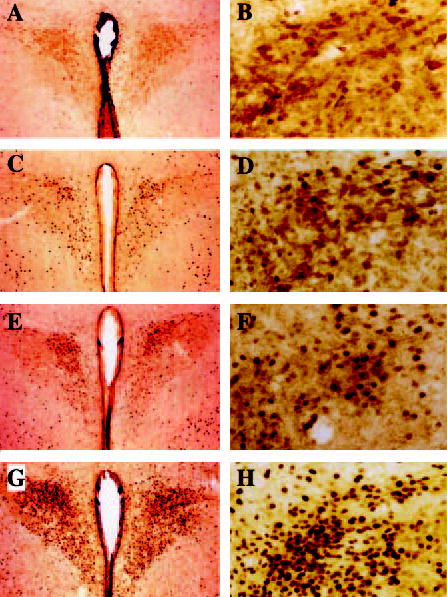
Fig. 3.

Representative photomicrographs of the parvocellular and magnocellular region of the PVN. Neuronal cell bodies were immu-nostained for CRH (indicated as light brown) and c-Fos (indicated as dark nuclear staining) of rats treated i3cv with saline (A and B), leptin (3.0 μg, C and D), and MTII (0.05 μg, E and F; 0.5 μg, G and H).
