Abstract
Proteinases have been proposed to play important roles in pathogenesis and various biologic actions in Acanthamoeba. Although genetic characteristics of several proteases of Acanthamoeba have been reported, the intracellular localization and trafficking of these enzymes has yet to be studied. In the present study, we analyzed the intracellular localization and trafficking of two proteinases, AhSub and AhCP, of Acanthamoeba healyi by transient transfection. Full-length AhSub-enhanced green fluorescent protein (EGFP) fusion protein was found in intracellular vesicle-like structures of transfected amoebae. Time-lapse photographs confirmed the secretion of the fluorescent material of the vesicle toward the extracellular space. The mutated AhSub, of which the pre or prepro region was deleted, was found to localize diffusely throughout the cytoplasm of the amoeba rather than concentrated in the secretory vesicle. Transfection of the construct containing the pre region only showed the same localization and trafficking of the full-length AhSub. A cysteine proteinase AhCP-EGFP fusion protein showed similar localization in the vesicle-like structure in the amoeba. However, using Lyso Tracker analysis, these vesicular structures of AhCP were confirmed to be lysosomes rather than secretory vesicles. The AhCP construct with a deletion of the prepro region showed a dispersed distribution of fluorescence in the cytoplasm of the cells. These results indicated that AhSub and AhCP would play different roles in Acanthameoba biology and that the pre region of AhSub and pro region of AhCP are important for proper intracellular localization and trafficking of each proteinase.
The genus Acanthamoeba, one of the amphizoic amoebae with ubiquitous distribution in human environments, has been known to cause granulomatous amoebic encephalitis in immune-compromised hosts and sight-threatening amebic keratitis, especially in contact lens users (15).
Proteinases have been known to play important roles in the metabolism, development, or survival of protozoa (16). Considering the high phagocytic activity of Acanthamoeba as a free-living organism and tissue-invasive behavior as a parasite, proteases would be involved in various processes of Acanthamoeba, such as pathogenesis, nutrient obtainment, and host tissue destruction. To date, several reports on the serine, cysteine, and metalloproteinases of Acanthamoeba were published (9, 12, 18, 19). Serine proteinase purified from culture supernatant was reported to play roles in host tissue invasion because of its strong activity against a broad spectrum of extracellular matrix and serum proteins of humans (12). The gene of the purified serine proteinase was characterized and named AhSub based on high homology with bacterial subtilisins (3). Cathepsin L-like cysteine proteinase genes were cloned from Acanthamoeba healyi and Acanthamoeba culbertsoni (3, 29). However, the specific function of these cysteine proteinases in Acanthamoeba is still unclear. The intracellular localization and trafficking of the enzymes may suggest their possible role in Acanthamoeba.
The sequence data of the subtilisin (AhSub) and cysteine proteinase (AhCP) of A. healyi suggested that they would be produced as a prepro enzyme. Pre and pro domains of subtilisin and cysteine proteinases were reported to play roles in localization or trafficking of the enzymes in several protozoa and bacteria (14, 17, 24).
In this paper, we demonstrate the intracellular localization, trafficking, and secretion of a subtilisin-like serine proteinase, AhSub, and cysteine proteinase, AhCP, by transient transfection as an enhanced green fluorescent protein (EGFP) fusion protein in live cells and determine the important domain of the proteinase for the proper localization, trafficking, and secretion of the proteinase by deletion mutation analyses.
MATERIALS AND METHODS
Amoeba.
A. healyi OC-3A strain (ATCC 30866) isolated from the brain of a granulomatous amoebic encephalitis patient (20) was obtained from ATCC and cultured axenically at 25°C in Proteose peptone-yeast extract-glucose liquid medium, containing 20 g/liter Proteose peptone, 1 g/liter yeast extract, 0.1 M glucose, 4 mM MgSO4, 0.4 mM CaCl2, 3.4 mM sodium citrate, 0.05 mM Fe(NH4)2(SO4)2, and 2.5 mM (each) Na2HPO4 and KH2PO4. The final pH of the medium was adjusted to 6.5.
Expression vector construction.
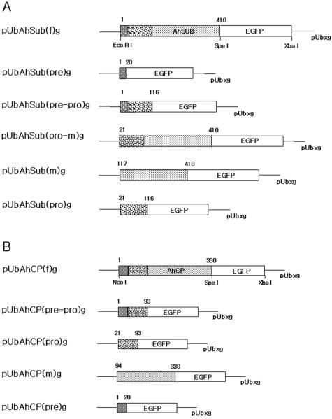
The pUbg vector with the Acanthamoeba ubiquitin promoter and EGFP as a reporter gene (13) was used to construct the expression vectors for this study. AhSub is composed of prepeptide of 20 amino acid residues, propeptide of 96 residues, and mature protein of 293 residues (3). AhCP is composed of prepropeptide of 93 amino acid residues and mature protein of 237 amino acid residues (4). As shown in Fig. 1, a total of six different expression vectors of full-length AhSub and five deletion mutants of AhSub and a total of five different expression vectors of full-length AhCP and four deletion mutants of AhCP were designed for transient transfection. Primer sequences for PCR amplification for all constructs are shown in Table 1. Each PCR product of AhSub was ligated into the vector by the EcoRI and SpeI sites, and each PCR product of AhCP was ligated into the vector by the NcoI and SpeI sites with C-terminal EGFP. Two amino acids coding for the SpeI enzyme site, threonine and serine, were inserted between the AhSub/AhCP and EGFP. Plasmid DNAs of 12 different constructs were prepared using the Miniprep kit (iNtRON, Korea).
FIG. 1.
Schematic representation of different expression vectors for transient transfection of AhSub-EGFP and AhCP-EGFP fusion proteins in A. healyi. (A) pUbAhSub(f)g, full-length AhSub; pUbAhSub(pre)g, pre region of AhSub; pUbAhSub(pre-pro)g, prepro region of AhSub; pUbAhSub(pro-m)g, promature region of AhSub; pUbAhSub(m)g, mature region of AhSub; pUbAhSub(pro)g, pro region of AhSub. (B) pUbAhCP(f)g, full-length AhCP; pUbAhCP(pre-pro)g, prepro region of AhCP; pUbAhCP(pro)g, pro region of AhCP; pUbAhCP(m)g, for mature region of AhCP; pUbAhCP(pre)g, pre region of AhCP.
TABLE 1.
Primer sequences for amplication of AhSub and AhCP inserted into expression vector
| Construct | Primer sequencea |
|---|---|
| pUbAhSub(f)g | F: CCGCCATGGCCATGCGCGCCGTCACCCCTC |
| R: CCGACTAGTGGCGGTGGGGTACGAAGCAGC | |
| pUbAhSub(pre)g | F: CCGCCATGGCCATGCGCGCCGTCACCCCTC |
| R: ATAACTAGTAGCGAGAGCGCTAGCGCAGAG | |
| pUbAhSub(pre-pro)g | F: CCGCCATGGCCATGCGCGCCGTCACCCCTC |
| R: CCGACTAGTAGCAAGACCCTTGAAGAGGCG | |
| pUbAhSub(pro-m)g | F: AATCCATGGCCACGCACGACCCGCTCACGG |
| R: CCGACTAGTGGCGGTGGGGTACGAAGCAGC | |
| pUbAhSub(m)g | F: AATCCATGGACTTCGACTACTCCAAGCACG |
| R: CCGACTAGTGGCGGTGGGGTACGAAGCAGC | |
| pUbAhSub(pro)g | F: AATCCATGGCCACGCACGACCCGCTCACGG |
| R: CCGACTAGTAGCAAGACCCTTGAAGAGGCG | |
| pUbAhCP(f)g | F: ATTGAATTCATGCGTGCCTACTTCGTGGGT |
| R: TATACTAGTGCACCGGGCGGAGTAAAGCAG | |
| pUbAhCP(pre-pro)g | F: ATTGAATTCATGCGTGCCTACTTCGTGGGT |
| R: ATAACTAGTCTGCGCGCTGGAGATGGAGAC | |
| pUbAhCP(pro)g | F: ATTGAATTCATGTCTCTTGCCCCTCTTCACAGG |
| R: ATAACTAGTCTGCGCGCTGGAGATGGAGAC | |
| pUbAhCP(m)g | F: ATTGAATTCATGAACTGCCTGTCCCAGAGCGGC |
| R: TATACTAGTGCACCGGGCGGAGTAAAGCAG | |
| pUbAhCP(pre)g | F: ATTGAATTCATGCGTGCCTACTTCGTGGGT |
| R: ATAACTAGTAGCGAGAGCGCTAGCGCAGAG |
F, forward; R, reverse. Restriction enzyme sites are underlined.
Transfection.
Trophozoites of A. healyi grown to mid-log phase in a 25°C incubator were washed twice with phosphate-buffered saline (PBS) and resuspended in Proteose peptone-yeast extract-glucose culture medium. Cells were cultured overnight at 25°C in a six-well culture plate with 4 × 10⁵ cells per well in 3 ml of culture medium. Four micrograms of plasmid DNA in 100 μl of amoeba culture medium mixed with 20 μl of Superfect (QIAGEN) was incubated for 10 min at room temperature and then diluted with 600 μl of culture medium. Adherent amoebae in a six-well plastic culture plate were washed once with PBS at room temperature. After removal of most of the PBS, DNA-Superfect mixture was added dropwise onto the cells. Cells were incubated at 25°C for 3 h to allow uptake of DNA-Superfect complexes. Cells were washed once with PBS, resuspended in 3 ml fresh growth medium, and incubated at 25°C for 24 to 48 h. Expression of the EGFP fusion protein was checked by fluorescence microscopy.
Flow cytometry.
Forty hours after transfection, cells were washed twice and resuspended in 2 ml of PBS. The number of transfected cells and the fluorescence of expressed EGFP among 1 × 10⁶ cells were measured with a FACScalibur (Becton-Dickinson) to determine the transfection efficiency.
Fluorescence microscopy.
Transfected cells expressing EGFP fusion protein were selected and allowed to adhere to a glass-bottom cell culture dish (BD Falcon). The cells were analyzed using an Olympus IX 70 fluorescent microscope with cooled charge-coupled device camera (Roper Scientific). Alternatively, transfected amoebae were flattened with an agar overlay to observe detailed intracellular structures (27). EGFP fluorescence was achieved with a 500- to 530-nm band-pass filter. Images and time-lapse photographs were captured and analyzed through the Metamorph imaging system (Universal Imaging Corp.).
Labeling of endocytic compartments with Lyso Tracker.
Transfected cells were grown on a cell culture dish, rinsed with 1× PBS, and stained with 1 nM Lyso Tracker Red DND-99 (Molecular Probes, Leiden, The Netherlands) in medium without serum for 30 min at room temperature. Thereafter, cells were washed with PBS, fixed with 70% ethanol in PBS for 2 min at room temperature, and flattened on an agar overlay.
RESULTS
As described by Kong and Pollard (13), 20 ml of Superfect and 4 μg of plasmid DNA were transfected, and the transfection efficiency obtained ranged from 1.5 to 2.2%. There was no significant difference in the efficiency among constructs analyzed in this study. The amoebae transfected with the constructs began to show fluorescence 12 h after transfection and retained fluorescence for several days. We examined the amoebae with a fluorescent microscope from 36 h to 40 h after transfection. Also, the agar overlay method was employed to flatten and minimize the movement of transfected amoeba cells during the acquisition of images and time-lapse photographs (28).
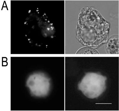
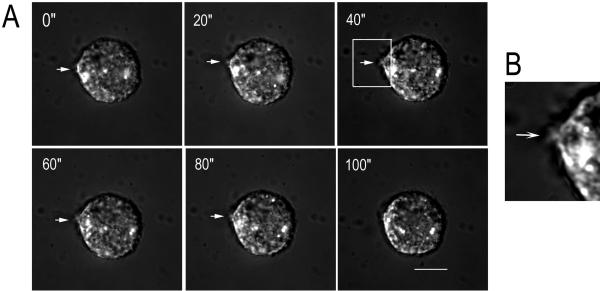
The amoeba expressing full-length AhSub as an EGFP fusion protein showed fluorescent vesicle-like structures which were distributed mainly at the periphery of the cytoplasm (Fig. 2A). In contrast, the fluorescence of EGFP alone showed dispersed distribution in the cytoplasm (Fig. 2B). Careful time-lapse examination of these fluorescent vesicle-like structures revealed that the fluorescent material was secreted toward the outside of the amoeba (Fig. 3A). The excretion of the fluorescent material at the surface of the amoeba taken at 40 s was magnified and shown in Fig. 3B. The entire secretory process of the fluorescent material occurred in less than 2 min.
FIG. 2.
(A) Fluorescence (left) and light (right) micrographs of a live Acanthamoeba expressing the full-length AhSub-EGFP fusion protein. Small fluorescent vesicle-like structures were distributed at the periphery of the cytoplasm. (B) Two amoebae with the fluorescence of EGFP alone showed dispersed distribution in the cytoplasm. Bar, 10 μm.
FIG. 3.
(A) Fluorescence micrographs of Acanthamoeba secreting AhSub-EGFP fusion protein photographed at 20-s time interval. The numbers in the upper left corners denote time in seconds. The arrows indicate the excretion of a secretory vesicle containing the AhSub-EGFP fusion protein. (B) Magnification of the amoeba expelling the fluorescent material taken at 40 s (white box in 40-s panel). Bar, 10 μm.
To determine the region of AhSub that plays an important role in the intracellular localization and trafficking of the proteinase, 5 different deletion mutants were constructed and transfected as EGFP fusion proteins (Fig. 4). Amoebae expressing the pre or prepro domain as an EGFP fusion protein displayed small fluorescent vesicle-like structures in the cytoplasm (Fig. 4A and B). The results of these two constructs were very similar to that of full-length AhSub. Amoebae transfected with the other three constructs without the pre region of AhSub demonstrated dispersed distribution of fluorescence in the cytoplasm (Fig. 4C, D, and E). The premature construct without propeptide failed to secrete the proteinase (data not shown).
FIG. 4.
Fluorescence (left) and light (right) micrographs of live amoeba expressing each deletion mutant construct of AhSub-EGFP fusion protein. (A) Pre region of AhSub-EGFP; (B) prepro region of AhSub-EGFP; (C) promature region of AhSub-EGFP; (D) mature region of AhSub-EGFP; (E) pro region of AhSub-EGFP. Small fluorescent vesicle-like structures were observed in cells expressing the prepro region and pre region, whereas amoeba cells expressing the other 3 constructs demonstrated dispersed localization of fluorescence in the cytoplasm. Bar, 10 μm.
The fluorescent vesicles in the cell expressing EGFP fused to the pre region of AhSub also disappeared within 5 min through the time-lapse examination, indicating the secretion of the EGFP fusion protein as seen in the amoeba transfected with full-length AhSub-EGFP (data not shown). From these results, it is possible to conclude that the pre domain of AhSub is the most important part for proper intracellular localization and secretion of the proteinase in Acanthamoeba.
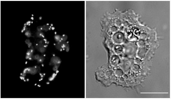
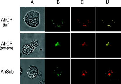
The amoeba transfected to express full-length AhCP as EGFP fusion protein showed fluorescent vesicles in the cytoplasm (Fig. 5). To analyze the localization of cysteine proteinase in A. healy (AhCP), amoebae transfected with the full-length AhCP were stained with Lyso Tracker Red DND-99 (Fig. 6). The position of green-colored fluorescent vesicle-like structures of AhCP (Fig. 6B) completely overlapped with the red-colored lysosomes stained with Lyso Tracker Red DND-99 (Fig. 6C), producing a yellow-colored signal (Fig. 6D). As shown in Fig. 6, all of green vesicle-like structures exhibited yellow signals after overlapping them with the red-colored lysosomes. In contrast to AhCP, AhSub-EGFP fusion protein didn't demonstrate localization in the lysosome, confirming that AhCP is the lysosomal enzyme of Acanthamoeba. The amoeba transfected with the prepro region of AhCP showed a yellow signal, indicating its localization in the lysosomes of the amoeba. This result confirms that the prepro region is sufficient for the proper localization of AhCP in Acanthamoeba.
FIG. 5.
Fluorescence (left) and light (right) micrographs of a live Acanthamoeba expressing the full-length AhCP-EGFP fusion protein. Small fluorescent vesicle-like structures were randomly distributed in the cytoplasm of the amoeba. Bar, 10 μm.
FIG. 6.
Localization of AhCP-EGFP in A. healyi. (A) Light micrograph; (B) expressed AhCP-EGFP and AhSub-EGFP fusion proteins (green signal); (C) lysosomes of Acanthamoeba stained with Lyso Tracker Red DND-99 (red signal); (D) overlapped micrograph images of panels B and C (yellow signal). The presence of the yellow signals was demonstrated only in the cells expressing AhCP but not in the cells transfected with AhSub. Bar, 10 μm.
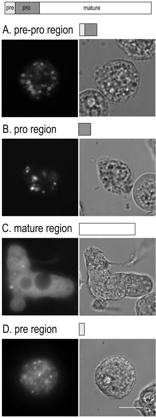
Similarly, to determine the region of AhCP that plays an important role in the intracellular localization and trafficking of the proteinase, four different deletion mutants fused with EGFP were transfected (Fig. 7). Amoebae transfected with the constructs of AhCP(prepro)-EGFP and AhCP(pro)-EGFP showed small fluorescent vesicle-like structures in the cytoplasm (Fig. 7A and B), which was very similar to that of full-length AhCP. However, the amoebae transfected with the mature region or pre region alone of AhCP showed dispersed distribution of fluorescence in the cytoplasm (Fig. 7C and D). These results suggest that the prepro region is required for proper intracellular localization of AhCP in Acanthamoeba.
FIG. 7.
Fluorescence (left) and light (right) micrographs of live amoeba expressing each deletion mutant construct of AhCP-EGFP. (A) Prepro region of AhCP-EGFP; (B) pro region of AhCP-EGFP; (C) mature region of AhCP-EGFP; (D) pre region of AhCP-EGFP. Amoeba cells expressing prepro region and pro region displayed the fluorescent vesicle-like structures. In contrast, dispersed distribution of fluorescence in the cytoplasm was observed in cells transfected with mature region and pre region. Bar, 10 μm.
DISCUSSION
In this study, we have demonstrated the intracellular localization, trafficking, and secretory processes of proteinases AhSub and AhCP of Acanthamoeba healyi by transient transfection. The amoeba expressing full-length AhSub-EGFP showed the fluorescent vesicle-like structures in the cytoplasm and secreted the fluorescent material outside the cell. In the case of the amoeba transfected with full-length AhCP-EGFP, the fluorescent vesicle-like structures were identified as lysosome by Lyso Tracker staining. Various deletion mutant analyses revealed that the pre domain of AhSub and the prepro domain of AhCP are essential for appropriate intracellular localization and trafficking of these proteinases.
Subtilisins are usually synthesized as prepro enzymes, which are later posttranslationally activated to the active enzymes by cleavage of the pre- and propeptides (11). The roles of pre- and propeptides of subtilisin trafficking in bacteria have been studied by biochemical and molecular biological investigations. The prepeptide functions as the signal sequence required for protein secretion across the cytoplasmic membrane, and the propeptide has been proposed to function as an intramolecular chaperone for production of active subtilisin (1, 7, 22, 26). The role of pre- or propeptide in the protozoan subtilases was analyzed in PfSUB-1 (21). However, subtilases of apicomplexan protozoa, including PfSUB-1 of Plasmodium falciparum, PbSUB2 of Plasmodium berghei, and TgSUB2 of Toxoplasma gondii, are transmembrane proteins rather than secretory proteins (17, 23). Therefore, the pre region of PfSUB-1 cleaved at the endoplasmic reticulum during secretory transport may not affect the localization of the proteinase at secretory organelles (21). In the case of AhSub, the pre region-EGFP fusion protein was localized in the secretory vesicles and secreted toward outside of the amoeba. This result indicates that the pre region of AhSub would be essential for proper intracellular trafficking and secretion of the proteinase. This role of the pre domain of AhSub is more similar to that of bacterial subtilisins than that of apicomplexan subtilisins. Furthermore, the sequence analysis of AhSub indicated that AhSub is more closely related to bacterial than apicomplexan subtilisins (3).
Proteinase has been hypothesized as a sorting receptor/chaperone within the secretory pathway (2). In several protease families, propeptides function as intramolecular chaperones that are essential for correct folding of the catalytic domain during secretory transport (6). The prosequence of subtilisins may be required for the association of the proenzyme with the cell before the release of the mature active enzyme into the medium and/or for guiding the protein to the appropriate folding for the active conformation (5). We found that the premature construct without propeptide failed to secrete the proteinase, substantiating the possible role of propeptide. Compared to the small sized prepeptide alone (259 amino acid residues including EGFP), this improper trafficking of the premature construct could have resulted from masking of prepeptide due to the improper folding of the mature domain (533 amino acid residues including EGFP). As reported for other proteinases, the propeptide of AhSub may act as a chaperonin for appropriate protein folding of AhSub (2). The role of propeptide in the AhSub trafficking would be the next step to understand the full process of intracellular trafficking of AhSub in the Acanthamoeba.
The vesicle-like structures of the full-length AhCP-EGFP fusion protein exhibited the same localization with lysosome in the transfected amoeba. This result could clarify the biological function of AhCP originally suggested by Hong et al. (4). They reported that AhCP in Acanthamoeba may play a role in digestion of phagocytosed material rather than pathogenesis because the Northern blot analysis revealed higher expression of AhCP in a soil isolate than in a clinical isolate (4). The majority of the cysteine proteinases of the C1 family are known to be lysosomal enzymes that function in intracellular protein degradation. AhCP showed perfect conservation of amino acid residues in the motif, like mammalian cathepsin L (4).
The amoebae expressing the prepro region of AhCP-EGFP showed localization similar to that of the full-length AhCP-EGFP fusion protein. However, the amoebae transfected with the mature region of AhCP-EGFP showed the dispersed fluorescence in the cytoplasm. In the case of amoebae expressing the pro region, some cells exhibited similar localization with the cells expressing the prepro region. However, some of these cells also demonstrated dispersed localization in the cytoplasm, similar to the amoebae expressing only the mature region. The classical trafficking mechanism of lysosomal enzymes in mammals involves mannose-6-phosphate receptors (8). However, alternative targeting mechanisms have also been described for Saccharomyces cerevisiae and Leishmania (10, 14). In the case of carboxypeptidase Y of S. cerevisiae, the involvement of sequences in the pro region was reported previously (24, 25). The Leishmania showed the lack of a role for N-glycosylation on targeting to the lysosome (14). The pro region (from amino acids 21 to 93) of AhCP containing the N-glycosylation site (from amino acids 67 to 70) may play some role in localization and trafficking. Further studies on the functional role and mechanism of the pre region and N-glycosylation site of the pro region are recommended.
Acknowledgments
We thank Joanna Alafag for assistance in editing the manuscript.
This work was supported by a Korea Research Foundation grant (KRF-2001-042-F00031).
REFERENCES
- 1.Chang, Y. C., H. Kadokura, K. Yoda, and M. Yamasaki. 1996. Secretion of active subtilisin YaB by a simultaneous expression of separate pre-pro and pre-mature polypeptides in Bacillus subtilis. Biochem. Biophys. Res. Commun. 219:463-468. [DOI] [PubMed] [Google Scholar]
- 2.Cool, D. R., E. Normant, F. Shen, H. C. Chen, L. Pannell, Y. Zhang, and Y. P. Loh. 1997. Carboxypeptidase E is a regulated secretory pathway sorting receptor: genetic obliteration leads to endocrine disorders in Cpe (fat) mice. Cell 88:73-83. [DOI] [PubMed] [Google Scholar]
- 3.Hong, Y. C., H. H. Kong, M. S. Ock, I. S. Kim, and D. I. Chung. 2000. Isolation and characterization of a cDNA encoding a subtilisin-like serine proteinase (AHSUB) from Acanthamoeba healyi. Mol. Biochem. Parasitol. 111:441-446. [DOI] [PubMed] [Google Scholar]
- 4.Hong, Y. C., M. Y. Hwang, H. C. Yun, H. S. Yu, H. H. Kong, T. S. Yonh, and D. I. Chung. 2002. Isolation and characterization of a cDNA encoding a mammalian cathepsin L-like cysteine proteinase from Acanthameoba healyi. Korean J. Parasitol. 40:17-24. [DOI] [PMC free article] [PubMed] [Google Scholar]
- 5.Ikemura, H., H. Takagi, and M. Inouye. 1987. Requirement of pro-sequence for production of active subtilisin E in Escherichia coli. J. Biol. Chem. 262:7859-7864. [PubMed] [Google Scholar]
- 6.Jain, S. C., U. Shinde, U. Li, M. Inouye, and H. M. Berman. 1998. The crystal structure of an autoprocessed Ser221Cys-subtilisin E-propeptide complex at 2.0 A resolution. J. Mol. Biol. 284:137-144. [DOI] [PubMed] [Google Scholar]
- 7.Kaneko, R., N. Koyama, Y. C. Tsai, R. Y. Juang, K. Yoda, and M. Yamasaki. 1989. Molecular cloning of the structural gene for alkaline elastase YaB, a new subtilisin produced by an alkalophilic Bacillus strain. J. Bacteriol. 171:5232-5236. [DOI] [PMC free article] [PubMed] [Google Scholar]
- 8.Kaplan, A., D. Fischer, D. Achord, and W. Sly. 1977. Phosphohexosyl recognition is a general characteristic of pinocytosis of lysosomal glycosidases by human fibroblasts. J. Clin. Investig. 60:1088-1093. [DOI] [PMC free article] [PubMed] [Google Scholar]
- 9.Kim, H. K., Y. R. Ha, H. S. Yu, H. H. Kong, and D. I. Chung. 2003. Purification and characterization of a 33 kDa serine protease from Acanthamoeba lugdunensis KA/E2 isolated from a Korean keratitis patient. Korean J. Parasitol. 41:189-196. [DOI] [PMC free article] [PubMed] [Google Scholar]
- 10.Klionsky, D. J. 1998. Nonclassical protein sorting to the yeast vacuole. J. Biol. Chem. 273:10807-10810. [DOI] [PubMed] [Google Scholar]
- 11.Kluskens, L. D., W. G. Voorhorst, R. J. Siezen, R. M. Schwerdtfeger, G. Antranikian, J. van der Oost, and W. M. de Vos. 2002. Molecular characterization of fervidolysin, a subtilisin-like serine protease from the thermophilic bacterium Fervidobacterium pennivorans. Extremophiles 6:185-194. [DOI] [PubMed] [Google Scholar]
- 12.Kong, H. H., T. H. Kim, and D. I. Chung. 2000. Purification and characterization of a secretory serine proteinase of Acanthamoeba healyi isolated from GAE. J. Parasitol. 86:12-17. [DOI] [PubMed] [Google Scholar]
- 13.Kong, H. H., and T. H. Pollard. 2002. Intracellular localization and dynamics of myosin-II and myosin-IC in live Acanthamoeba by transient transfection of EGFP fusion proteins. J. Cell Sci. 115:4993-5002. [DOI] [PubMed] [Google Scholar]
- 14.Linda, K. B., C. P. Diamar, J. S. Maurilio, M. P. Diane, and M. T. C. Yara. 2000. Trafficking of cysteine proteinase to Leishmania lysosomes: lack of involvement glycosylation. Mol. Biochem. Parasitol. 107:321-325. [DOI] [PubMed] [Google Scholar]
- 15.Marciano-Cabral, F., and G. Cabral. 2003. Acanthamoeba spp. as agents of disease in humans. Clin. Microbiol. Rev. 16:273-307. [DOI] [PMC free article] [PubMed] [Google Scholar]
- 16.McKerrow, J. H., E. Sun, P. J. Rosenthal, and J. Bouvier. 1993. The protease and pathogenicity of parasitic protozoa. Annu. Rev. Microbiol. 47:821-853. [DOI] [PubMed] [Google Scholar]
- 17.Miller, S. A., V. Thathy, J. W. Ajioka, M. J. Blackman, and K. Kim. 2003. TgSUB2 is a Toxoplasma gondii rhoptry organelle processing proteinase. Mol. Microbiol. 49:883-894. [DOI] [PubMed] [Google Scholar]
- 18.Mitra, M. M., H. Alizadeh, R. D. Gerard, and J. Y. Niederkorn. 1995. Characterization of a plasminogen activator produced by Acanthamoeba castellanii. Mol. Biochem. Parasitol. 73:157-164. [DOI] [PubMed] [Google Scholar]
- 19.Mitro, K., A. Bhagavathiammai, O. M. Zhou, G. Bobbett, J. H. Mckerrow, R. Chokshi, B. Chokshi, and E. R. James. 1994. Partial characterization of the proteolytic secretions of Acanthamoeba polyphaga. Exp. Parasitol. 78:377-385. [DOI] [PubMed] [Google Scholar]
- 20.Moura, H., S. Wallace, and G. S. Visvesvara. 1992. Acanthamoeba healyi n. sp. and the isoenzyme and immunoblot profiles of Acanthamoeba spp., groups 1 and 3. J. Protozool. 39:573-583. [DOI] [PMC free article] [PubMed] [Google Scholar]
- 21.Sajid, M., C. Withers-Martinez, and M. J. Blackman. 2000. Maturation and specificity of Plasmodium falciparum subtilisin-like protease-1, a malaria merozoite subtilisin-like serine protease. J. Biol. Chem. 275:631-641. [DOI] [PubMed] [Google Scholar]
- 22.Stahl, M. L., and E. Ferrari. 1984. Replacement of the Bacillus subtilis subtilisin structural gene with an in vitro-derived deletion mutation. J. Bacteriol. 158:411-418. [DOI] [PMC free article] [PubMed] [Google Scholar]
- 23.Uzureau, P., J. C. Barale, C. J. Janse, A. P. Waters, and C. B. Breton. 2004. Gene targeting demonstrates that the Plasmodium berghei subtilisin PbSUB2 is essential for red cell invasion and reveals spontaneous genetic recombination events. Cell. Microbiol. 6:65-78. [DOI] [PubMed] [Google Scholar]
- 24.Valls, L. A., C. P. Hunter, J. H. Rothman, and Y. H. Stevens. 1987. Protein sorting in yeast; the localization determinant of yeast vacuolar carboxypeptidase Y resides in the propeptide. Cell 48:887-897. [DOI] [PubMed] [Google Scholar]
- 25.Valls, L. A., J. R. Winther, and T. H. Stevens. 1990. Yeast carboxypeptidase Y vacuolar targeting signal is defined by four propeptide amino acids. J. Cell Biol. 111:361-368. [DOI] [PMC free article] [PubMed] [Google Scholar]
- 26.Wells, J. A., E. Ferrari, D. J. Henner, D. A. Estell, and E. Y. Chen. 1983. Cloning, sequencing, and secretion of Bacillus amyloliquefaciens subtilisin in Bacillus subtilis. Nucleic Acids Res. 11:7911-7925. [DOI] [PMC free article] [PubMed] [Google Scholar]
- 27.Yumura, S., H. Mori, and Y. Fukui. 1984. Localization of actin and myosin for the study of ameboid movement in Dictyostelium using improved immunofluorescence. J. Cell Biol. 99:894-899. [DOI] [PMC free article] [PubMed] [Google Scholar]
- 28.Yumura, S., and T. Kitanishi-Yumura. 1992. Release of myosin II from the membrane-cytoskeleton of Dictyostelium discoideum mediated by heavy-chain phosphorylation at the foci within the cortical actin network. J. Cell Biol. 117:1231-1239. [DOI] [PMC free article] [PubMed] [Google Scholar]
- 29.Yun, H. C., K. Y. Kim, S. Y. Park, S. K. Park, H. Park, U. W. Hwang, K. M. Hong, J. S. Ryu, and D. Y. Min. 1999. Cloning of a cysteine proteinase gene from Acanthamoeba culbertsoni. Mol. Cell 31:491-496. [PubMed] [Google Scholar]


