Abstract
A series of four large deletions that removed a total of ca. 36 kb of DNA from the ca. 70-kb Yersinia pestis pCD1 virulence plasmid were constructed using lambda Red-mediated recombination. Escherichia coli hha deletion mutants carrying the virulence plasmid with the deletions expressed a functional calcium-regulated type III secretion system. The E. coli hha/pCD1 system should facilitate molecular studies of the type III secretion process.
Yersinia pestis is the etiologic agent of plague, an often fatal disease of both animals and humans (6). The pathogenicty of Y. pestis results largely from its ability to evade the defenses of its mammalian hosts. This ability is dependent upon the presence of a 70-kb plasmid designated pCD1 in Y. pestis KIM (18, 28). Plasmid pCD1 and related plasmids (2) in the enteropathogenic yersiniae (Yersinia enterocolitica and Yersinia pseudotuberculosis) encode a set of secreted antihost proteins termed Yersinia outer proteins (Yops) and a unique delivery system, classified as a type III secretion system (T3SS) (13). The type III secretion apparatus, or “injectisome” as it is sometimes called, spans the bacterial cell envelope and allows extracellular yersiniae to inject Yops directly into host phagocytic cells (15, 29, 32). Injected Yops prevent bacterial engulfment and block production of proinflammatory cytokines (26, 27, 34).
The type III secretion process is activated by contact between a bacterium and the surface of a eukaryotic cell (33). Following cell contact, effector Yops are injected into the eukaryotic cell and are not found in substantial amounts in the extracellular environment, indicating that the type III secretion process is activated only at the point of contact between the two cells. In vitro, Yop secretion is blocked in the presence of millimolar amounts of extracellular calcium and is activated during growth at 37°C in the absence of calcium (23, 35). Secretion of Yops in vitro is accompanied by cessation of bacterial growth, an event termed growth restriction (3).
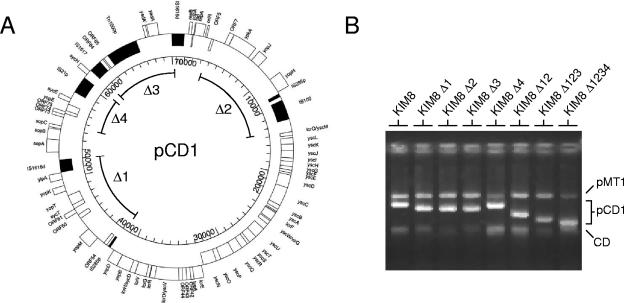
To determine the minimum number of pCD1 genes required to express a functional calcium-regulated T3SS, we created four large deletions in plasmid pCD1 that together removed more than one-half of the genetic material carried on the plasmid (Fig. 1A). The four deletions eliminated pCD1 sequences at positions 41,640 to 52,022 (Δ1), 3,740 to 15,602 (Δ2), 61,430 to 70,331 (Δ3), and 55,568 to 60,495 (Δ4). In general, the deletions were designed to remove the genes encoding the six effector Yops (yopE, yopH, yopJ, yopM, yopT, and ypkA), two chaperones (sycE and sycT), ylpA, yopK, yadA, all uncharacterized open reading frames, and essentially all transposable elements. The four deletions did not disrupt the low-calcium response (Lcr) region (positions 15,801 to 41,540), the origin of replication, the partitioning region, or the sycH gene.
FIG. 1.
Construction of plasmid pCD1-Δ1234. (A) Map of the whole pCD1 plasmid of Y. pestis KIM5. The outer circle shows the positions of identified pCD1 genes and open reading frames (open boxes). Genes located on the outside of the circle are transcribed in a clockwise direction, whereas genes located on the inside of the circle are transcribed in a counterclockwise direction. Transposable elements are indicated by solid boxes. The pCD1 sequences removed by deletions Δ1, Δ2, Δ,3 and Δ4 are indicated by the labeled lines. Reprinted from reference 28 with permission of the publisher. (B) Agarose gel electrophoretogram of plasmid pCD1 and derivatives of this plasmid. Plasmid DNA was isolated from Y. pestis KIM8, KIM8 Δ1, KIM8 Δ2, KIM8 Δ3, KIM8 Δ4, KIM8 Δ12, KIM8 Δ123, and KIM8 Δ1234 by the procedure of Kado and Liu (21), separated on 0.7% agarose gels, and stained with ethidium bromide. CD, chromosomal DNA.
Lambda Red-mediated recombination was used to delete each specified region of pCD1 and to simultaneously insert an FLP recognition target-flanked kanamycin resistance (kan) cassette or a dhfr cassette essentially as described by Datsenko and Wanner (8). PCR products used to construct the gene replacements were generated using plasmid pKD4 (kan) or dhfr (EZ::TN<DHFR>; Epicentre, Madison, WI) as a template for PCR. The oligonucleotide primers used in this study are listed in Table 1. Deletions were confirmed by PCR analysis. Each individual deletion was initially constructed in plasmid pCD1 of Y. pestis KIM8 (pMT1+, pCD1+, and pPCP1−), which generated kanamycin-resistant (Kmr) strains KIM8 Δ1, KIM8 Δ2, and KIM8 Δ3, as well as trimethoprim-resistant (Tmr) strain KIM8 Δ4. Plasmid pCP20, which encodes the FLP recombinase (8), was electroporated into KIM8 Δ1 to facilitate removal of the FLP recognition target-flanked kan cassette, generating strain KIM8 Δ1s. Plasmids pKD46 and pCP20, which carry temperature-sensitive origins of replication, were cured from the kanamycin-sensitive strain by overnight growth at 39°C (8). The presence of the deletions and the absence of pKD46 and pCP20 were confirmed by PCR analysis and by agarose gel electrophoresis of plasmids isolated by the method of Kado and Liu (21).
TABLE 1.
Oligonucleotides used for lambda Red-mediated recombination
| Deletion | Template | Primer | Oligonucleotide sequence (5′ to 3′) |
|---|---|---|---|
| Δ1::kan | pKD4 (kan) | Δ1-P1 | TGATAGAGCTTATCTATATAAGGTATAAGGTGCTGAAAATGTGTAGGCTGGAGCTGCTTC |
| Δ1-P2 | TTACATGTTGAATTTGAAGGCGAGTGGACCGGACCTTATCATATGAATATCCTCCTTAGT | ||
| Δ2::kan | pKD4 (kan) | Δ2-P1 | AGGTGAAATATGACGACACGGTATTCTCGTAGTGAACGGTGTGTAGGCTGGAGCTGCTTC |
| Δ2-P2 | GAGTCTGCTCCTCATATAAATTGAGAGAATTAGGATGAACATATGAATATCCTCCTTAGT | ||
| Δ3::kan | pKD4 (kan) | Δ3-P1 | GTTTAATTCTATAAAAGAAAAACGTACGATATCCATTAATGTGTAGGCTGGAGCTGCTTC |
| Δ3-P2 | CAAAGATTTGATGGGGAACTCGCTCATCAACACGGCTGCCATATGAATATCCTCCTTAGT | ||
| Δ4::dhfr | EZ::TN<DHFR>a | Δ4-P1 | TTTTTGATAGCGATATCCATCCCGCCAGGTTGGATACGGATAGACGGCATGCACGATTTG |
| Δ4-P2 | TAACTTTTACTGAGCGAAATCTGATATTACTGGCACCACCGCATCCAATGTTTCCGCCAC | ||
| Δ2B::kan | pKD4 (kan) | Δ2B-P1 | AGGTGAAATATGACGACACGGTATTCTCGTAGTGAACGGCGCTGCCGCAAGCACTCAGGG |
| Δ2B-P2 | GAGTCTGCTCCTCATATAAATTGAGAGAATTAGGATGAAGTCCCGCTCAGAAGAACTCGT | ||
| Δ3B::cat | pKD3 (cat) | Δ3B-P1 | TGACTTCGGGCAAATTCTGCCGGAGTCAGGTTATTTAACCTTCGGAATAGGAACTTCATT |
| Δ3B-P2 | CGTTTAATCTGCGCCCTGAGCGACTGGATTTCACGTTGCGGCGCGCCTACCTGTGACGGA | ||
| Δhha | pKD4 (kan) | Δhha-P1 | TATTTAATGCGTTTACGTCGTTGCCAGACAATTGACACGTGTGTAGGCTGGAGCTGCTTC |
| Δhha-P2 | ATTCATGGTCAATTCGGCGAGGCGGTGATCTGCGGCTGACATATGAATATCCTCCTTAGT |
Obtained from Epicentre.
To construct a pCD1 variant that had all four deletions, we used lambda Red-mediated recombination (8) to sequentially delete three additional regions (Δ2, Δ3, and Δ4) from pCD1 of KIM8 Δ1s. PCR products used to construct the sequential gene replacements were generated using pKD4 (kan), pKD3 (cat), or dhfr as the template for PCR. The Δ2::kan, Δ3::cat, and Δ4::dhfr insertional deletions were confirmed by PCR analysis and by agarose gel electrophoresis of plasmids isolated by the method of Kado and Liu (21). The resultant multiple-deletion pCD1 plasmids pCD1-Δ12 (kan), pCD1-Δ123 (kanΔ2 catΔ3), and pCD1-Δ1234 (kanΔ2 catΔ3 dhfrΔ4), as well as the original single-deletion plasmids pCD1-Δ1 (kan), pCD1-Δ2 (kan), pCD1-Δ3 (kan), and pCD1-Δ4 (dhfr), were isolated and electroporated into Y. pestis KIM10 (pMT1+, pCD1−, pPCP1−), generating Y. pestis KIM8 Δ1 (Kmr), KIM8 Δ2 (Kmr), KIM8 Δ3 (Kmr), KIM8 Δ4 (Tmr), KIM8 Δ12 (Kmr), KIM8 Δ123 (Kmr Cmr), and KIM8 Δ1234 (Kmr Cmr Tmr) in a clean genetic background (Fig. 1B). The resultant strains were used in all subsequent studies.
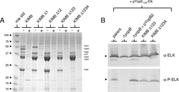
Secretion of Yops by Y. pestis KIM8, KIM8 Δ1, KIM8 Δ12, KIM8 Δ123, and KIM8 Δ1234 was measured following growth of the bacteria in TMH medium for 5 h at 37°C in the presence and absence of calcium. Concentrated culture supernatant proteins were separated by sodium dodecyl sulfate (SDS)-polyacrylamide gel electrophoresis (PAGE) and stained with Coomassie blue R-250 (Fig. 2A). All five strains secreted Yops in the absence of calcium but not in the presence of calcium. Y. pestis KIM8 Δ1234, which had all four deletions, expressed and secreted YopB, YopD, YopN, and LcrV but none of the effector Yops. In addition to the deletions in plasmid pCD1 mentioned above, a deletion (Δ5) eliminating the pCD1 sequence at positions 55,568 to 70,331 from pCD1 of Y. pestis KIM8 was constructed using primers Δ4-P1 and Δ3-P2. Y. pestis KIM8 carrying plasmid pCD1-Δ5, in which the sycH gene was deleted, expressed and secreted only low levels of Yops (data not shown), confirming that SycH is required for efficient T3SS gene expression (4). These studies confirmed that with the exception of sycH, all of the genes required to express a functional calcium-regulated T3SS are encoded in the Lcr region of plasmid pCD1. Similar results were obtained by Trulzsch et al. (36), who inserted the entire Lcr region of the Y. enterocolitica pYV virulence plasmid into a cosmid vector (mini-pYV). In this case, the mini-pYV plasmid directed secretion and translocation of effector Yops encoded on expression plasmids provided in trans. Although the mini-pYV plasmid encoded a functional T3SS, it did not carry the sycH gene; therefore, LcrQ (YscM)-dependent regulation of T3SS gene transcription was not reconstituted with this construct. SycH, the chaperone for YopH and LcrQ, is required for efficient export of the negative regulatory component LcrQ in the absence of calcium (4, 37). Deletion of sycH prevents secretion of LcrQ and results in repression of T3SS gene transcription (30, 31).
FIG. 2.
Secretion and translocation of Yops by Y. pestis strains carrying plasmid pCD1 or versions of this plasmid with deletions. (A) Y. pestis KIM8, KIM8 Δ1, KIM8 Δ12, KIM8 Δ123, and KIM8 Δ1234 were grown in TMH medium with (lanes +) or without (lanes −) 2.5 mM CaCl2 for 5 h at 37°C. Volumes of trichloroacetic acid-precipitated culture supernatant proteins corresponding to equal numbers of bacteria were resolved by SDS-PAGE and stained with Coomassie blue. The positions of different Yops are indicated on the right, and the molecular masses (in kilodaltons) of protein standards are indicated on the left. (B) Translocation of YopE129-ELK into HeLa cells. Y. pestis KIM8 Δ123, KIM8 Δ1234, KIM8-3002.P40 (ΔyopE ΔyopJ; parent), KIM8-3002.41 (ΔyopE ΔyopJ ΔyopB), and KIM8-3002.41 complemented with pYopB2, all carrying pYopE129-ELK, were used to infect HeLa cell monolayers at a multiplicity of infection of 30. After 3 h of incubation at 37°C, infected monolayers were solubilized with SDS-PAGE sample buffer and analyzed by SDS-PAGE and immunoblotting with anti-Elk antipeptide antibodies (α-ELK) and phosphospecific anti-Elk antipeptide antibodies (α-P-ELK). The position of YopE129-ELK is indicated by arrowheads.
The injection of Yops into a eukaryotic cell is essentially a three-step process that involves (i) attachment of the bacterium to a eukaryotic cell via specific bacterial adhesins, (ii) secretion of effector Yops across the bacterial membranes, and (iii) translocation of Yops across the eukaryotic membrane. The ability of Y. pestis KIM8 Δ123 and KIM8 Δ1234 to inject Yops into a eukaryotic cell was evaluated using the ELK tag reporter system described previously (9). Y. pestis KIM8 Δ123, KIM8 Δ1234, KIM8-3002.P40 (ΔyopE ΔyopJ), KIM8-3002.41 (ΔyopE ΔyopJ ΔyopB), and KIM8-3002.41 carrying pYopB2 were transformed with plasmid pYopE129-ELK (9). The reporter plasmid encodes a truncated YopE protein with a C-terminal ELK tag that is phosphorylated upon translocation into a eukaryotic cell. Cultured HeLa cells were infected at a multiplicity of infection of 30 for 3 h at 37°C, and the infected monolayers were analyzed by SDS-PAGE and immunoblot analysis (Fig. 2B). The total YopE129-ELK expressed during the infection was detected with anti-ELK antipeptide antibodies, whereas YopE129-ELK translocated into the HeLa cells was phosphorylated and detected with phospho-specific antipeptide antibodies (Cell Signaling Technology).
Immunoblot analysis with anti-ELK antipeptide antibodies demonstrated that all the strains expressed comparable amounts of the YopE129-ELK protein (Fig. 2B, top panel). Y. pestis KIM8-3002.P40 (parent), KIM8 Δ123, and KIM8 Δ1234 translocated approximately equal amounts of YopE129-ELK into the HeLa cells (Fig. 2B, bottom panel). In contrast, the translocation-defective ΔyopB strain was unable to inject the YopE129-ELK protein into the HeLa cells. Providing complementing plasmid pYopB2 in trans completely restored the ability of the yopB deletion mutant to translocate the YopE129-ELK protein. These studies confirmed that the pCD1-Δ1234 plasmid encodes a functional T3SS capable of injecting Yops into eukaryotic cells.
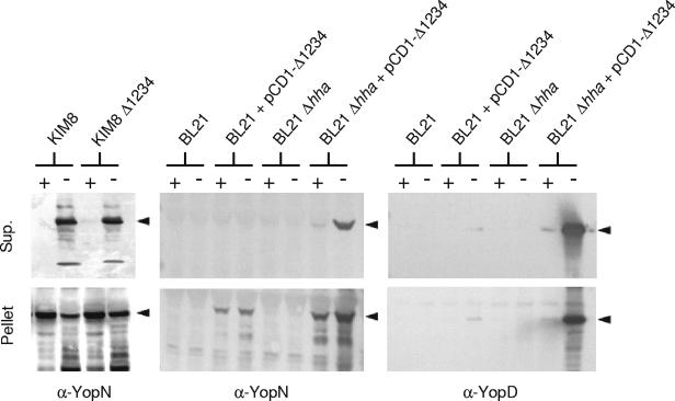
The calcium-regulated secretion of Yops by Yersinia spp. is dependent upon at least 30 different pCD1 genes; however, the role of chromosomal genes in this process is not fully understood. Recently, several chromosomal genes required for efficient expression of the Yersinia spp. T3SS have been identified (7, 10, 19, 20); however, the gene products identified thus far do not appear to be intimately involved in the type III secretion process per se. To determine if the Y. pestis chromosome carries genes that are essential for expression of a functional calcium-regulated T3SS, we moved the pCD1-Δ1234 construct into E. coli DH5α and BL21. Only low levels of Yop expression and no Yop secretion were observed in E. coli DH5α or BL21 carrying plasmid pCD1-Δ1234, indicating that pCD1 T3SS genes are not efficiently expressed in E. coli (17) (Fig. 3 and data not shown).
FIG. 3.
Expression and secretion of Yops by an E. coli hha deletion mutant carrying pCD1-Δ1234. E. coli BL21 and BL21 hha with and without plasmid pCD1-Δ1234 were grown in TMH medium with (lanes +) or without (lanes −) 2.5 mM CaCl2 for 5 h at 37°C. Cultures of Y. pestis KIM8 and KIM8 Δ1234 were grown under identical conditions. Volumes of culture supernatant (Sup.) proteins and cell pellet (Pellet) fractions corresponding to equal numbers of bacteria were analyzed by SDS-PAGE and immunoblotting with antisera specific for YopN or YopD. The locations of YopN and YopD are indicated by arrowheads.
Transcription of T3SS genes in Yersinia spp. is negatively regulated by a small histone-like protein designated YmoA (7). E. coli strains express a protein homologous to YmoA, designated Hha (24), which functions with H-NS to thermoregulate the transcription of several virulence-related genes (22, 25). To examine the role of Hha in regulating the expression of pCD1-Δ1234 T3SS genes in E. coli DH5α and BL21, we used lambda Red-mediated recombination to delete the hha locus and to insert a kan cassette at the site of the deletion. The deletion of hha in both DH5α and BL21 was confirmed by PCR analysis. Plasmid pKD46 was cured from the hha deletion mutants, and the pCD1-Δ1234 plasmid was electroporated into the pKD46-cured hha deletion strains.
Expression and secretion of YopN and YopD by Y. pestis KIM8 and KIM8 Δ1234 and by E. coli BL21, BL21 Δhha, DH5α, and DH5α Δhha with and without plasmid pCD1-Δ1234 were determined following growth of the bacteria at 37°C in TMH medium in the presence and absence of calcium (Fig. 3). The E. coli BL21 and DH5α hha deletion strains carrying plasmid pCD1-Δ1234 secreted YopN and YopD in the absence of calcium but not in the presence of calcium. The amounts of YopN and YopD secreted by these strains were comparable to or slightly less than the amounts secreted by Y. pestis KIM8 or KIM8 Δ1234. These studies confirmed that plasmid pCD1-Δ1234 contains the complete complement of genes unique to the yersiniae that are required to express a functional T3SS. Interestingly, the E. coli hha strains carrying pCD1-Δ1234 secreted high levels of Yops but did not exhibit growth restriction (data not shown), suggesting that the physiological determinants that link growth and secretion are unique to the yersiniae.
E. coli DH5α and BL21 are avirulent bacteria that do not express the surface adhesins required for attachment to the surface of eukaryotic cells (5). Therefore, the E. coli strains that express a functional T3SS should not be able to attach to HeLa cells and inject Yops. Indeed, E. coli DH5α Δhha and BL21 Δhha carrying pCD1-Δ1234 and pYopE129-ELK expressed the YopE129-ELK protein but did not translocate the protein into cultured HeLa cells (data not shown).
Previous studies have suggested that Hha and YmoA, which exhibit 82% amino acid sequence identity, are interchangeable (1, 11). Our results indicate that Hha in E. coli, but not YmoA in Y. pestis, prevents essentially all pCD1 T3SS gene expression at 37°C. The reason for the difference is unclear; however, we hypothesize that it could be related to the amount of YmoA or Hha present in the relevant bacterial cells at 37°C. We have demonstrated previously that YmoA is rapidly degraded by the Lon protease at 37°C in Y. pestis, a process that is essential to trigger T3SS gene transcription in Y. pestis (20). It is possible that Hha is more resistant to Lon-mediated proteolysis and is not rapidly degraded at 37°C in E. coli. To begin to examine this possibility, we constructed plasmid pFLAG-Hha, which encodes the Hha protein with an N-terminal 14-amino-acid tag (N-MDYKDDDDKVKLLE-Hha). Plasmid pFLAG-Hha was electroporated into E. coli DH5α and BL21, whereas plasmid pFLAG-YmoA was electroporated into Y. pestis KIM5-3001 (20). The stability of FLAG-Hha and FLAG-YmoA at 37°C was determined by determining the amounts of these proteins after new synthesis was blocked by addition of chloramphenicol (40 μg/ml) to the cultures, as described previously (20). Samples corresponding to equal numbers of bacteria were removed at specific times and analyzed by SDS-PAGE and immunoblot analysis with the FLAG M2 antibody (Sigma, St. Louis, MO) (Fig. 4). As previously demonstrated, FLAG-YmoA was rapidly degraded at 37°C in Y. pestis. FLAG-Hha was stable at 37°C in lon-deficient E. coli BL21 (14), but it was degraded in E. coli DH5α. These results suggest that Hha, like YmoA, is degraded in a Lon-dependent manner; however, the half-life of FLAG-YmoA in Y. pestis (19.3 min) was significantly less than the half-life of FLAG-Hha in DH5α (40.3 min). These results suggest that the degradation of Hha in E. coli and the degradation of YmoA in Y. pestis are to some extent different, and the difference may account for the ability of Hha to suppress pCD1 gene transcription at 37°C in E. coli. Alternate explanations are also possible, and further studies are required to determine the mechanism by which Hha represses T3SS gene transcription at 37°C in E. coli.
FIG. 4.
Stability of FLAG-Hha in E. coli DH5α and BL21. E. coli DH5α and BL21, both carrying pFLAG-Hha, as well as Y. pestis KIM5-3001 carrying pFLAG-YmoA, were grown in heart infusion broth containing 2.5 mM CaCl2 for 3 h at 37°C. Chloramphenicol (final concentration, 40 μg/ml) was added to block protein synthesis. An aliquot of each culture was removed at the times indicated at the top (15m, 15 min; 30m, 30 min). Whole-cell protein extracts from equivalent numbers of bacteria were separated by SDS-PAGE, transferred to Immobilon-P membranes, and probed with antibodies specific for the FLAG epitope tag.
The E. coli Δhha/pCD1-Δ1234 strain-plasmid combination provides a system to study the Yersinia spp. type III secretion process in a heterologous host. Previously, only the Erwinia chrysanthemi T3SS has been shown to function in a heterologous bacterial host (16). The combination of the pCD1-Δ1234 virulence plasmid lacking the genes encoding all six translocated effector Yops with an avirulent E. coli host provides a reliable and safe system to analyze the molecular events involved in the Y. pestis type III secretion process. The procedures for preparation and manipulation of membrane vesicles suitable for in vitro secretion studies have been developed and fine-tuned in E. coli (12). Thus, the E. coli Δhha/pCD1-Δ1234 system should facilitate the development of in vitro secretion assays that are required to dissect the molecular events involved in the type III secretion process.
Acknowledgments
This work was supported by Public Health Service grants AI 50552 and AI 39575 from the National Institutes of Health to G.V.P.
Editor: J. B. Bliska
REFERENCES
- 1.Balsalobre, C., A. Juarez, C. Madrid, M. Mourino, A. Prenafeta, and F. J. Munoa. 1996. Complementation of the hha mutation in Escherichia coli by the ymoA gene from Yersinia enterocolitica: dependence on the gene dosage. Microbiology 142:1841-1846. [DOI] [PubMed] [Google Scholar]
- 2.Ben-Gurion, R., and A. Shafferman. 1981. Essential virulence determinants of different Yersinia species are carried on a common plasmid. Plasmid 5:183-187. [DOI] [PubMed] [Google Scholar]
- 3.Brubaker, R. R. 1983. The Vwa+ virulence factor of yersiniae: the molecular basis of the attendant nutritional requirement for Ca++. Rev. Infect. Dis. 5:748-758. [DOI] [PubMed] [Google Scholar]
- 4.Cambronne, E. D., L. W. Cheng, and O. Schneewind. 2000. LcrQ/YscM1, regulators of the Yersinia yop virulon, are injected into host cells by a chaperone-dependent mechanism. Mol. Microbiol. 37:263-273. [DOI] [PubMed] [Google Scholar]
- 5.Chart, H., H. R. Smith, R. M. La Ragione, and M. J. Woodward. 2000. An investigation into the pathogenic properties of Escherichia coli strains BLR, BL21, DH5α and EQ1. J. Appl. Microbiol. 89:1048-1058. [DOI] [PubMed] [Google Scholar]
- 6.Cornelis, G. R. 2000. Molecular and cell biology aspects of plague. Proc. Natl. Acad. Sci. USA 97:8778-8783. [DOI] [PMC free article] [PubMed] [Google Scholar]
- 7.Cornelis, G. R., C. Sluiters, I. Delor, D. Geib, K. Kaniga, C. Lambert de Rouvroit, M. P. Sory, J. C. Vanooteghem, and T. Michiels. 1991. ymoA, a Yersinia enterocolitica chromosomal gene modulating the expression of virulence functions. Mol. Microbiol. 5:1023-1034. [DOI] [PubMed] [Google Scholar]
- 8.Datsenko, K. A., and B. L. Wanner. 2000. One-step inactivation of chromosomal genes in Escherichia coli K-12 using PCR products. Proc. Natl. Acad. Sci. USA 97:6640-6645. [DOI] [PMC free article] [PubMed] [Google Scholar]
- 9.Day, J. B., F. Ferracci, and G. V. Plano. 2003. Translocation of YopE and YopN into eukaryotic cells by Yersinia pestis yopN, tyeA, sycN, yscB and lcrG deletion mutants measured using a phosphorylatable peptide tag and phosphospecific antibodies. Mol. Microbiol. 47:807-823. [DOI] [PubMed] [Google Scholar]
- 10.DeBord, K. L., N. S. Galanopoulos, and O. Schneewind. 2003. The ttsA gene is required for low-calcium-induced type III secretion of Yop proteins and virulence of Yersinia enterocolitica W22703. J. Bacteriol. 185:3499-3507. [DOI] [PMC free article] [PubMed] [Google Scholar]
- 11.de la Cruz, F., M. Carmona, and A. Juarez. 1992. The Hha protein from Escherichia coli is highly homologous to the YmoA protein from Yersinia enterocolitica. Mol. Microbiol. 6:3451-3452. [DOI] [PubMed] [Google Scholar]
- 12.Douville, K., A. Price, J. Eichler, A. Economou, and W. Wickner. 1995. SecYEG and SecA are the stoichiometric components of preprotein translocase. J. Biol. Chem. 270:20106-20111. [DOI] [PubMed] [Google Scholar]
- 13.Ghosh, P. 2004. Process of protein transport by the type III secretion system. Microbiol. Mol. Biol. Rev. 68:771-795. [DOI] [PMC free article] [PubMed] [Google Scholar]
- 14.Gottesman, S. 1989. Genetics of proteolysis in Escherichia coli. Annu. Rev. Genet. 23:163-198. [DOI] [PubMed] [Google Scholar]
- 15.Grosdent, N., I. Maridonneau-Parini, M. P. Sory, and G. R. Cornelis. 2002. Role of Yops and adhesins in resistance of Yersinia enterocolitica to phagocytosis. Infect. Immun. 70:4165-4176. [DOI] [PMC free article] [PubMed] [Google Scholar]
- 16.Ham, J. H., D. W. Bauer, D. E. Fouts, and A. Collmer. 1998. A cloned Erwinia chrysanthemi Hrp (type III protein secretion) system functions in Escherichia coli to deliver Pseudomonas syringae Avr signals to plant cells and to secrete Avr proteins in culture. Proc. Natl. Acad. Sci. USA 95:10206-10211. [DOI] [PMC free article] [PubMed] [Google Scholar]
- 17.Heesemann, J., U. Gross, N. Schmidt, and R. Laufs. 1986. Immunochemical analysis of plasmid-encoded proteins released by enteropathogenic Yersinia sp. grown in calcium-deficient media. Infect. Immun. 54:561-567. [DOI] [PMC free article] [PubMed] [Google Scholar]
- 18.Hu, P., J. Elliott, P. McCready, E. Skowronski, J. Garnes, A. Kobayashi, R. R. Brubaker, and E. Garcia. 1998. Structural organization of virulence-associated plasmids of Yersinia pestis. J. Bacteriol. 180:5192-5202. [DOI] [PMC free article] [PubMed] [Google Scholar]
- 19.Jackson, M. W., and G. V. Plano. 1999. DsbA is required for stable expression of outer membrane protein YscC and for efficient Yop secretion in Yersinia pestis. J. Bacteriol. 181:5126-5130. [DOI] [PMC free article] [PubMed] [Google Scholar]
- 20.Jackson, M. W., E. Silva-Herzog, and G. V. Plano. 2004. The ATP-dependent ClpXP and Lon proteases regulate expression of the Yersinia pestis type III secretion system via regulated proteolysis of YmoA, a small histone-like protein. Mol. Microbiol. 54:1364-1378. [DOI] [PubMed] [Google Scholar]
- 21.Kado, C. I., and S. T. Liu. 1981. Rapid procedure for detection and isolation of large and small plasmids. J. Bacteriol. 145:1365-1373. [DOI] [PMC free article] [PubMed] [Google Scholar]
- 22.Madrid, C., J. M. Nieto, and A. Juarez. 2002. Role of the Hha/YmoA family of proteins in the thermoregulation of the expression of virulence factors. Int. J. Med. Microbiol. 291:425-432. [DOI] [PubMed] [Google Scholar]
- 23.Michiels, T., P. Wattiau, R. Brasseur, J. M. Ruysschaert, and G. Cornelis. 1990. Secretion of Yop proteins by yersiniae. Infect. Immun. 58:2840-2849. [DOI] [PMC free article] [PubMed] [Google Scholar]
- 24.Nieto, J. M., M. Carmona, S. Bolland, Y. Jubete, F. de la Cruz, and A. Juarez. 1991. The hha gene modulates haemolysin expression in Escherichia coli. Mol. Microbiol. 5:1285-1293. [DOI] [PubMed] [Google Scholar]
- 25.Nieto, J. M., C. Madrid, E. Miquelay, J. L. Parra, S. Rodriguez, and A. Juarez. 2002. Evidence for direct protein-protein interaction between members of the enterobacterial Hha/YmoA and H-NS families of proteins. J. Bacteriol. 184:629-635. [DOI] [PMC free article] [PubMed] [Google Scholar]
- 26.Orth, K., L. E. Palmer, Z. Q. Bao, S. Stewart, A. E. Rudolph, J. B. Bliska, and J. E. Dixon. 1999. Inhibition of the mitogen-activated protein kinase kinase superfamily by a Yersinia effector. Science 285:1920-1923. [DOI] [PubMed] [Google Scholar]
- 27.Palmer, L. E., S. Hobbie, J. E. Galan, and J. B. Bliska. 1998. YopJ of Yersinia pseudotuberculosis is required for the inhibition of macrophage TNF-alpha production and downregulation of the MAP kinases p38 and JNK. Mol. Microbiol. 27:953-965. [DOI] [PubMed] [Google Scholar]
- 28.Perry, R. D., S. C. Straley, J. D. Fetherston, D. J. Rose, J. Gregor, and F. R. Blattner. 1998. DNA sequencing and analysis of the low-Ca2+-response plasmid pCD1 of Yersinia pestis KIM5. Infect. Immun. 66:4611-4623. [DOI] [PMC free article] [PubMed] [Google Scholar]
- 29.Persson, C., R. Nordfelth, A. Holmstrom, S. Hakansson, R. Rosqvist, and H. Wolf-Watz. 1995. Cell-surface-bound Yersinia translocate the protein tyrosine phosphatase YopH by a polarized mechanism into the target cell. Mol. Microbiol. 18:135-150. [DOI] [PubMed] [Google Scholar]
- 30.Pettersson, J., R. Nordfelth, E. Dubinina, T. Bergman, M. Gustafsson, K. E. Magnusson, and H. Wolf-Watz. 1996. Modulation of virulence factor expression by pathogen target cell contact. Science 273:1231-1233. [DOI] [PubMed] [Google Scholar]
- 31.Rimpilainen, M., A. Forsberg, and H. Wolf-Watz. 1992. A novel protein, LcrQ, involved in the low-calcium response of Yersinia pseudotuberculosis shows extensive homology to YopH. J. Bacteriol. 174:3355-3363. [DOI] [PMC free article] [PubMed] [Google Scholar]
- 32.Rosqvist, R., A. Forsberg, and H. Wolf-Watz. 1991. Intracellular targeting of the Yersinia YopE cytotoxin in mammalian cells induces actin microfilament disruption. Infect. Immun. 59:4562-4569. [DOI] [PMC free article] [PubMed] [Google Scholar]
- 33.Rosqvist, R., K. E. Magnusson, and H. Wolf-Watz. 1994. Target cell contact triggers expression and polarized transfer of Yersinia YopE cytotoxin into mammalian cells. EMBO J. 13:964-972. [DOI] [PMC free article] [PubMed] [Google Scholar]
- 34.Schesser, K., A. K. Spiik, J. M. Dukuzumuremyi, M. F. Neurath, S. Pettersson, and H. Wolf-Watz. 1998. The yopJ locus is required for Yersinia-mediated inhibition of NF-kappaB activation and cytokine expression: YopJ contains a eukaryotic SH2-like domain that is essential for its repressive activity. Mol. Microbiol. 28:1067-1079. [DOI] [PubMed] [Google Scholar]
- 35.Straley, S. C., G. V. Plano, E. Skrzypek, P. L. Haddix, and K. A. Fields. 1993. Regulation by Ca2+ in the Yersinia low-Ca2+ response. Mol. Microbiol. 8:1005-1010. [DOI] [PubMed] [Google Scholar]
- 36.Trulzsch, K., A. Roggenkamp, M. Aepfelbacher, G. Wilharm, K. Ruckdeschel, and J. Heesemann. 2003. Analysis of chaperone-dependent Yop secretion/translocation and effector function using a mini-virulence plasmid of Yersinia enterocolitica. Int. J. Med. Microbiol. 293:167-177. [DOI] [PubMed] [Google Scholar]
- 37.Wattiau, P., B. Bernier, P. Deslee, T. Michiels, and G. R. Cornelis. 1994. Individual chaperones required for Yop secretion by Yersinia. Proc. Natl. Acad. Sci. USA 91:10493-10497. [DOI] [PMC free article] [PubMed] [Google Scholar]


