Abstract
Although γδ T cells are a common feature of many pathogen-induced immune responses, the factors that influence, promote, or regulate the response of individual γδ T-cell subsets to infection is unknown. Here we show that in the absence of Vγ1+ T cells, novel subsets of γδ T cells, expressing T-cell receptor (TCR)-Vγ chains that normally define TCRγδ+ dendritic epidermal T cells (DETCs) (Vγ5+), intestinal intraepithelial lymphocytes (iIELs) (Vγ7+), and lymphocytes associated with the vaginal epithelia (Vγ6+), are recruited to the spleen in response to bacterial infection in TCR-Vγ1−/− mice. By comparison of phenotype and structure of TCR-Vγ chains and/or -Vδ chains expressed by these novel subsets with those of their epithelium-associated counterparts, the Vγ6+ T cells elicited in infected Vγ1−/− mice were shown to be identical to those found in the reproductive tract, from where they are presumably recruited in the absence of Vγ1+ T cells. By contrast, Vγ5+ and Vγ7+ T cells found in infected Vγ1−/− mice were distinct from Vγ5+ DETCs and Vγ7+ iIELs. Functional analyses of the novel γδ T-cell subsets identified for infected Vγ1−/− mice showed that whereas the Vγ5+ and Vγ7+ subsets may compensate for the absence of Vγ1+ T cells by producing similar cytokines, they do not possess cytocidal activity and they cannot replace the macrophage homeostasis function of Vγ1+ T cells. Collectively, these findings identify novel subsets of γδ T cells, the recruitment and activity of which is under the control of Vγ1+ T cells.
The two lineages of T lymphocytes, αβ and γδ, are distinguished by their T-cell receptor (TCR) expression and have nonoverlapping but complementary roles in immune responses. Whereas αβ T cells respond to peptide antigens presented in the context of major histocompatibility complex class I and II molecules and are pivotal in the sterile elimination and generation of long-lived immunity to pathogens, the nature of antigen recognition by γδ T cells and their biological function is uncertain (reviewed in reference 8). γδ T cells appear to have both proinflammatory and regulatory functions: they can act as a bridge between innate and adaptive immunity early in responses and can down-modulate inflammatory responses once the infection is cleared (reviewed in references 1 and 8). γδ T cells are found in a number of different anatomical sites, with localization often associated with the expression of distinct TCR-Vγ chains. Of the commonly expressed Vγ chains, Vγ7+ (TCR-Vγ nomeclature used is that of Heilig and Tonegawa [16]) T cells are resident within the intestinal epithelium (35) and contain a high proportion of extrathymically generated cells (5, 29). Vγ5+ and Vγ6+ T cells are found in the skin and reproductive mucosa, respectively (3, 22), and are among the first γδ T cells to be generated in the fetal thymus (15, 21, 23). Fetal thymic-derived γδ T cells coexpress identical Vδ1 chains and differ from later thymic emigrants in that they express invariant TCRs composed of canonical Vγ5Jγ1Cγ1 or Vγ6Jγ1Cγ1 and Vδ1Dδ2Jδ2Cδ TCRs with no junctional (N-region) diversity (3, 26). Vγ1+ and Vγ4+ T cells, which are produced later in the thymus, around birth (23, 41), are found primarily in the peripheral lymphoid tissues and the lung, respectively, and are prominent among γδ T cells responding to microbial infection (reviewed in reference 8).
The requirement for γδ T cells in the response to microbial pathogens has been shown by a number of studies of TCRγδ-deficient mice, where exacerbated pathology, due to both increased susceptibility and exaggerated inflammatory responses, occurs during infection with viruses (40, 43, 44), bacteria (11, 14, 30), and parasites (31, 38, 39). The murine model of listeriosis has been extensively used to study the role of γδ T cells in pathogen-induced immune responses (8). The γδ T-cell response to Listeria monocytogenes occurs in two temporally distinct phases (17, 33). Prior to the induction of the adaptive αβ T-cell response (∼2 days postinfection), responding γδ T cells are predominantly proinflammatory, characterized by gamma interferon production (1, 13, 18). Later during infection, coincident with bacterial clearance (6), γδ T cells are required to down-modulate the inflammatory response and eliminate activated macrophages (9, 12, 19). In C57BL/6 mice, both the early- and late-responding γδ T cells are dominated by the Vγ1+ T-cell subset.
Listeria infection of TCR-Vγ1-deficient (Vγ1−/−) mice results in the accumulation of activated macrophages in sites of bacterial infection, consistent with the nonredundant role of Vγ1+ T cells in macrophage homeostasis (2). An unexpected observation with Listeria-infected Vγ1−/− mice was the accumulation in the spleen late in infection of γδ T cells expressing Vγ chains normally associated with epithelium-associated γδ T cells found in the skin (Vγ5+), small intestine (Vγ7+), and reproductive tract (Vγ6+). In this study, we have examined these unusual populations of γδ cells in more detail, characterizing their phenotype and origins and determining their potential functional significance.
MATERIALS AND METHODS
Mice and infection.
Male and female mice were used at 6 to 8 weeks of age, with three to six mice per group. C57BL/6 TCR-Vγ1−/− mice were generated as described previously (2) and backcrossed onto a C57BL/6 background for at least six generations, C57BL/6 TCRδ−/− mice were obtained from the Jackson Laboratory (Bar Harbor, ME), wild-type (WT) C57BL/6 mice were obtained from Harlan (Bicester, United Kingdom), and all were housed in the animal facility at the University of Leeds. Mice were infected intraperitoneally with 1.5 × 10⁴ bacterial CFU Listeria monocytogenes (strain 10403S).
Cell preparation.
Standard protocols were used to isolate lymphocytes from the spleen, thymus, and liver (25). Tissues were homogenized, contaminating erythrocytes were lysed with 0.84% (wt/vol) ammonium chloride solution, and the cell suspension was washed and passed through a 0.7-μm nylon filter before use. For intestinal intraepithelial lymphocyte (iIELs) isolation, whole small intestines were flushed through with phosphate-buffered saline, and the mesentery and Peyer's patches were removed, opened longitudinally, and cut into 1- to 2-cm fragments. The intestinal fragments were then incubated with dithioerythritol-EDTA solution (0.3 mg/ml dithioerythritol, 5 mM EDTA, 10% fetal bovine serum in Hanks balanced salt solution [HBSS]) at 37°C for 30 min to remove the epithelial layer. Cells were then pelleted at 500 × g for 5 min at 20°C, resuspended in 5 ml 80% Percoll (Amersham Biosciences), overlaid with 9 ml 40% Percoll, and centrifuged at 800 × g for 20 min at 20°C. iIELs were isolated from the 40/80 interface and washed in HBSS-10% fetal calf serum. Peritoneal exudate cells (PECs) were collected by peritoneal lavage with HBSS containing 10 U/ml heparin.
RT-PCR.
Cells (107) were pelleted and RNA extracted using Tri-reagent (Sigma, Poole, United Kingdom) following the manufacturer's instructions. The resultant RNA was subjected to reverse transcription-PCR (RT-PCR) using ImpromII reverse transcriptase (Promega, Southampton, United Kingdom) and the cDNA amplified with Reddy-mix Taq polymerase (Abgene, Epsom, United Kingdom), using primers for TCR Vγ chains as described previously (2). Products were run out on 2% agarose gels and visualized with ethidium bromide.
Spectratyping and DNA sequencing.
PCR products (1 μl) were used as templates in a 10-cycle runoff reaction using Reddy-mix polymerase with a 6-carboxyfluorescein-labeled J-region-specific primer (for Jγ1, CTTAGTTCCTTCTGCAAATACC; for Jγ2, ATGAGCTTTGTTCCTTCTGC; or for Jγ4, TACGAGCTTTGTCCCTTTG). Products were electrophoretically separated (Lark Technologies, Inc., Takeley, United Kingdom) and analyzed using Genescanview4 software (CRIBI, Padua, Italy). For sequencing, PCR-amplified TCR-Vγ products were excised from agarose gel slices following electrophoresis by use of a Genelute gel purification kit (Sigma, Poole, United Kingdom) and ligated into the T-cloning vector pGEM-T Easy (Promega, Southampton, United Kingdom). DNA, prepared using a Genelute miniprep kit (Sigma, Poole, United Kingdom), was then sequenced using universal cloning vector primers (Lark Technologies, Inc., Takeley, United Kingdom).
Flow cytometry.
Antibodies for surface staining were F(ab)2 fragments of antibodies specific for TCRδ (clone GL3), intact antibody specific for TCR-Vγ7 (clone F2.67 provided by Pablo Pereira [Institut Pasteur, Paris, France]), the anti-Vγ5/Vδ1 clonotype antibody 17D1 (provided by Adrian Hayday, Kings College, London, United Kingdom), and commercial preparations of anti-TCRγδ (GL3), -CD3 (145-2C11), -TCR-Vγ2 (Vγ4; UC3-10A6), -TCR-Vγ3 (Vγ5; 536), -F4/80, -CD8α (CT-CD8a), -CD103 (M290), -NKG2D (CX5), -Fas (Jo2), and -FasL (MFL3), obtained from Caltag-Medsystems (Towcester, United Kingdom) or PharMingen (Oxford, United Kingdom). Streptavidin conjugates of phycoerythrin, fluorescein isothiocyanate (FITC) (Caltag), or Alexa Fluor 633 (Molecular Probes, Eugene, OR) were used as secondary reagents. To block nonspecific antibody binding, cells were preincubated with an anti-Fc-receptor antibody cocktail (anti-CD16/32; Caltag). Isotype-matched antibodies of irrelevant specificity were used to determine the level of nonspecific staining. Stained cells were analyzed with a FACSCalibur flow cytometer by use of Cellquest software (Becton Dickinson, Oxford, United Kingdom).
Mononuclear cell killing assay.
Peritoneal exudate-derived macrophages or splenocytes (5 × 10⁵) from day 8 L. monocytogenes-infected TCRδ−/− mice were incubated on 8-well chamber slides (ICN Pharmaceuticals, Ltd., Basingstoke, United Kingdom) with Live/Dead cell reagent (Molecular Probes) containing fluorescent dyes that identify intracellular esterase activity of viable cells (calcein AM) or are incorporated in the nuclei of dead cells (ethidium bromide homodimer 1), as previously described (9). Splenocytes (1 × 10⁶) from day 8 L. monocytogenes-infected TCR-Vγ1−/− or wild-type mice were added and cultures were incubated at 37°C in RPMI (Sigma-Aldrich) with 10% fetal bovine serum for 1 h. Viable and dead cells were visualized and quantitated under UV illumination by using a Zeiss Axiovert 200 M microscope (Welwyn Garden City, Herts, United Kingdom) and Axiovison image analysis software (Imaging Associates, Ltd., Bicester, United Kingdom), counting at least 100 cells in four separate fields of view.
Intracellular-cytokine analysis.
Intracellular cytokines were detected by cytoplasmic staining of cells cultured in the presence of brefeldin A (10 μg/ml) (Sigma). The cells were stained for surface markers, fixed in 1% paraformaldehyde, and permeabilized with 0.5% of saponin (Sigma) before being stained with phycoerythrin-conjugated anti-mouse cytokine monoclonal antibodies to interleukin 2 (IL-2), IL-4, IL-5, IL-6, IL-10, gamma interferon, and tumor necrosis factor alpha, from Caltag-Medsystems (Towcester, United Kingdom) or PharMingen (Oxford, United Kingdom), or FITC-conjugated polyclonal antibodies to monocyte chemoattractant protein 1 (Sigma). FITC (Sigma-Aldrich, Dorset, United Kingdom) was conjugated to the antibodies by standard procedures.
RESULTS
Novel γδ T-cell subsets accumulate in spleens of Vγ1−/− mice following Listeria infection.
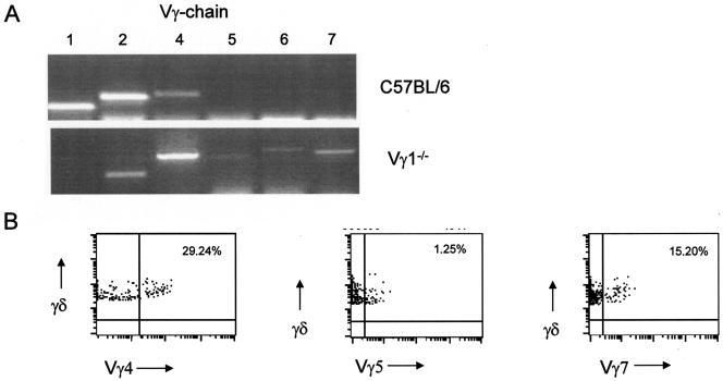
We have previously shown that the recruitment of γδ T-cell subsets to the spleen during Listeria infection is regulated by Vγ1+ T cells and that, in the absence of these cells, γδ subsets which are epithelium associated and thought to be resident only in the skin (Vγ5), reproductive tract (Vγ6), and intestine (Vγ7) appear in the spleen in the late stage of infection (2). This unusual finding raised the questions of whether these cells had been recruited from their epithelial sites, what their functional significance might be in the spleen at a time when the infection was being cleared, and when in wild-type mice the inflammatory response would be downmodulated by Vγ1+ T cells (2, 9, 12). After verifying the expression of Vγ chain mRNA (Fig. 1A) and the presence of γδ T cells which stained antibody positive for these subsets (Fig. 1B) in the spleen 8 days after Listeria infection, the phenotypic and functional characteristics of splenic Vγ subsets and the spectratypes of PCR-derived TCR-Vγ transcripts were compared with those from epithelial sites to identify similarities or differences that might shed light on their origins and function.
FIG. 1.
Appearance of unusual subsets of γδ T cells elicited by microbial infection in the absence of Vγ1+ T cells. (A) Profile of TCR-Vγ chain expression in the spleens of wild-type (C57BL/6) and TCR-Vγ1−/− mice 8 days after infection with Listeria, as determined by RT-PCR analysis. The results shown are representative of those obtained from more than 10 mice of each strain. (B) Representative flow cytometric staining profiles of γδ T cells expressing Vγ4, Vγ5, or Vγ7 receptors among spleen cells recovered from TCR-Vγ1−/− mice 8 days after Listeria infection (n = 10). Percentages of γδ cells positive for each marker are shown on the dot plots.
Pathogen-elicited γδ T cells in Vγ1−/− mice express distinct TCRs.
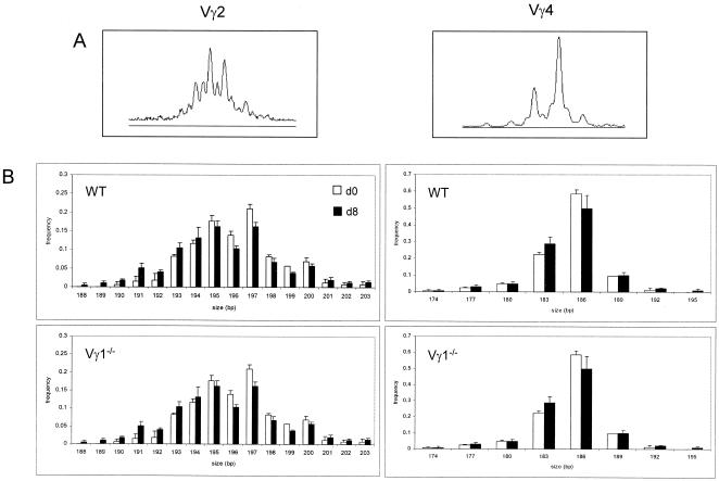
Transcripts of Vγ2 and Vγ4 occurred in both wild-type and Vγ1−/− spleens before and 8 days after infection (Fig. 1A). Their TCR spectratypes were therefore compared to identify any differences in these commonly expressed Vγ chains that were expressed prior to and after Listeria infection in these two strains of mice, and if differences were found, to determine if the differences might be attributable to the presence or absence of Vγ1+ T cells (Fig. 2). The Vγ2 spectratype showed a number of peaks separated by only 1 bp, indicative of a high proportion of out-of-frame transcripts, as previously reported (36). By contrast, there were fewer peaks for the Vγ4 transcripts, all of which were separated by 3 bp, consistent with in-frame products. There were no obvious differences in range or frequency of transcript size of either Vγ2 or Vγ4 between both strains of mice before or after infection, suggesting that the absence of Vγ1+ T cells does not affect the repertoire of these subsets in noninfected or Listeria-infected mice.
FIG. 2.
Structural analysis of CDR3 region of TCR-Vγ chains expressed by Listeria-elicited γδ T cells in wild-type and Vγ1−/− mice. (A) TCR CDR3 spectraype analysis of Vγ2 and Vγ4 transcripts expressed in the spleens of WT and Vγ1−/− mice prior to (d0) and 8 days after (d8) Listeria infection. (B) Collated profiles of two or more mice of each strain.
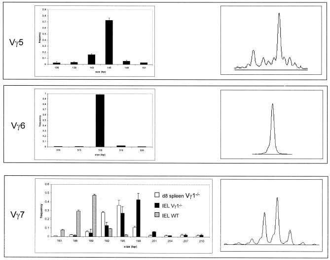
The spectratype analysis of Vγ5, Vγ6, and Vγ7 TCRs expressed in the spleens of Listeria-infected Vγ1−/− mice (Fig. 3) revealed a limited number of peaks, dominated by a single peak. In particular, the Vγ6 spectratype consisted of a single peak, which would be expected of a population of γδ T cells expressing an invariant or canonical TCR-Vγ chain (8, 26). The Vγ5 TCR transcripts detected in the spleens of Listeria-infected Vγ1−/− mice showed that in addition to the predominant peak corresponding to the expected canonical sequence at 145 bp, a number of other productively rearranged transcripts were detected. Comparisons of Vγ7 TCR spectratypes expressed in the spleens of Vγ1−/− mice on day 8 after Listeria infection with Vγ7+ iIELs from infected wild-type mice showed that the size distribution of each population was distinct (Fig. 3). The spectratype profile of Vγ7 TCRs expressed in the intestines of noninfected Vγ1−/− mice was also distinct from that of Vγ7+ iIELs in wild-type mice, with smaller-sized CDR3 regions being seen among Vγ7-encoded TCRs of wild-type mice.
FIG. 3.
Structural analysis of epithelium-associated TCR-Vγ chains expressed by splenic γδ T cells in infected Vγ1−/− mice. TCR CDR3 spectratypes were generated from Vγ5 (top), Vγ6 (middle), and Vγ7 (bottom) transcripts expressed in the spleens of Vγ1−/− mice infected 8 days previously with Listeria. Representative profiles obtained from cDNA samples of a single mouse are shown in the plots on the right side of the figure, and the cumulative profiles of more than three mice are shown in the bar graphs on the left side of the figure. The spectratypes of Vγ7 transcripts expressed in the spleens of day 8 (d8) infected Vγ1−/− mice are compared to those of iIELs from noninfected WT and Vγ1−/− mice.
The relationship of Vγ5 and Vγ6 TCR transcripts expressed in the spleens of Listeria-infected Vγ1−/− mice with those of epithelium-associated Vγ5+ and Vγ6+ T cells in wild-type mice was established by DNA sequencing of individual, cloned TCR cDNAs. Among the Vγ5 cDNAs cloned from day 8 spleens of Listeria-infected Vγ1−/− mice, the canonical dendritic epidermal T-cell (DETC) sequence was only a minor constituent (2/12) among those obtained (Table 1), with the majority being unique, consistent with the majority of the Vγ5+ T cells elicited by Listeria infection in Vγ1−/− mice being distinct from Vγ5+ DETCs. The characteristic canonical Vγ6 TCR sequence expressed by Vγ6+ T cells in the reproductive epithelium was detected in all (12/12) of the sequences generated from splenocytes of Vγ1−/− mice at day 8 following infection (Table 1).
TABLE 1.
Sequence analysis of Vγ5 and Vγ6 clonesa
| Clone and Sequence type | Sequence of:
|
n | Translation | |
|---|---|---|---|---|
| Clone | Jγ1 | |||
| Vγ5 | ||||
| Germ line | GCC TGC TGG GAT CT | AT AGC TCA GGT TTT | ||
| Canonical | GCC TGC TGG GAT | AGC TCA GGT TTT | ||
| GCC TGC TGG TAT | AGC TCA GGT TTT | 2 | ACWYSSGF | |
| GCC TGC TGG GTA | AGC TCA GGT TTT | ACWVSSGF | ||
| GCC TGC TGG GAT | AGC TCA GGT TTT | 2 | ACWDSSGF | |
| GCC TGC T | AT AGC TCA GGT TTT | ACYSSGF | ||
| GCC TGC TGG GTA | AGC TCA GGT TTT | ACWVSSGF | ||
| GCC TAT | AGC TCA GGT TTT | 5 | AYSSGF | |
| GCC TAT TGG GT | AGC TCA GGT TTT | NCb | ||
| GCC TGC TGG ATC ATA | T AGC TCA GGT TTT | NC | ||
| GCC TGC TGG GAT CT | AGC TCA GGT TTT | NC | ||
| GCC TGC TGG GAT | AT AGC TCA GGT TTT | NC | ||
| GCC TGC TGG GAT CAT | AT AGC TCA GGT TTT | NC | ||
| Vγ6 | ||||
| Germ line | GCA TGC TGG GAT AA | T AGC TCA GGT TTT | ||
| Canonical | GCA TGC TGG GAT | AGC TCA GGT TTT | ||
| GCA TGC TGG GAT | AGC TCA GGT TTT | 12 | ACWDSSGF | |
Boldface type indicates cannonical, invariant receptor sequences.
NC, noncoding.
Vγ5+ and Vγ7+ T cells do not conform to DETC or IEL phenotypes.
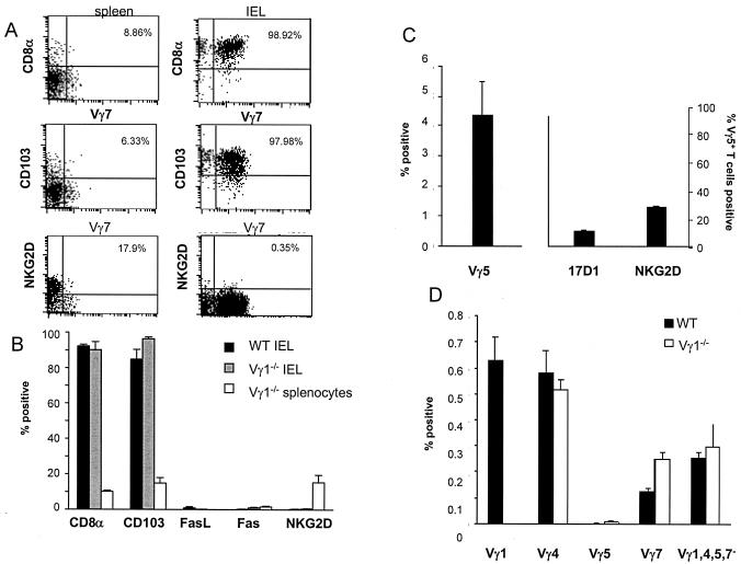
The relationship between Listeria-elicited Vγ5+ and Vγ7+ T cells in Vγ1−/− mice and γδ DETCs and iIELs in wild-type mice was further investigated by comparing their phenotypes by using a panel of antibodies that distinguish iIELs and DETCs. Whereas iIELs from both WT and Vγ1−/− mice characteristically express a CD8α homodimer and the αEβ7 integrin CD103 (27, 28), Vγ7+ T cells from spleens of Listeria-infected Vγ1−/− mice expressed very low levels of CD8α and CD103 (Fig. 4A). By contrast, expression of the killing activatory receptor NKG2D, which is generally very low in iIELs (24), was substantially higher among spleen Vγ7+ T cells from Listeria-infected Vγ1−/− mice (Fig. 4B). Similarly to populations of Vγ7+ iIELs in both WT and Vγ1−/− mice, Listeria-elicited splenic Vγ7+ cells in Vγ1−/− mice did not appear to express Fas or FasL (Fig. 4B). On balance, therefore, these results suggest that Vγ7+ T cells present in the spleens of Listeria-infected Vγ1−/− mice are distinct from Vγ7+ iIELs.
FIG. 4.
Phenotype of Listeria-elicited γδ T-cell subsets in Vγ1−/− mice. (A and B) Splenocytes and iIELs from Vγ1−/− mice 8 days after Listeria infection and iIELs from WT mice were analyzed by flow cytometry for expression of cell surface antigens CD8α, CD103, FasL, Fas, and NKG2D. The dot plots in panel A are examples of individual staining profiles for positive populations showing percentages of double-positive cells from day 8 Vγ1−/− infected spleen (left) and day 0 WT IEL (right), and the bar graph in panel B summarizes the data obtained from the analysis of iIELs and splenocytes of eight mice. (C) Distribution (left) and phenotype (right) of Vγ5+ T cells in the spleens of Vγ1−/− mice 8 days postinfection with Listeria. The data show the mean values obtained from more than eight mice. (D) Distribution of different γδ T-cell subsets in the thymuses of adult Vγ1−/− mice 8 days after Listeria infection, as determined by flow cytometric analysis of more than five mice.
Although Vγ5+ T cells represented only a small proportion of splenocytes (<5%) in Listeria-infected Vγ1−/− mice, it was possible to compare their phenotype to that of Vγ5+ DETCs. Whereas Vγ5/Vδ1+ DETCs can be identified by reactivity with the anticlonotype antibody 17D1 (42), only a small proportion (<10%) of the Vγ5+ T cells present in the spleens of day 8 Listeria-infected Vγ1−/− mice were 17D1+ (Fig. 4C). This together with the nonoverlapping Vγ5 CDR3 sequences (Table 1) suggests that Listeria-elicited Vγ5+ cells in Vγ1−/− mice are not DETCs, with further evidence of this being provided by the expression of NKG2D by a significant proportion (∼35%) of these cells, which is normally found with all DETCs (24).
The possibility that the Listeria-elicited Vγ5+ and Vγ7+ T cells in Vγ1−/− mice were thymically derived was investigated by attempting to demonstrate their presence in the thymuses of Vγ1−/− mice. The thymuses of adult wild-type and Vγ1−/− mice contained a small population of Vγ7+ cells but very few if any Vγ5+ cells (Fig. 4D). This raises the possibility that the source of the unusual Vγ7+ T cells present in the spleens of infected Vγ1−/− mice may be the thymus.
Pathogen-elicited γδ subsets in Vγ1−/− mice produce anti-inflammatory cytokines but play no role in macrophage homeostasis.
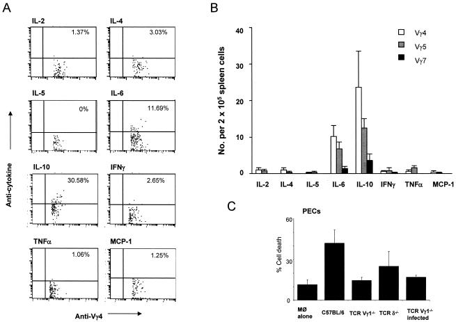
During the late stage of Listeria infection, Vγ1+ cells are a major source of γδ T-cell-derived cytokines (IL-10, IL-6, and IL-2) (2) and via a Fas-FasL mechanism eliminate pathogen-elicited macrophages (10). We therefore determined whether the Vγ5+, Vγ6+, Vγ7+, and Vγ4+ T cells recruited to sites of infection in the absence of Vγ1+ T cells in Vγ1−/− mice could compensate for or replace Vγ1+ T-cell function. The only cytokines synthesized by Vγ4, Vγ5, or Vγ7 subsets during the late stage of the infection in Vγ1−/− mice were IL-10, IL-2, and IL-6 (summarized in Fig. 5B, with representative plots shown in Fig. 5A), which is similar to the profile of cytokines produced by Vγ1+ T cells in response to Listeria infection (2). Among Vγ4+ T cells, IL-10 was the most prominent cytokine synthesized, with approximately 20% of cells being positive and with a smaller proportion (∼10%) synthesizing IL-6. This pattern of expression was similar to that of Vγ5+ cells, though a small number of Vγ5+ cells also produced tumor necrosis factor alpha. Together, these two subsets of γδ T cells might compensate for the absence of anti-inflammatory Vγ1+ cells that display a similar profile of cytokine production late during the course of Listeria infection in wild-type mice (2). Of note was the finding that Vγ7+ T cells do not appear to produce any of the cytokines analyzed (Fig. 5A), and they did not express either Fas or FasL (Fig. 4B).
FIG. 5.
Functional properties of γδ T cells responding to Listeria infection in Vγ1−/− mice. Cytokine synthesis by Vγ4+, Vγ5+, and Vγ7+ T cells elicited in response to Listeria infection was determined by staining splenocytes with TCRγδ-, CD3-, and TCR-Vγ-specific antibodies in conjunction with anticytokine antibodies and flow cytometric analysis as described in Materials and Methods. Representative dot plots are shown (A), with percentages of Vγ4 T cells positive for each marker indicated, and results for Vγ4, Vγ5, and Vγ7 subsets were compiled from three independent experiments (B). The ability of splenocytes from Listeria-infected Vγ1−/− mice (TCR-Vγ1−/− infected) enriched for Vγ4+, Vγ5+, Vγ6+, and Vγ7+ T cells to kill target peritoneal macrophage obtained from day 8 Listeria-infected TCRδ−/− mice was determined using a fluorescent-based cytotoxicity assay as described in Materials and Methods (C). As controls, target PECs were cultured alone (Mφ alone), and splenocytes from noninfected wild-type (C57BL/6), Vγ1−/−, or TCRδ−/− mice were used as additional sources of effector cells. IFNγ, gamma interferon; TNFα, tumor necrosis factor alpha; MCP-1, monocyte chemoattractant protein 1; No., number.
In contrast to the nonredundant cytokine production by Listeria-elicited Vγ1+ cells, there was no evidence of any cytotoxic activity or killing of pathogen-elicited macrophages by splenocytes from day 8 Listeria-infected Vγ1−/− mice (Fig. 5B). As seen previously (9, 10), macrophage cytocidal activity was restricted to splenocytes containing γδ T cells (wild-type mice) and was absent in mice deficient in all γδ T cells (TCRδ−/−). Killing was also absent in cells from the spleens of both noninfected and Listeria-infected Vγ1−/− mice by use of peritoneal macrophages from Listeria-infected TCRδ−/− mice as target cells. These findings are consistent with macrophage cytocidal activity being a unique property of Vγ1+ T cells.
DISCUSSION
Although γδ T cells are a common feature of many pathogen-induced immune responses, the factors that influence, promote, or regulate the response of individual populations of γδ T cells to infection is largely unknown. In addition to providing more evidence of the immunoregulatory properties of the Vγ1+ T-cell population of γδ T cells, the study described here provides evidence for their involvement in regulating the response of other, novel γδ T-cell populations to microbial infection. While the absence of Vγ1+ T cells has little impact on γδ T-cell repertoires in primary lymphoid tissues of naïve, noninfected, adult mice, their absence has a profound effect on γδ T cells during the course of infection. This is characterized by the appearance of γδ T cells expressing Vγ-encoded TCRs normally found among epithelium-associated γδ T cells (Vγ5, Vγ6, and Vγ7), although detailed structural analyses of their Vγ chains have shown that most of them are unique and distinct from epithelium-associated γδ T cells.
The expression of Vγ5-encoded TCRs has been reported only for the neonatal thymuses and skin of adult mice (34). DETC Vγ5 transcripts are invariant and are generated only in the fetal thymus, from which they migrate early as a single wave to populate the epidermis (15). The majority of Vγ5 CDR3 sequences expressed in the spleens of infected Vγ1−/− mice are by comparison noncanonical and display a degree of N-region diversity, which would suggest they are generated later. These cells can be further distinguished from DETCs by the absence of expression of the DETC-associated integrin CD103, required for epithelial localization (28), their lack of reactivity with a DETC Vγ5/Vδ1 TCR anticlonotype-specific antibody (17D1), and low to moderate levels of expression of the killing receptor NKG2D, which is generally expressed by the majority of tissue-associated γδ cells (24). The inability to detect any Vγ5+ T cells in thymuses of 6- to 8-week-old Vγ1−/− mice that express noncanonical sequences suggests that these cells either are of extrathymic origin or represent a population of Vγ5+ cells which leaves the thymus at other times during thymic development and after DETC progenitors have left to occupy an as-yet-unidentified tissue.
Vγ6 cells expressing an invariant γ-chain are usually associated with the epithelia of the reproductive tract and tongue (22). They have also been identified at sites of inflammation, among Vγ6+ hybridomas generated from Listeria-evoked and autoimmune orchiditis (32), and from the livers and kidneys of Listeria-infected wild-type mice (20, 37). Structural analyses of Listeria-elicited Vγ6+ T cells in Vγ1−/− mice revealed that these cells express a single, fetal-type, invariant Vγ6 chain, consistent with them being related to and perhaps being mobilized from the tissues in which cells bearing these TCRs normally reside. The absence of a specific monoclonal antibody for this receptor restricted any further phenotypic characterization and analysis of the potential functional significance of these cells.
Vγ7+ T cells form the major component of the iIELs compartment in the small intestine and have been shown to express high levels of the CD8αα homodimer (27). As shown by the expression of NKG2D and the absence of CD8αα expression, Vγ7+ cells found in the spleens of day 8 Listeria-infected Vγ1−/− mice are distinct from Vγ7+ iIELs, which show similar expression patterns with both WT and Vγ1−/− mice. The possibility remains, however, that the cells found in spleens of infected Vγ1−/− mice are related to the CD8αα− fraction of the gut-associated iIELs. Whatever their relationship to Vγ7+ iIELs, their presence in the thymus suggests they may be thymic in origin.
The functional properties of these unusual populations of γδ T cells in the spleens of Vγ1−/− mice are unclear, although these cells do appear to be distinct from the known effector functions of epithelium-associated γδ T cells that express the same Vγ chains. Their cytokine profile is different from that of iIELs or DETCs, and they do not appear to be cytotoxic, which is a characteristic feature of both DETCs and iIELs. This does not, however, exclude the possibility that these unusual Vγ5+ and Vγ7+ T cells produce cytokines other than those assayed for here and/or possess cytotoxic activity that is directed at target cells other than peritoneal macrophages. The mechanism by which these unusual γδ T-cell populations are recruited to the spleens of Vγ1−/− mice during the late phase of infection with Listeria is also not known. Since these mice can resolve bacterial infection in the absence of any tissue pathology and liver necrosis (2), the alteration in γδ T-cell homeostasis seen with these animals may be a result of the failure of Vγ1−/− mice to eliminate activated macrophages which can elicit (e.g., via specific chemokines) the recruitment of γδ T cells. Alternatively, it may be a direct consequence of the absence of Vγ1+ T cells, which would normally restrict or prevent the mobilization of other γδ T-cell populations to the spleen. The recent description of γδ T-cell homeostasis being controlled in part by γδ T-cell-specific factors (4) is consistent with this interpretation. In characterizing the aberrant recruitment of γδ subtypes following Listeria infection, we have shown a central, nonredundant role for Vγ1 cells in γδ T-cell recruitment, although the mechanism by which this is achieved requires further study.
Editor: J. D. Clements
REFERENCES
- 1.Andrew, E. M., and S. R. Carding. 2005. Murine gammadelta T cells in infections: beneficial or deleterious? Microbes Infect. 7:529-536. [DOI] [PubMed] [Google Scholar]
- 2.Andrew, E. M., D. J. Newton, J. E. Dalton, C. E. Egan, S. J. Goodwin, D. Tramonti, P. Scott, and S. R. Carding. 2005. Delineation of the function of a major gamma delta T cell subset during infection. J. Immunol. 175:1741-1750. [DOI] [PubMed] [Google Scholar]
- 3.Asarnow, D. M., W. A. Kuziel, M. Bonyhadi, R. E. Tigelaar, P. W. Tucker, and J. P. Allison. 1988. Limited diversity of gamma delta antigen receptor genes of Thy-1+ dendritic epidermal cells. Cell 55:837-847. [DOI] [PubMed] [Google Scholar]
- 4.Baccala, R., D. Witherden, R. Gonzalez-Quintial, W. Dummer, C. D. Surh, W. L. Havran, and A. N. Theofilopoulos. 2005. Gamma delta T cell homeostasis is controlled by IL-7 and IL-15 together with subset-specific factors. J. Immunol. 174:4606-4612. [DOI] [PubMed] [Google Scholar]
- 5.Bandeira, A., S. Itohara, M. Bonneville, O. Burlen-Defranoux, T. Mota-Santos, A. Coutinho, and S. Tonegawa. 1991. Extrathymic origin of intestinal intraepithelial lymphocytes bearing T-cell antigen receptor gamma delta. Proc. Natl. Acad. Sci. USA 88:43-47. [DOI] [PMC free article] [PubMed] [Google Scholar]
- 6.Belles, C., A. K. Kuhl, A. J. Donoghue, Y. Sano, R. L. O'Brien, W. Born, K. Bottomly, and S. R. Carding. 1996. Bias in the gamma delta T cell response to Listeria monocytogenes. V delta 6.3+ cells are a major component of the gamma delta T cell response to Listeria monocytogenes. J. Immunol. 156:4280-4289. [PubMed] [Google Scholar]
- 7.Carding, S. R., and P. J. Egan. 2000. The importance of gamma delta T cells in the resolution of pathogen-induced inflammatory immune responses. Immunol. Rev. 173:98-108. [DOI] [PubMed] [Google Scholar]
- 8.Carding, S. R., and P. J. Egan. 2002. Gammadelta T cells: functional plasticity and heterogeneity. Nat. Rev. Immunol. 2:336-345. [DOI] [PubMed] [Google Scholar]
- 9.Dalton, J. E., J. Pearson, P. Scott, and S. R. Carding. 2003. The interaction of gamma delta T cells with activated macrophages is a property of the V gamma 1 subset. J. Immunol. 171:6488-6494. [DOI] [PubMed] [Google Scholar]
- 10.Dalton, J. E., G. Howell, J. Pearson, P. Scott, and S. R. Carding. 2004. Fas-Fas ligand interactions are essential for the binding to and killing of activated macrophages by gamma delta T cells. J. Immunol. 173:3660-3667. [DOI] [PubMed] [Google Scholar]
- 11.D'Souza, C. D., A. M. Cooper, A. A. Frank, R. J. Mazzaccaro, B. R. Bloom, and I. M. Orme. 1997. An anti-inflammatory role for gamma delta T lymphocytes in acquired immunity to Mycobacterium tuberculosis. J. Immunol. 158:1217-1221. [PubMed] [Google Scholar]
- 12.Egan, P. J., and S. R. Carding. 2000. Downmodulation of the inflammatory response to bacterial infection by gammadelta T cells cytotoxic for activated macrophages. J. Exp. Med. 191:2145-2158. [DOI] [PMC free article] [PubMed] [Google Scholar]
- 13.Ferrick, D. A., M. D. Schrenzel, T. Mulvania, B. Hsieh, W. G. Ferlin, and H. Lepper. 1995. Differential production of interferon-gamma and interleukin-4 in response to Th1- and Th2-stimulating pathogens by gamma delta T cells in vivo. Nature 373:255-257. [DOI] [PubMed] [Google Scholar]
- 14.Fu, Y. X., C. E. Roark, K. Kelly, D. Drevets, P. Campbell, R. O'Brien, and W. Born. 1994. Immune protection and control of inflammatory tissue necrosis by gamma delta T cells. J. Immunol. 153:3101-3115. [PubMed] [Google Scholar]
- 15.Havran, W. L., and J. P. Allison. 1988. Developmentally ordered appearance of thymocytes expressing different T-cell antigen receptors. Nature 335:443-445. [DOI] [PubMed] [Google Scholar]
- 16.Heilig, J. S., and S. Tonegawa. 1986. Diversity of murine gamma genes and expression in fetal and adult T lymphocytes. Nature 322:836-840. [DOI] [PubMed] [Google Scholar]
- 17.Hiromatsu, K., G. Matsuzaki, Y. Tauchi, Y. Yoshikai, and K. Nomoto. 1992. Sequential analysis of T cells in the liver during murine listerial infection. J. Immunol. 149:568-573. [PubMed] [Google Scholar]
- 18.Hiromatsu, K., Y. Yoshikai, G. Matsuzaki, S. Ohga, K. Muramori, K. Matsumoto, J. A. Bluestone, and K. Nomoto. 1992. A protective role of gamma/delta T cells in primary infection with Listeria monocytogenes in mice. J. Exp. Med. 175:49-56. [DOI] [PMC free article] [PubMed] [Google Scholar]
- 19.Hsieh, B., M. D. Schrenzel, T. Mulvania, H. D. Lepper, L. Molfetto-Landon, and D. A. Ferrick. 1996. In vivo cytokine production in murine listeriosis. Evidence for immunoregulation by gamma delta+ T cells. J. Immunol. 156:232-237. [PubMed] [Google Scholar]
- 20.Ikebe, H., H. Yamada, M. Nomoto, H. Takimoto, T. Nakamura, K. H. Sonoda, and K. Nomoto. 2001. Persistent infection with Listeria monocytogenes in the kidney induces anti-inflammatory invariant fetal-type gammadelta T cells. Immunology 102:94-102. [DOI] [PMC free article] [PubMed] [Google Scholar]
- 21.Ito, K., M. Bonneville, Y. Takagaki, N. Nakanishi, O. Kanagawa, E. G. Krecko, and S. Tonegawa. 1989. Different gamma delta T-cell receptors are expressed on thymocytes at different stages of development. Proc. Natl. Acad. Sci. USA 86:631-635. [DOI] [PMC free article] [PubMed] [Google Scholar]
- 22.Itohara, S., A. G. Farr, J. J. Lafaille, M. Bonneville, Y. Takagaki, W. Haas, and S. Tonegawa. 1990. Homing of a gamma delta thymocyte subset with homogeneous T-cell receptors to mucosal epithelia. Nature 343:754-757. [DOI] [PubMed] [Google Scholar]
- 23.Itohara, S., N. Nakanishi, O. Kanagawa, R. Kubo, and S. Tonegawa. 1989. Monoclonal antibodies specific to native murine T-cell receptor gamma delta: analysis of gamma delta T cells during thymic ontogeny and in peripheral lymphoid organs. Proc. Natl. Acad. Sci. USA 86:5094-5098. [DOI] [PMC free article] [PubMed] [Google Scholar]
- 24.Jamieson, A. M., A. Diefenbach, C. W. McMahon, N. Xiong, J. R. Carlyle, and D. H. Raulet. 2002. The role of the NKG2D immunoreceptor in immune cell activation and natural killing. Immunity 17:19-29. [DOI] [PubMed] [Google Scholar]
- 25.Kruisbeek, A. M. 2000. Isolation and fractionation of mononuclear cell populations, p. 311-315. In J. E. Coligan, A. M. Kruisbeek, D. H. Marguiles, E. M. Shevach, and W. Strober (ed.), Current protocols in immunology. John Wiley & Sons, Inc., New York, N.Y.
- 26.Lafaille, J. J., A. DeCloux, M. Bonneville, Y. Takagaki, and S. Tonegawa. 1989. Junctional sequences of T cell receptor gamma delta genes: implications for gamma delta T cell lineages and for a novel intermediate of V-(D)-J joining. Cell 59:859-870. [DOI] [PubMed] [Google Scholar]
- 27.LeFrancois, L. 1991. Intraepithelial lymphocytes of the intestinal mucosa: curiouser and curiouser. Semin. Immunol. 3:99-108. [PubMed] [Google Scholar]
- 28.LeFrancois, L., T. A. Barrett, W. L. Havran, and L. Puddington. 1994. Developmental expression of the alpha IEL beta 7 integrin on T cell receptor gamma delta and T cell receptor alpha beta T cells. Eur. J. Immunol. 24:635-640. [DOI] [PubMed] [Google Scholar]
- 29.LeFrancois, L., R. LeCorre, J. Mayo, J. A. Bluestone, and T. Goodman. 1990. Extrathymic selection of TCR gamma delta + T cells by class II major histocompatibility complex molecules. Cell 63:333-340. [DOI] [PubMed] [Google Scholar]
- 30.Mombaerts, P., J. Arnoldi, F. Russ, S. Tonegawa, and S. H. Kaufmann. 1993. Different roles of alpha beta and gamma delta T cells in immunity against an intracellular bacterial pathogen. Nature 365:53-56. [DOI] [PubMed] [Google Scholar]
- 31.Moretto, M., B. Durell, J. D. Schwartzman, and I. A. Khan. 2001. Gamma delta T cell-deficient mice have a down-regulated CD8+ T cell immune response against Encephalitozoon cuniculi infection. J. Immunol. 166:7389-7397. [DOI] [PubMed] [Google Scholar]
- 32.Mukasa, A., M. Lahn, E. K. Pflum, W. Born, and R. L. O'Brien. 1997. Evidence that the same gamma delta T cells respond during infection-induced and autoimmune inflammation. J. Immunol. 159:5787-5794. [PubMed] [Google Scholar]
- 33.Ohga, S., Y. Yoshikai, Y. Takeda, K. Hiromatsu, and K. Nomoto. 1990. Sequential appearance of gamma/delta- and alpha/beta-bearing T cells in the peritoneal cavity during an i.p. infection with Listeria monocytogenes. Eur. J. Immunol. 20:533-538. [DOI] [PubMed] [Google Scholar]
- 34.Payer, E., A. Elbe, and G. Stingl. 1991. Circulating CD3+/T cell receptor V gamma 3+ fetal murine thymocytes home to the skin and give rise to proliferating dendritic epidermal T cells. J. Immunol. 146:2536-2543. [PubMed] [Google Scholar]
- 35.Pereira, P., D. Gerber, S. Y. Huang, and S. Tonegawa. 1995. Ontogenic development and tissue distribution of V gamma 1-expressing gamma/delta T lymphocytes in normal mice. J. Exp. Med. 182:1921-1930. [DOI] [PMC free article] [PubMed] [Google Scholar]
- 36.Pereira, P., D. Gerber, A. Regnault, S. Y. Huang, V. Hermitte, A. Coutinho, and S. Tonegawa. 1996. Rearrangement and expression of Vzeta1, Vzeta2 and Vzeta3 TCR zeta genes in C57BL/6 mice. Int. Immunol. 8:83-90. [DOI] [PubMed] [Google Scholar]
- 37.Roark, C. E., M. K. Vollmer, P. A. Campbell, W. K. Born, and R. L. O'Brien. 1996. Response of a gamma delta+ T cell receptor invariant subset during bacterial infection. J. Immunol. 156:2214-2220. [PubMed] [Google Scholar]
- 38.Roberts, S. J., A. L. Smith, A. B. West, L. Wen, R. C. Findly, M. J. Owen, and A. C. Hayday. 1996. T-cell αβ+ and γδ+ deficient mice display abnormal but distinct phenotypes toward a natural, widespread infection of the intestinal epithelium. Proc. Natl. Acad. Sci. USA 93:11774-11779. [DOI] [PMC free article] [PubMed] [Google Scholar]
- 39.Seixas, E. M., and J. Langhorne. 1999. gammadelta T cells contribute to control of chronic parasitemia in Plasmodium chabaudi infections in mice. J. Immunol. 162:2837-2841. [PubMed] [Google Scholar]
- 40.Selin, L. K., P. A. Santolucito, A. K. Pinto, E. Szomolanyi-Tsuda, and R. M. Welsh. 2001. Innate immunity to viruses: control of vaccinia virus infection by gamma delta T cells. J. Immunol. 166:6784-6794. [DOI] [PubMed] [Google Scholar]
- 41.Takagaki, Y., N. Nakanishi, I. Ishida, O. Kanagawa, and S. Tonegawa. 1989. T cell receptor-gamma and -delta genes preferentially utilized by adult thymocytes for the surface expression. J. Immunol. 142:2112-2121. [PubMed] [Google Scholar]
- 42.Tigelaar, R. E., and J. M. Lewis. 1995. Immunobiology of mouse dendritic epidermal T cells: a decade later, some answers, but still more questions. J. Investig. Dermatol. 105:43S-49S. [DOI] [PubMed] [Google Scholar]
- 43.Wallace, M., M. Malkovsky, and S. R. Carding. 1995. Gamma/delta T lymphocytes in viral infections. J. Leukoc. Biol. 58:277-283. [DOI] [PubMed] [Google Scholar]
- 44.Wang, T., E. Scully, Z. Yin, J. H. Kim, S. Wang, J. Yan, M. Mamula, J. F. Anderson, J. Craft, and E. Fikrig. 2003. IFN-gamma-producing gamma delta T cells help control murine West Nile virus infection. J. Immunol. 171:2524-2531. [DOI] [PubMed] [Google Scholar]


