Abstract
Epizootic subtype IAB and IC Venezuelan equine encephalitis viruses (VEEV) readily infect the epizootic mosquito vector Aedes taeniorhynchus. The inability of enzootic subtype IE viruses to infect this mosquito species provides a model system for the identification of natural viral determinants of vector infectivity. To map mosquito infection determinants, reciprocal chimeric viruses generated from epizootic subtype IAB and enzootic IE VEEV were tested for mosquito infectivity. Chimeras containing the IAB epizootic structural gene region and, more specifically, the IAB PE2 envelope glycoprotein E2 precursor gene demonstrated an efficient infection phenotype. Introduction of the PE2 gene from an enzootic subtype ID virus into an epizootic IAB or IC genetic backbone resulted in lower infection rates than those of the epizootic parent. The finding that the E2 envelope glycoprotein, the site of epitopes that define the enzootic and epizootic subtypes, also encodes mosquito infection determinants suggests that selection for efficient infection of epizootic mosquito vectors may mediate VEE emergence.
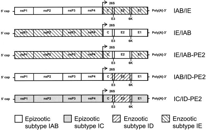
Venezuelan equine encephalitis viruses (VEEV; Togaviridae family, Alphavirus genus) are single-stranded, enveloped, message-sense RNA viruses with genomes containing approximately 11,400 nucleotides (5, 9). Structural genes found in a 26S subgenomic message encode the capsid protein and the E2 and E1 envelope glycoproteins. During virus maturation, a PE2 precursor protein is cleaved into the E3 and E2 proteins; the E2 protein forms spikes on the surface of the virion (16), while the E1 protein lies parallel to the envelope (12). E2 is the site of the major antigenic determinants and probably interacts with cellular receptors. The nonstructural proteins responsible for viral replication are encoded by the nsP1 through nsP4 genes found in the 5′ two-thirds of the genomic RNA (9) (Fig. 1).
FIG. 1.
Genetic organization of chimeric VEEV constructs used for experimental mosquito infections.
VEEV occur in two epidemiological settings: (i) a continuous, sylvatic enzootic cycle maintained between Culex (Melanoconion) spp. mosquito vectors and rodent reservoir hosts and (ii) an epidemic and/or epizootic cycle that involves several different mammalophilic mosquito species and exploits equines as highly efficient amplification hosts, resulting in extensive equine and human disease (29, 32). Aedes (Ochlerotatus) taeniorhynchus (Reinert [18a] proposed that the subgenus Ochlerotaus be elevated to generic status; we do not believe that this change is justified until a more rigorous analysis is conducted) has been implicated as an epizootic vector during several VEE outbreaks (20, 23, 32, 34).
Of the six VEE antigenic subtypes (I through VI), only IAB and IC are etiologic agents of epidemics and/or epizootics because they generate equine viremia sufficient for efficient amplification (29, 32). Phylogenetic analyses indicate that epizootic VEEV have emerged repeatedly through mutations in enzootic subtype ID VEEV progenitors that enhance equine replication (18, 32). The recent isolation from encephalitic horses during Mexican epizootics of subtype IE VEEV indicates that this subtype may also have epizootic potential (14). Mutations in the E2 glycoprotein that increase positive charge on the virion surface are putative determinants of the phenotypic changes that accompany all of these VEE emergences (3).
Adaptation to equine replication clearly accompanies the emergence of epizootic VEEV from enzootic progenitors, which replicate poorly in equine hosts (8, 13, 28, 31). However, adaptation to epizootic mosquito vectors like A. taeniorhynchus may also mediate epizootic emergence (32). Previous experiments demonstrating that epizootic subtype IAB (11, 26) and IC (25) VEEV readily infect the epizootic mosquito vector A. taeniorhynchus, while subtype IE VEEV fails to efficiently infect this mosquito species (11), support this hypothesis. However, the IE viruses are not direct ancestors of the epizootic IAB and IC strains, so adaptation is not implied by these previous findings.
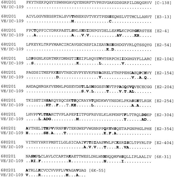
To further test the hypothesis that VEEV adapts to epizootic mosquito vectors and to investigate the viral genetic determinants of adaptation, previously (10, 17) and newly constructed infectious cDNA clones were used to generate chimeric VEEV containing genes from both enzootic and epizootic strains. Structural and nonstructural chimeras (Fig. 1) between the epizootic IAB strain Trinidad donkey (10) and the enzootic subtype IE Guatemalan strain 68U201 were described previously (17). The IE/IAB-PE2 chimera (PE2 glycoprotein precursor gene of the IAB virus in the IE backbone; Fig. 1) was generated from pIE.AA by using SwaI and SgrAI sites at nucleotide positions 8139 and 10003, respectively. This fragment encoded the C-terminal 87 amino acids of the capsid protein, all of the E3 protein, all of the E2 glycoprotein, and the N-terminal 55 amino acids of the 6,000-kDa protein (6K protein) that, like E3, is not present in virions (Fig. 2). The missing SwaI site in VE/IC-109 was introduced using PCR mutagenesis.
FIG. 2.
Amino acid alignment of subtype IE (strain 68U201, clone pIE.AA) and IAB (strain Trinidad donkey, clone VE/IC-109) viral C-E3-E2-6K gene sequences used for chimera production. •, same amino acid as the 68U201 sequence. A total of 71 amino acids differ between the IE/IAB-PE2 construct and the IE.AA clone from which it was constructed. Numbers on the right indicate amino acid positions within the capsid or E3, E2, or 6K protein gene for the last residue on each line.
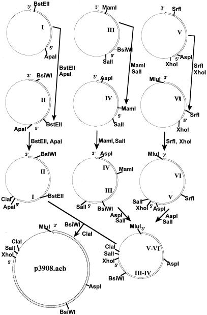
An epizootic VEEV subtype IC human isolate, strain 3908 (34), was chosen for construction of the new clone p3908.acb because of its low passage history (single C6/36 mosquito cell passage) and its available genomic sequence (4). PCR amplification of six overlapping genome regions was performed using Pfu Turbo high fidelity polymerase (Stratagene, La Jolla, Calif.) as previously described (4). All amplicons were blunt-end ligated into EcoRV-pBS-SKII(+) (Stratagene, La Jolla, Calif.). Incorporation of strain 3908 amplicons was performed in a 3′ to 5′ antisense orientation (in relation to the viral genome and T7 promoter) through the digestion of vector sequences with the corresponding unique overlapping viral and pBluescript II SK(+) restriction sites (Fig. 3). The production of chimeras IC/ID-PE2 and IAB/ID-PE2 (Fig. 1) involved cloning the PE2 gene of enzootic subtype ID strain 66637 (30) into the IAB or IC backbone (pVE/IC-109 and p3908.acb, respectively) by using AflII and SrfI (positions 8876 and 9806, respectively).
FIG. 3.
Construction of infectious cDNA clone p3908.acb from VEEV subtype IC strain 3908. All six amplicons generated by reverse transcsription-PCR were ligated into the pBS-SKII shuttle plasmid. The full-length clone was constructed by the addition of subclones to the 3′-most fragment, using a 3′ conserved unique restriction site and a unique site found within the pBS plasmid at the 5′ end.
Clones were linearized with MluI, and in vitro transcription was performed using standard conditions (17). RNA was electroporated into BHK-21 cells, and cultures were harvested 48 to 72 h later when cytopathic effect was evident. Virus titers were determined by plaque assay on Vero cells. The antigenic subtype of viruses was confirmed by an immunofluorescence assay with IAB/IC-specific (1A3A-5) and ID/IE-specific (1A1B-9) monoclonal antibodies as described previously (21). Immunofluorescence results have been described previously for the IE and IAB reciprocal chimeras (17). The IE/IAB-PE2 chimera had an IAB/IC-specific antibody (1A3A-5) reactivity only. Virus from the IC clone (p3908acb) was also reactive with the IAB/IC-specific antibody (1A3A-5) only (data not shown). Conversely, IAB/ID-PE2 and IC/ID-PE2 were reactive with the ID/IE-specific antibody 1A1B-9 and were negative with the IAB/IC-specific antibody 1A3A-5, confirming the expected antigenic phenotypes based on the E2 sequence content.
To confirm that all rescued clones maintained their wild-type mouse-virulent phenotype, 1,000 PFU of virus from clones 3908.acb, IC/ID-PE2, and IAB/ID-PE2, as well as virus strains IC-3908, ID-66637, and IAB-TRD, was subcutaneously inoculated into (n = 6) 12-week-old Swiss NIH mice. The wild-type mouse virulence of virus rescued from the IAB clone VEE/IC-109 has been described previously (10), as well as that of the IAB/IE and IE/IAB chimeras (17). The IC and IE viruses rescued from clones as well as all recombinant viruses were also virulent for mice; the IC (p3908.acb) clone-derived virus resulted in paralysis and death of mice within 5 to 7 days, and the two chimeras resulted in paralysis and death on days 6 to 9. The average survival times for mice infected with the p3908.acb were 5.6 ± 1.1 days and 7.2 ± 1.3 and 7.5 ± 1.4 days for the IAB/ID-PE2 and IC/ID-PE2 chimeras, respectively. All viruses yielded titers similar to those of the parent viruses in a variety of cell cultures (data not shown).
A. taeniorhynchus mosquitoes were collected in Galveston, Tex.(latitude, N 29° 13′ 28″; longitude, W 94° 56′ 63″), and F1 offspring were reared under standard conditions (6). Hanging blood droplets were prepared from 0.33 volume of packed sheep erythrocytes, 33% fetal bovine serum, and approximately 7 log10 PFU of recombinant virus, and mosquitoes were allowed to feed at room temperature for 1 h. Engorged females were incubated at 27°C for 14 days and then were assayed for cytopathic effect on BHK-21 cells, followed by plaque assay on Vero cells. A chi-square (χ2) 2-way test of independence analysis was used with 1 degree of freedom to determine statistical differences between infection rates among cohorts; significance was assigned at P ≤ 0.1, and infection rates for mosquito cohorts infected with epizootic strains were generally compared with those of equal- or higher-dose cohorts for enzootic strains to interpret the results in a conservative manner. Titers of viruses from infected mosquitoes did not differ significantly between parental IE and IAB viruses and therefore were not measured for any of the chimeric viruses.
The IAB/IE chimeric virus containing the structural genes from subtype IE strain 68U201 infected 16% of A. taeniorhynchus exposed to 6.5 log10 PFU/ml of blood meal (Table 1). This rate was similar to that of the 68U201 parental virus. The mosquito infection rate for the IE/IAB chimera was 56% with 6.9 log10 PFU/ml of blood meal (Table 1). This infection rate was higher (P < 0.1) than that of the 68U201 parental virus, even though 68U201 had a higher blood meal infectivity of 7.3 log10 PFU/ml. In addition, the IE/IAB-PE2 construct had a higher (P < 0.1) infection rate (54% at 6.8 log10 PFU/ml of blood meal) than the IE parent (Table 1).
TABLE 1.
Infection rates for parental and chimeric VEEV in A. taeniorhynchus mosquitoes
| Virus | Antigenic subtype | Blood meal titer (log10 PFU/ml on Vero cells) | Fraction of mosquitoes infected | % Mosquitoes infected |
|---|---|---|---|---|
| IE.AA (IE parent) | IE | 4.3 | 0/19 | 0 |
| 5.9 | 0/9 | 0 | ||
| 7.3 | 2/21 | 10 | ||
| 9.1 | 7/33 | 21 | ||
| VE/IC-109 (IAB parent) | IAB | 4.2 | 0/16 | 0 |
| 5.9 | 5/16 | 31 | ||
| 7.7 | 20/30 | 67 | ||
| IE/IAB | IAB | 6.9 | 14/25 | 56 |
| IAB/IE | IE | 6.5 | 4/25 | 16 |
| IE/IAB-PE2 | IAB | 6.8 | 15/28 | 54 |
| 3908.acb (IC parent) | IC | 7.1 | 21/25 | 84 |
| 66637 (ID parent) | ID | 6.6 | 11/29 | 38 |
| IAB/ID-PE2 | ID | 6.8 | 5/16 | 31 |
| IC/ID-PE2 | ID | 7.0 | 4/11 | 36 |
Because subtype ID and IC VEEV represent ancestral and derived phenotypes related to VEE emergence (18, 19, 30), the role of their PE2 genes in infection of A. taeniorhynchus was also investigated. The epizootic IAB virus (infection rates of 31 and 67% at 5.9 and 7.7 log10 PFU/ml blood meal, respectively; Table 1) did not infect significantly (P = 0.12) more efficiently than subtype ID strain 66637 (38% at 6.6 log 10 PFU/ml blood meal) (Table 1) when the lower dosage rates were compared. Interestingly, the infection rate for theIAB/IDE2 construct did differ significantly (P = 0.1) from that of the IAB parent. The lack of statistical significance between parental IAB and ID viruses could be the result of small sample sizes or extensive cell culture passaging (6-Vero and 1-BHK) of the IAB virus used to generate the VEE/IC-109 clone; cell culture passage is known to reduce mosquito infectivity by subtype IAB VEEV (27). In contrast, virus from the subtype IC clone p3908acb (generated from a virus that had been passaged only once in mosquito cells) infected mosquitoes at a higher (P = 0.08) rate, 84% at 7.1 log10 PFU/ml (Table 1), than the enzootic ID virus, also of low passage (once in suckling mice and once in Vero cells). The IAB/ID-PE2 chimera infected 31% of mosquitoes exposed to 6.8 log10 PFU/ml. The VEE/IC-109 IAB parental clone infected 67% of mosquitoes exposed to 7.7 log10 PFU/ml. Virus from the IC/ID-PE2 construct demonstrated a 48% reduction (P < 0.1) in infection compared to that of its parental IC virus; the IC/ID-PE2 infection rate was 36% at 7.0 log 10 PFU/ml of blood meal compared to the rate for parental IC virus (84% at 7.1 log10 PFU/ml).
Subtype IE strains differ from subtype IAB, IC, and ID VEEV by approximately 11% of their amino acid sequences, precluding analysis and discussion of potential amino acid determinants of vector infection. Among the more closely related subtype I viruses, there was a total of nine E2 glycoprotein-deduced amino acid differences between IAB and IC viruses (Table 2), with no net charge difference. E2 amino acid differences between the IAB or IC and ID VEEV were 11 and 8, respectively, with the IAB and IC strains exhibiting higher net charges of +3 and +2. One amino acid position (E2 position 117 [E2-117]) was different among the three subtypes (Asp [IAB], Gly [IC], and Asn [ID]). The IC virus, with the greatest mosquito infectivity, had unique E2 residues at positions 179 and 201. The E2-201 residue was positively charged in the IC virus and has been demonstrated through phylogenetic analysis to be associated with the 1992 IC epizootic emergence from an enzootic ID progenitor (30). No capsid but two E3 amino acids also differed among the IAB, IC, and ID strains, but only position 41 was consistent with respect to the mosquito infection phenotype (Ser for enzootic strain 66637 and Pro for the epizootic IAB and IC strains). These sequence comparisons imply that the E2 glycoprotein differences responsible for enhanced mosquito infection by the epizootic strains may include charge differences on the surface of the virion that were previously implicated in VEE emergence (3).
TABLE 2.
Amino acid differences between the subtype IAB, IC, and ID PE2 chimerasa
| Nucleotide position | Amino acid position | VE/IC-109 (IAB) | p3908.acb (IC) | Strain 66637 (ID) |
|---|---|---|---|---|
| 8438 | E3-18 | Gln | Glu | Gln |
| 8507 | E3-41 | Pro | Pro | Ser |
| 8786 | E2-75 | Lys | Glu | Glu |
| 8912 | E2-117 | Asp | Gly | Asn |
| 8915 | E2-118 | Ser | Ser | Leu |
| 9002 | E2-147 | Val | Ala | Ala |
| 9098 | E2-179 | Val | Ile | Val |
| 9137 | E2-192 | Val | Val | Ala |
| 9164 | E2-201 | Glu | Lys | Glu |
| 9200 | E2-213 | Lys | Lys | Thr |
| 9203 | E2-214 | Thr | Ala | Ala |
| 9695 | E2-378 | Ala | Val | Val |
| 9734 | E2-391 | Arg | Lys | Lys |
| 9746 | E2-395 | Ala | Ser | Ser |
Boldface residues represent positively charged amino acids found in the epizootic strains but not in the enzootic subtype ID virus strain 66637.
Our results demonstrate that genetic determinants within the PE2 envelope glycoprotein precursor gene are responsible for the increased efficiency of VEEV infection of the epizootic mosquito vector, A. taeniorhynchus. Insertion of the PE2 gene from enzootic ID and IE strains into the highly infectious IC and to a lesser degree the IAB backbone reduced infection rates, demonstrating that the epizootic PE2 gene is necessary for efficient infection. More importantly, insertion of the IAB PE2 gene into the enzootic IE backbone significantly increased infectivity compared to that of the enzootic parent, demonstrating that the PE2 gene alone is sufficient to confer the epizootic high infectivity phenotype. Since the IAB, IC, and ID genome regions swapped do not include any capsid or E1 protein amino acid differences, the PE2 gene itself is probably responsible for most or all of the mosquito infection phenotype differences among the enzootic and epizootic strains we evaluated.
Subtype IAB epizootic VEEV strains efficiently infect and replicate in the midgut of the enzootic mosquito vector Culex (Melanoconion) taeniopus following intrathoracic inoculation but are incapable of efficient infection via the oral route (35). The previous data indicate that the mosquito infection block does not involve virion maturation, as would be expected if the E3 protein were involved. Although the E3 protein is not present in VEEV virions (9) and infection of the midgut reflects the initial interaction of the virus with the apical surface of epithelial cells (33), we cannot rule out an in vivo mosquito infection role for E3 amino acid substitutions. E3 protein mutations that affect E2 cleavage during polyprotein processing can affect infectivity for mosquito cells (7), suggesting their possible role in vector infection in vivo. More detailed mutagenesis experiments are needed to assess the role of specific proteins and amino acids as determinants of vector infection.
Our findings represent the first identification of natural vector infection determinants for an alphavirus. Previous experiments implicated envelope glycoproteins of LaCrosse and snowshoe hare viruses in Aedes triseriatus infection (1, 2, 24). Insertion of the 26S structural genome region from a dissemination-competent Malaysian Sindbis virus strain into an incompetent strain was sufficient to enhance dissemination from the midgut of Aedes aegypti (15). In addition, the use of a monoclonal antibody-resistant mutant VEEV demonstrated that a single amino acid substitution in the E2 glycoprotein could interfere with midgut infection of and dissemination within A. aegypti mosquitoes (36).
The results of these studies may have great epidemiological significance. We have hypothesized that adaptation of epizootic VEEV to epizootic mosquito vectors like A. taeniorhynchus contributes to VEE emergence by enhancing transmission among equines rather than or in addition to elevated equine viremia levels, as has been previously demonstrated (8, 13, 28, 31). Such adaptation to epizootic vectors could be responsible for a loss of fitness in the enzootic (ancestral) Culex (Melanoconion) vectors due to the host specificity of the adaptation. The lack of susceptibility to infection with epizootic VEEV of Culex (Melanoconion) taeniopus, the enzootic vector for VEE subtype IE viruses in Central America, has been implicated as a factor in the inability of subtype IAB viruses to persist in Central America after the 1969 outbreak (22). Although this vector is readily infected with subtype IE viruses, highly infectious blood meals of epizootic subtype IAB VEEV fail to efficiently establish midgut infections (35). Determination of infection rates for C. (Melanoconion) taeniopus and other enzootic VEEV vectors using these new infectious constructs could provide further evidence for this hypothesis.
Acknowledgments
We thank Richard Kinney and John Roehrig from the Centers for Disease Control and Prevention (Fort Collins, Colo.) for providing the pVE/IC-109 infectious cDNA clone and VEEV monoclonal antibodies, respectively.
A.C.B. and A.M.P. were supported by the James L. McLaughlin Infection and Immunity Fellowship Fund and by NIH Emerging and Tropical Diseases T32 Training grants AI-107526 and AI-07536. This research was supported by National Institutes of Health grants AI-39800, AI48807, and AI-10984 and by the National Aeronautics and Space Administration.
REFERENCES
- 1.Beaty, B. J., M. Holterman, W. Tabachnick, R. E. Shope, E. J. Rozhon, and D. H. Bishop. 1981. Molecular basis of bunyavirus transmission by mosquitoes: role of the middle-sized RNA segment. Science 211:1433-1435. [DOI] [PubMed] [Google Scholar]
- 2.Beaty, B. J., B. R. Miller, R. E. Shope, E. J. Rozhon, and D. H. Bishop. 1982. Molecular basis of bunyavirus per os infection of mosquitoes: role of the middle-sized RNA segment. Proc. Natl. Acad. Sci. USA 79:1295-1297. [DOI] [PMC free article] [PubMed] [Google Scholar]
- 3.Brault, A. C., A. M. Powers, E. C. Holmes, C. H. Woelk, and S. C. Weaver. 2002. Positively charged amino acid substitutions in the E2 envelope glycoprotein are associated with the emergence of Venezuelan equine encephalitis virus. J. Virol. 76:1718-1730. [DOI] [PMC free article] [PubMed] [Google Scholar]
- 4.Brault, A. C., A. M. Powers, G. Medina, E. Wang, W. Kang, R. A. Salas, J. De Siger, and S. C. Weaver. 2001. Potential sources of the 1995 Venezuelan equine encephalitis subtype IC epidemic. J. Virol. 75:5823-5832. [DOI] [PMC free article] [PubMed] [Google Scholar]
- 5.Calisher, C. H., and N. Karabatsos. 1988. Arbovirus serogroups: definition and geographic distribution, p. 19-57. In T. P. Monath (ed.), The arboviruses: epidemiology and ecology, vol. 1. CRC Press, Boca Raton, Fla. [Google Scholar]
- 6.Gerberg, E. J., D. R. Barnard, and R. A. Ward. 1994. Manual for mosquito rearing and experimental techniques, bulletin no. 5. American Mosquito Control Association, Lake Charles, La.
- 7.Heidner, H. W., T. A. Knott, and R. E. Johnston. 1996. Differential processing of Sindbis virus glycoprotein PE2 in cultured vertebrate and arthropod cells. J. Virol. 70:2069-2073. [DOI] [PMC free article] [PubMed] [Google Scholar]
- 8.Johnson, K. M., and D. H. Martin. 1974. Venezuelan equine encephalitis. Adv. Vet. Sci. Comp. Med. 18:79-116. [PubMed] [Google Scholar]
- 9.Johnston, R. E., and C. J. Peters. 1996. Alphaviruses, p. 843-898. In B. N. Fields, D. M. Knipe, and P. M. Howley (ed.), Virology, 3rd ed. Lippincott-Raven, New York, N.Y.
- 10.Kinney, R. M., G. J. Chang, K. R. Tsuchiya, J. M. Sneider, J. T. Roehrig, T. M. Woodward, and D. W. Trent. 1993. Attenuation of Venezuelan equine encephalitis virus strain TC-83 is encoded by the 5′-noncoding region and the E2 envelope glycoprotein. J. Virol. 67:1269-1277. [DOI] [PMC free article] [PubMed] [Google Scholar]
- 11.Kramer, L. D., and W. F. Scherer. 1976. Vector competence of mosquitoes as a marker to distinguish Central American and Mexican epizootic from enzootic strains of Venezuelan encephalitis virus. Am. J. Trop. Med. Hyg. 25:336-346. [DOI] [PubMed] [Google Scholar]
- 12.Lescar, J., A. Roussel, M. W. Wien, J. Navaza, S. D. Fuller, G. Wengler, and F. A. Rey. 2001. The Fusion glycoprotein shell of Semliki Forest virus: an icosahedral assembly primed for fusogenic activation at endosomal pH. Cell 105:137-148. [DOI] [PubMed] [Google Scholar]
- 13.Martin, D. H., W. H. Dietz, O. Alvaerez, Jr., and K. M. Johnson. 1982. Epidemiological significance of Venezuelan equine encephalomyelitis virus in vitro markers. Am. J. Trop. Med. Hyg. 31:561-568. [DOI] [PubMed] [Google Scholar]
- 14.Oberste, M. S., M. Fraire, R. Navarro, C. Zepeda, M. L. Zarate, G. V. Ludwig, J. F. Kondig, S. C. Weaver, J. F. Smith, and R. Rico-Hesse. 1998. Association of Venezuelan equine encephalitis virus subtype IE with two equine epizootics in Mexico. Am. J. Trop. Med. Hyg. 59:100-107. [DOI] [PubMed] [Google Scholar]
- 15.Olson, K. E., K. M. Myles, R. C. Seabaugh, S. Higgs, J. O. Carlson, and B. J. Beaty. 2000. Development of a Sindbis virus expression system that efficiently expresses green fluorescent protein in midguts of Aedes aegypti following per os infection. Insect Mol. Biol. 9:57-65. [DOI] [PubMed] [Google Scholar]
- 16.Pletnev, S. V., W. Zhang, S. Mukhopadhyay, B. R. Fisher, R. Hernandez, D. T. Brown, T. S. Baker, M. G. Rossmann, and R. J. Kuhn. 2001. Locations of carbohydrate sites on alphavirus glycoproteins show that E1 forms an icosahedral scaffold. Cell 105:127-136. [DOI] [PMC free article] [PubMed] [Google Scholar]
- 17.Powers, A. M., A. C. Brault, R. M. Kinney, and S. C. Weaver. 2000. The use of Chimeric Venezuelan equine encephalitis viruses as an approach for the molecular identification of natural virulence determinants. J. Virol. 74:4258-4263. [DOI] [PMC free article] [PubMed] [Google Scholar]
- 18.Powers, A. M., M. S. Oberste, A. C. Brault, R. Rico-Hesse, S. M. Schmura, J. F. Smith, W. Kang, W. P. Sweeney, and S. C. Weaver. 1997. Repeated emergence of epidemic/epizootic Venezuelan equine encephalitis from a single genotype of enzootic subtype ID virus. J. Virol. 71:6697-6705. [DOI] [PMC free article] [PubMed] [Google Scholar]
- 18a.Reinert, J. F. 2000. New classification for the composite genus Aedes (Diptera: Culicidae: Aedini), elevation of subgenus Ochlerotatus to generic rank, reclassification of the other subgenera, and notes on certain subgenera and species. J. Am. Mosq. Control Assoc. 16:175-188. [PubMed] [Google Scholar]
- 19.Rico-Hesse, R., S. C. Weaver, J. de Siger, G. Medina, and R. A. Salas. 1995. Emergence of a new epidemic/epizootic Venezuelan equine encephalitis virus in South America. Proc. Natl. Acad. Sci. USA 92:5278-5281. [DOI] [PMC free article] [PubMed] [Google Scholar]
- 20.Rivas, F., L. A. Diaz, V. M. Cardenas, E. Daza, L. Bruzon, A. Alcala, O. De la Hoz, F. M. Caceres, G. Aristizabal, J. W. Martinez, D. Revelo, F. De la Hoz, J. Boshell, T. Camacho, L. Calderon, V. A. Olano, L. I. Villarreal, D. Roselli, G. Alvarez, G. Ludwig, and T. Tsai. 1997. Epidemic Venezuelan equine encephalitis in La Guajira, Colombia, 1995. J. Infect. Dis. 175:828-832. [DOI] [PubMed] [Google Scholar]
- 21.Roehrig, J. T., and R. A. Bolin. 1997. Monoclonal antibodies capable of distinguishing epizootic from enzootic varieties of subtype I Venezuelan equine encephalitis viruses in a rapid indirect immunofluorescence assay. J. Clin. Microbiol. 35:1887-1890. [DOI] [PMC free article] [PubMed] [Google Scholar]
- 22.Scherer, W. F., S. C. Weaver, C. A. Taylor, and E. W. Cupp. 1986. Vector incompetency: its implication in the disappearance of epizootic Venezuelan equine encephalomyelitis virus from Middle America. J. Med. Entomol. 23:23-29. [DOI] [PubMed] [Google Scholar]
- 23.Sudia, W. D., V. F. Newhouse, and B. E. Henderson. 1971. Experimental infection of horses with three strains of Venezuelan equine encephalomyelitis virus. II. Experimental vector studies. Am. J. Epidemiol. 93:206-211. [DOI] [PubMed] [Google Scholar]
- 24.Sundin, D. R., B. J. Beaty, N. Nathanson, and F. Gonzalez-Scarano. 1987. A G1 glycoprotein epitope of La Crosse virus: a determinant of infection of Aedes triseriatus. Science 235:591-593. [DOI] [PubMed] [Google Scholar]
- 25.Turell, M. J. 1999. Vector competence of three Venezuelan mosquitoes (Diptera: Culicidae) for an epizootic IC strain of Venezuelan equine encephalitis virus. J. Med. Entomol. 36:407-409. [DOI] [PubMed] [Google Scholar]
- 26.Turell, M. J., G. V. Ludwig, and J. R. Beaman. 1992. Transmission of Venezuelan equine encephalomyelitis virus by Aedes sollicitans and Aedes taeniorhynchus (Diptera: Culicidae). J. Med. Entomol. 29:62-65. [DOI] [PubMed] [Google Scholar]
- 27.Turell, M. J., G. V. Ludwig, J. Kondig, and J. F. Smith. 1999. Limited potential for mosquito transmission of genetically engineered, live-attenuated Venezuelan equine encephalitis virus vaccine candidates. Am. J. Trop. Med. Hyg. 60:1041-1044. [DOI] [PubMed] [Google Scholar]
- 28.Walton, T. E., O. Alvarez, R. M. Buckwalter, and K. M. Johnson. 1973. Experimental infection of horses with enzootic and epizootic strains of Venezuelan equine encephalomyelitis virus. J. Infect. Dis. 128:271-282. [DOI] [PubMed] [Google Scholar]
- 29.Walton, T. E., and M. A. Grayson. 1988. Venezuelan equine encephalomyelitis, p. 203-231. In T. P. Monath (ed.), The arboviruses: epidemiology and ecology, vol. 4. CRC Press, Boca Raton, Fla. [Google Scholar]
- 30.Wang, E., R. Barrera, J. Boshell, C. Ferro, J. E. Freier, J. C. Navarro, R. Salas, C. Vasquez, and S. C. Weaver. 1999. Genetic and phenotypic changes accompanying the emergence of epizootic subtype IC Venezuelan equine encephalitis viruses from an enzootic subtype ID progenitor. J. Virol. 73:4266-4271. [DOI] [PMC free article] [PubMed] [Google Scholar]
- 31.Wang, E., R. A. Bowen, G. Medina, A. M. Powers, W. Kang, L. M. Chandler, R. E. Shope, and S. C. Weaver. 2001. Virulence and viremia characteristics of 1992 epizootic subtype IC Venezuelan equine encephalitis viruses and closely related enzootic subtype ID strains. Am. J. Trop. Med. Hyg. 65:64-69. [DOI] [PubMed] [Google Scholar]
- 32.Weaver, S. C. 1998. Recurrent emergence of Venezuelan equine encephalomyelitis, p. 27-42. In W. M. Scheld and J. Hughes (ed.), Emerging infections, vol. 1. ASM Press, Washington, D.C. [Google Scholar]
- 33.Weaver, S. C. 1997. Vector biology in viral pathogenesis, p. 329-352. In N. Nathanson (ed.), Viral pathogenesis. Lippincott-Raven, New York, N.Y.
- 34.Weaver, S. C., R. Salas, R. Rico-Hesse, G. V. Ludwig, M. S. Oberste, J. Boshell, and R. B. Tesh. 1996. Re-emergence of epidemic Venezuelan equine encephalomyelitis in South America. Lancet 348:436-440. [DOI] [PubMed] [Google Scholar]
- 35.Weaver, S. C., W. F. Scherer, E. W. Cupp, and D. A. Castello. 1984. Barriers to dissemination of Venezuelan encephalitis viruses in the Middle American enzootic vector mosquito Culex (Melanoconion) taeniopus. Am. J. Trop. Med. Hyg. 33:953-960. [DOI] [PubMed] [Google Scholar]
- 36.Woodward, T. M., B. R. Miller, B. J. Beaty, D. W. Trent, and J. T. Roehrig. 1991. A single amino acid change in the E2 glycoprotein of Venezuelan equine encephalitis virus affects replication and dissemination in Aedes aegypti mosquitoes. J. Gen. Virol. 72:2431-2435. [DOI] [PubMed] [Google Scholar]


