Abstract
The RNA genome of the vaccine strain 2024 of feline calicivirus was cloned as cDNA and analyzed by nucleotide sequencing. A full-length DNA copy of the viral genome was established and proved to be a source of infectious cRNA after in vitro transcription and RNA transfection. Virus could also be recovered when the DNA construct was introduced into cells containing phage T7 RNA polymerase that was provided by vaccinia virus MVA-T7. After insertion of the sequence encoding the green fluorescent protein into the structural protein-encoding region of the infectious cDNA clone, a defective replicon was recovered that was able to replicate autonomously and was packaged into virus particles when the structural proteins were provided in trans.
The family Caliciviridae comprises a variety of different pathogens of humans and animals (12). The human caliciviruses belong to the genera Norwalk-like virus and Sapporo-like virus, whereas the genus Lagovirus and the genus Vesivirus contain only animal pathogens. Caliciviruses generate nonenveloped virus particles of about 35 to 40 nm (determined by electron cryomicroscopy) that for most species share a characteristic morphology with 32 cup-shaped depressions in an icosahedral symmetry and contain a positive-sense single-stranded RNA genome of about 7.4 to 7.7 kb (12). The genomic RNA comprises a 3′ poly(A) tail and is linked to a protein of 12 to 14 kDa that in analogy to picornaviruses is called VPg (9, 13, 14, 17, 24). There is evidence that the calicivirus VPg is covalently linked to the 5′ end of the viral RNA, and tyrosine 21 was recently determined as the RNA binding residue for the lagovirus rabbit hemorrhagic disease virus (RHDV) (16).
A characteristic feature of all caliciviruses is the expression of a subgenomic mRNA of about 2.3 kb that is colinear with the 3′ third of the genome and codes for the ca. 60-kDa major capsid protein (VP60) (6, 12). The subgenomic mRNA is present in infected cells in molar excess compared to the genome (determined for FCV and RHDV by Northern blotting and phosphorimager analysis [G. Meyers, unpublished results]). It was shown for RHDV that the mRNA is also linked to VPg and packaged into virions (17). The finding that positive-sense as well as negative-sense mRNA is present within infected cells indicates that not only the genomic but also the subgenomic RNA is replicated (3).
In many aspects, the organization of the calicivirus genome with regard to the sequences coding for the nonstructural proteins is very similar to that of picornaviruses (7, 8, 14, 18, 31). However, the respective sequences are found in the 5′ region of the caliciviral genome, whereas the picornavirus RNA is organized the other way around, with the structural genes orientated to the 5′ end of the genome and the sequences coding for the nonstructural proteins located at the 3′ end. As another major difference, the calicivirus proteins are encoded in at least two separate open reading frames (ORFs). ORF1 starts close to the 5′ end of the genomic RNA and codes for the nonstructural proteins. For the genera Lagovirus and Sapporo-like virus, ORF1 also contains the sequence coding for the major capsid protein (6, 7, 12). In contrast, this protein is encoded in a separate reading frame in vesiviruses and Norwalk-like viruses. Close to the 3′ end, all calicivirus RNAs comprise a small ORF that is translated into a minor capsid protein of about 8.5 to 30 kDa as demonstrated for RHDV, Norwalk virus, and feline calicivirus (FCV) (11, 26, 31).
All human caliciviruses and the members of the genus Lagovirus cannot be propagated in standard tissue culture systems. In contrast, vesiviruses replicate to high titers in cell lines. The best-studied member of the genus Vesivirus is FCV, a major etiologic agent of upper respiratory tract illness in cats (10). The genomes of different FCV strains have been fully sequenced. Until now, the Urbana isolate of FCV represented the only calicivirus for which an infectious cDNA clone has been established (27). We report here on molecular cloning and nucleotide sequencing of the genome of the FCV vaccine strain 2024. The resulting cDNA clones and the sequence information served as a basis for construction of an infectious cDNA clone for this virus. For the first time, we demonstrate that infectious calicivirus can be recovered after transfection of DNA constructs. Moreover, an infectious clone was established that contains an insertion coding for the green fluorescent protein (GFP) and allows the recovery of a defective autonomous FCV replicon expressing GFP.
The FCV vaccine strain 2024 (kindly provided by K. Danner, Hoechst Roussel Vet GmbH) was propagated on Crandell Reese feline kidney (CRFK) cells (ATCC CCL 94) grown in Dulbecco's modified Eagle's medium (DMEM) supplemented with nonessential amino acids and 10% fetal calf serum (FCS). Virus particles were isolated by cesium chloride density centrifugation (31) and used for RNA isolation according to the guanidinium isothiocyanate method (5). The RNA was subsequently purified by cesium chloride centrifugation as described before (20). About 4 μg of virion RNA was used as template for cDNA synthesis primed with oligo(dT). After size selection for DNA fragments larger than 2.5 kb, a cDNA library was established in lambda ZAPII (Stratagene, Heidelberg, Germany) as described before (20). The library was screened using as a probe a 1-kb cDNA fragment amplified from the capsid gene by reverse transcription-PCR (RT-PCR) with primers Ol-FCV2 (ATG TCG ATT TGC CTG GAA GAC; reverse of nucleotides [nt] 6412 to 6432) and Ol-FCV3 (GTT TGA CCA TGG GCT CAA CCT GCG; nt 5306 to 5329) (conditions for RT-PCR were as described before [19]: 35 cycles for 30 s at 94°C; 30 s at 54°C and 60 s at 72°C). Positive phage clones were subjected to in vivo excision as recommended by the supplier (Stratagene). Bacterial clones were selected for further analyses after determination of the terminal sequences of their cDNA inserts. Using this approach, two cDNA clones (MP23 and MP9_3) were chosen for complete nucleotide sequencing (Fig. 1). Since no clones were found with inserts covering the 5′-terminal 2.0 kb of the genome, the respective sequence was cloned by RT-PCR (30 s at 94°C; 30 s at 58°C and 148 s at 72°C). The sense primer (Ol-FCV12, GGG GTA CCA ATA CGA CTC ACT ATA GTA AAA GAA ATT TGA GAC AAT G; non-FCV sequence underlined) contained a KpnI site, a T7 RNA polymerase promoter, and nt 1 to 22 of the FCV sequence. This region of the genome is strongly conserved between different isolates. The antisense primer (Ol-FCV 10, GGC TGC AGC AAG TGA CGC CTC GCT ACA TCG TG; reverse of nt 2283 to 2307) was complementary to a region of the FCV 2024 sequence present in cDNA clone pMP9_3 and corresponded to a position around nt 2300 in the viral genome. The amplified product was cloned into pBluescript SK(−) (Stratagene) via the KpnI and PstI sites according to standard procedures (23). Nucleotide sequencing was done with the ABI Prism BigDye Terminator Cycle Sequencing Ready Reaction Kit, using the ABI 377 System (Applied Biosystems). The sequence of 7681 nt without the poly(A) tail was determined from overlapping independent cDNA clones and RT-PCR fragments so that an overall coverage of more than 2 was reached for the entire genome (GenBank accession number AF479590). As already found for other FCV isolates, the 2024 genome contains three different ORFs, with ORF1 extending from nt 20 to 5311, ORF2 encompassing positions 5314 to 7320, and ORF3 covering residues 7317 to 7637. The sequence of strain 2024 shows a high degree of similarity with the other known FCV sequences (Table 1), and there is no region with obvious divergence that could be regarded as the basis for the attenuated phenotype of this virus strain.
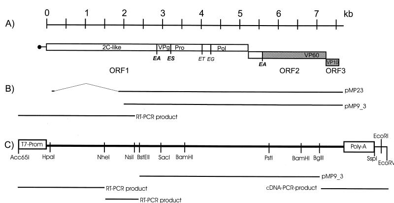
FIG. 1.
Schematic representation of FCV genome organization and the location of the different cDNA clones and PCR fragments used for sequencing and construction of the FCV infectious clone. (A) On top, a scale is given in kilobases of RNA sequence. Below, the FCV genome organization is shown with bars representing ORFs and a horizontal line showing the 5′ nontranslated region. VPg is represented by a black circle. Regions of the different ORFs coding for nonstructural proteins are presented as white bars, whereas bars drawn in grey indicate the regions encoding the known structural proteins. The location of the regions coding for the 2C-like protein, VPg, viral 3C-like protease, polymerase, and structural proteins VP60 and VP10 are designated. Published cleavage sites for the viral protease are indicated by vertical lines and the P1 and P1′ residues. Cleavage at the ET and EG sites presumably does not result in functional products (28-30). (B) The locations of the different cDNA clones and RT-PCR fragments used for sequencing are shown with regard to the FCV genome. As indicated, the cDNA clone pMP23 contains an internal deletion extending from about positions 340 to 1860. (C) Scheme of the infectious FCV cDNA clone pIK12; the different cDNA fragments used for construction of the full-length construct are indicated below the diagram. The upper line symbolizes the FCV cDNA present in pIK12 with the T7 promoter sequence (T7-Prom) and the oligo(A) tail (Poly-A) that were both introduced via PCR primers. The locations of selected restriction enzyme cleavage sites are indicated. The PCR fragments used for construction of the plasmid were all sequenced prior to cloning. The sequence of the full-length cDNA was verified after assembly of pIK12 by nucleotide sequencing.
TABLE 1.
Homology of nucleotide and deduced amino acid sequences of FCV 2024 and different full length FCV sequences
| FCV strain | Basis of comparison | Homology (%) of region (location)a
|
|||||
|---|---|---|---|---|---|---|---|
| 5′ NTR (1-19) | ORF1 (20-5311) | ORF2 (5314-7318) | ORF3 (7317-7634) | 3′ NTR (from 7635) | Complete genome | ||
| Urbana | nt | 100 | 80.3 | 78.1 | 84.4 | 85.4 | 80.0 |
| aa | 92.5 | 89.6 | 95.3 | ||||
| F9 | nt | 100 | 79.9 | 78.6 | 84.1 | 87.5 | 79.9 |
| aa | 92.1 | 88.1 | 92.5 | ||||
| F4 | nt | 100 | 79.0 | 79.0 | 85.0 | 87.5 | 79.4 |
| aa | 88.5 | 89.1 | 91.6 | ||||
| F65 | nt | 100 | 79.7 | 77.1 | 86.0 | 89.6 | 79.4 |
| aa | 89.7 | 88.1 | 95.3 | ||||
| CFI/68 | nt | 100 | 79.6 | 77.8 | 85.7 | 85.1 | 79.5 |
| aa | 91.0 | 89.6 | 95.3 | ||||
Based on information deposited at GenBank (accession numbers: strain Urbana, L40021; strain F9, M86379; strain F4, D31836; strain F65, AF109465; strain CFI/68, U13992). The results are shown for different functional regions of the genome and for the complete genomic sequence as indicated on top of the columns. 5′ NTR, 5′ nontranslated region; 3′ NTR, 3′ nontranslated region; nt, homology on the level of nucleotides; aa, homology on the level of amino acids. The numbers indicating the location of the different regions refer to the FCV 2024 sequence.
The insert of clone pMP9_3 was chosen as the starting point for establishing an infectious cDNA clone for FCV 2024. The 3′ as well as the 5′ regions of the construct were generated by PCR. Using oligonucleotides Ol-FCV42 (GCT CTA GAA GAT CTA TAG ATG TGT TCA ACT CTC, nt 7051 to 7069) and Ol-FCV43 (CGG AAT TCA ATA TT (T)30 CCC TGG GGT TAG GCG CTA GAG CGG C, reverse of nt 7665 to 7681) as primers, the 3′ region of the genome including a BglII site at position 7045 was amplified with cDNA clone pMP9_3 as a template. Ol-FCV43 was complementary to the 3′ end of the viral sequence and introduced an oligo(A) tail of 32 residues together with an SspI restriction site for linearization of the construct. The 5′ region of the genome was assembled from two different PCR products amplified with oligonucleotides Ol-FCV12 and OlFCV-10 or Ol-FCV9 (GGC TGC AGA TTC TTC CCA GAA GTG AAA AGA GCT C, reverse of nt 2795 to 2820) and Ol-FCV18 (GGG GTA CCG AAT TGG CTA AGA TCT TGC ATG, nt 1292 to 1313), respectively. As described before, Ol-FCV12 contained the 5′ end of the FCV sequence together with a promoter for the T7 phage RNA polymerase. By using standard cloning procedures, the different fragments were assembled in plasmid pACYC-MCSII, a derivative of pACYC177 (New England Biolabs, Schwalbach, Germany), with a synthetic polylinker sequence inserted after BamHI and NheI cleavage thereby deleting the kanamycin resistance gene. Further details of the cloning procedure of the construct named pIK12 are available from the authors upon request.
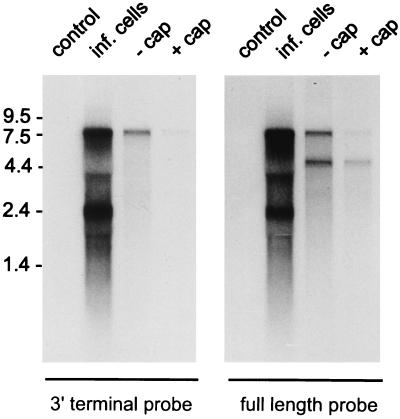
RNA was transcribed from clone pIK12 linearized with SspI. Two different reactions were run. For the first one, a cap structure analog was introduced during transcription with T7 RNA polymerase (reaction conditions were as described in reference 19, with the difference that the reaction mix contained 1 mM RNA cap structure analog [New England Biolabs] and only 0.1 mM GTP), whereas the second one was run without addition of the cap structure analog (reaction conditions were as described in reference 19). The RNA was purified by gel filtration and phenol extraction as described before (19). Analysis of the transcription products by denaturing agarose gel electrophoresis showed only a rather small amount of RNA with a size of more than 7 kb. The majority of the RNA molecules were smaller, with a strong band in the range of 5 kb, which presumably results from a stable secondary structure or cryptic T7 RNA polymerase stop signal within the FCV 2024 sequence around position 5000, since the 5-kb band did not hybridize with a probe directed against the 3′-terminal end of the viral RNA (Fig. 2). However, since full-length RNA was detectable in addition to the prematurely terminated products, transfection experiments were conducted to look for the recovery of infectious virus. CRFK cells were transfected at about 60% confluence in 3.5-cm dishes 1 day after seeding of the cells using the Lipofectin method (Invitrogen Life Technologies, Karlsruhe, Germany). Three microliters of in vitro-transcribed RNA (12.5% of a transcription reaction mixture) was mixed with 20 μl of Lipofectin in a total volume of 100 μl of Optimem medium (Invitrogen). Incubation and transfection procedures were as recommended by the supplier. The transfection mixture was removed after 4 to 6 h of incubation at 37°C, and the cells were further incubated in DMEM containing 10% FCS. Transfection of 2 μg of RNA derived from FCV-infected CRFK cells (isolated with guanidine isothiocyanate and cesium chloride centrifugation as described before [22]) served as a positive control, whereas a transfection without addition of RNA and a transfection of RNA from infected cells without addition of Lipofectin were used as negative controls. Neither the negative controls nor cells transfected with in vitro-transcribed RNA without cap structure showed a cytopathic effect (CPE). In contrast, first indications of CPE were detected after about 8 h when cells were transfected with RNA isolated from FCV-infected cells. Complete lysis of all cells in the dish was detectable after 16 to 24 h. Similarly, CPE was detectable after transfection of cells with the capped transcript derived from pIK12. However, only a few plaques were visible early after transfection and complete lysis was detectable only after about 36 to 48 h (data not shown). This result indicates a considerably reduced efficiency with regard to the generation of infectious virus from the synthetic RNA compared to that from FCV RNA.
FIG. 2.
Northern blot with RNA of FCV-infected cells (inf. cells) and RNA transcribed from the full-length cDNA construct pIK12 in vitro, with or without cap structure analogue (+ cap or −cap, respectively). RNA from noninfected cells (control) served as a negative control. As indicated, the blot on the left side was hybridized with a 3′ terminal probe (PstI/SspI fragment from pIK12), whereas the blot on the right side was hybridized with a full-length probe (full-length pIK12). On the left side the positions of RNA size marker bands are shown. Hybridization was carried out with DNA probes labeled with [α-32P]dCTP by nick translation as reported before (20). Note that the 5-kb band hybridizes only to the full-length probe (right) and thus represents a 3′-terminally truncated RNA that presumably results from an internal stop of transcription.
To prove that infectious virus had indeed been generated after RNA transfection, the supernatants of the dishes were harvested 24 h posttransfection and used for infection of CRFK cells. Again, CPE was visible for the samples derived from the dishes that were transfected with RNA from FCV-infected cells or the capped transcript (Fig. 3). No cell lysis was visible for the other samples that were previously negative. By using FCV-specific antisera, the presence of FCV antigen could be demonstrated in the cells infected with supernatant from either of the positively transfected dishes but not in the two negative ones (data not shown).
FIG. 3.
Crystal violet staining of CRFK control cells (A) or CRFK cells infected with the supernatants of cultures that were mock transfected (B) or transfected with either RNA of FCV-infected cells (C) or RNA transcribed in vitro from the full-length clone pIK12 (D). The supernatants of the transfected cells were harvested 24 h posttransfection. The infected cells were fixed with formaldehyde 50 h postinfection and stained with crystal violet as described previously (19).
The FCV cDNA present in construct pIK12 contains a silent mutation (G instead of A at position 1309 of the FCV sequence) that allows for discrimination of the in vitro-transcribed RNA from the wild-type FCV (wt FCV) genome. To provide formal proof for the recovery of infectious FCV after transfection of in vitro-transcribed RNA, the genome of the recovered virus was analyzed for the presence of this mutation. RT-PCR was conducted with RNA isolated from cells infected with the virus after the first passage posttransfection (oligonucleotides FCV-Seq-r 1720 [GGC TGC AGA ATT TGA GGT CAT TAT GAT ATA] and FCV-Seq-f 770 [GGC TTC CTT CCC AAG CTA ATT GG]; reaction conditions as described previously [19], with annealing for 55 s at 55°C, 35 cycles). Sequencing of the RT-PCR products revealed the presence of the mutation in the RNA of the virus derived from the in vitro-transcribed RNA and its absence from the genome of wt FCV recovered after transfection. Taken together, the results of our experiments prove that infectious FCV RNA can be transcribed from plasmid pIK12. The infectivity of the RNA is clearly dependent on the presence of a 5′ end cap structure, which apparently serves as a substitute for the VPg linked to the wt FCV genome. This result is in perfect accordance with the data published by Sosnovtsev and Green (27) and presumably reflects an important function of the calicivirus VPg with regard to translation initiation. Indications for such a function have also been found during in vitro translation studies that showed decreased translatability of FCV RNA after removal of VPg (13).
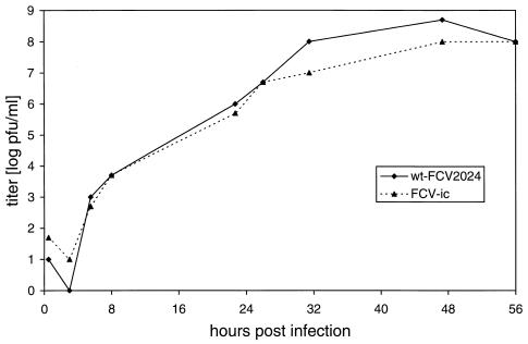
The growth characteristics of the virus recovered from the infectious FCV clone pIK12 were compared with those of wt FCV. Infection experiments with a multiplicity of infection of about 1 resulted in complete CPE within a few hours for both viruses. In order to avoid such early lysis of all the cells in the dish, growth curves were recorded after infection with a multiplicity of infection of about 0.0002. As can be seen in Fig. 4, both viruses showed very similar growth rates in these experiments. Thus, there was no indication for important functional differences between wt FCV and the recovered virus, even though the genome of the latter contained nine base exchanges with regard to our FCV 2024 consensus sequence, including the marker mutation at position 1309 (Table 2).
FIG. 4.
Growth curves of FCV 2024 and FCV-ic, the virus recovered from the infectious cDNA clone pIK12. Growth curves were recorded as described before (17).
TABLE 2.
Differences between the FCV 2024 consensus sequence and the full-length cDNA construct pIK12
| Position | Nucleotide of:
|
Amino acid | |
|---|---|---|---|
| Consensus | pFCV | ||
| 1309 | A | G | L |
| 1609 | T | A | F→L |
| 1807 | T | A | I |
| 1832 | G | A | Q→K |
| 2810 | C | A | L→M |
| 3190 | C | T | L |
| 6869 | A | G | E→G |
| 7505 | C | A | G |
| 7657 | G | C | 3′ NTRa |
NTR, nontranslated region.
The results of the RNA transfection experiments indicated a rather low efficiency of virus recovery after introduction of in vitro-transcribed RNA into the cells. To be able to obtain quantitative data with regard to this aspect, the amount of full-length transcription product was determined by a phosphorimager approach. RNA prepared by in vitro transcription was separated in a denaturing agarose gel, blotted to a nylon membrane, and hybridized with a cDNA probe labeled by nick translation (Nick Translation Kit; Amersham Pharmacia Biotech, Freiburg, Germany). A BamHI fragment corresponding to the region of nt 3648 to 6521 of the FCV genome was used as a probe. Only about 13% of the RNA represented full-length transcript as calculated from the ratio between the signal measured for the 7.5-kb region and the total counts of the lane. Based on this result, absolute values could be obtained after photometric determination of the total RNA content of the sample. Transfection experiments with different amounts of in vitro-transcribed RNA allowed us to determine that about 80 ng of the full-length transcript was necessary for reproducible induction of CPE. However, this value does not consider that much less than 100% of the transcripts can be expected to carry a 5′ cap, so that a specific infectivity of about 100 PFU per μg of capped full-length transcription product can be estimated from our experiments. Sosnovtsev and Green (27) published a specific infectivity of 300 PFU/μg of capped FCV RNA generated by in vitro transcription from their infectious FCV clone. This result is in the same range as the value deduced from our data, but it is not clear how these authors determined the amount of capped RNA.
To be able to directly compare the specific infectivity of the in vitro-transcribed RNA with that of the wt FCV genome, different dilutions of RNA from FCV-infected cells were separated by electrophoresis and subjected to Northern blotting and phosphorimager analysis. Based on a standard provided by the in vitro-transcribed RNA, the amount of FCV genomic RNA in the sample could be determined. Transfection experiments showed that about 15 pg of wt FCV RNA was sufficient for induction of cell lysis, resulting in a specific infectivity of more than 105 PFU/μg of RNA. Thus, in our hands the difference with regard to specific infectivity between wt FCV and in vitro-transcribed RNA can be estimated to be in the range of 102 to 103. It cannot be excluded that this clear difference somehow results from a not-yet-identified mutation or suboptimal feature of our infectious cDNA clone. However, this discrepancy can also be due to the fact that the cap analog present at the 5′ end of the synthetic RNA is not able to fully compensate for the absence of VPg. The calicivirus VPg probably serves different functions not only for translation initiation but also, for example, for RNA replication. Better knowledge of the activities of the protein during virus replication is necessary to finally assess the differences in specific infectivity between synthetic and viral RNA.
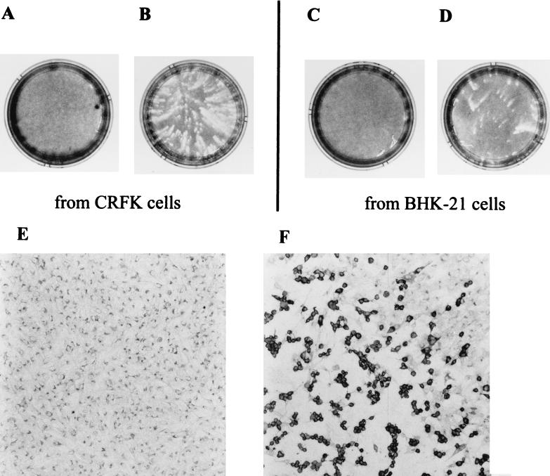
The generation of infectious FCV 2024 transcripts in vitro was rather inefficient, which in part can be attributed to the observed premature termination of transcription and the inefficient introduction of a 5′ cap structure. As an alternative, intracellular transcription of viral RNA after transfection of DNA constructs could also result in infectious viruses. Such approaches were successfully employed, for example, for picornaviruses and a variety of negative-strand RNA viruses (1, 8). The easiest way to achieve intracellular transcription of the viral RNA is to coexpress an RNA polymerase of a bacteriophage so that transcription can occur in the cytoplasm to avoid problems with splicing or a failure in nuclear export of the transcripts. As a first approach, we tried to determine whether infectious FCV could be recovered after transfection of pIK12 into cells infected with vaccinia virus MVA-T7 that expresses the T7 RNA polymerase (32). MVA-T7 is known to exhibit a narrow host range so that efficient infection of CRFK cells could not be expected. Baby hamster kidney (BHK-21) cells (kindly provided by T. Rümenapf, University of Giessen) represent well-suited host cells for MVA-T7, but these cells cannot be infected with FCV and it is not known whether viral replication and production of infectious particles can be achieved in BHK cells. Because of these uncertainties, we tried to recover FCV after DNA transfection of both of these cell lines. The cells were seeded to 80% confluence in 3.5-cm dishes, infected with MVA-T7 (kindly provided by B. Moss, National Institutes of Health, Bethesda, Md.), and transfected with pIK12 DNA essentially as described before for transient expression experiments (18). The cells were incubated with the transfection mixture for 4 h and incubated for another 16 h in DMEM with 10% FCS. After this incubation time, a pronounced CPE was visible that was at least mostly due to MVA-T7, since it was also detectable in mock-transfected cells (data not shown). The supernatant of the dishes was harvested and passed through a 0.1-μm-pore-size sterile filter (MILLEX-VV; Millipore Products, Bedford, Mass.) to eliminate the vaccinia virus (25). One hundred fifty microliters of filtrate from each transfection reaction mixture was used to infect CRFK cells. After 20 h of incubation at 37°C, cells infected with supernatant of the mock transfections were confluent and showed no CPE (Fig. 5A and C). In contrast, plaques were visible in both dishes that were infected with the supernatant of cells which had been transfected with pIK12 (Fig. 5B and D). To prove that these plaques were due to FCV infection, viral protein expression was demonstrated by staining with an FCV-specific hyperimmune serum (antiserum raised in rabbits by immunization with purified FCV; kindly provided by M. Büttner, Federal Research Centre for Virus Diseases of Animals, Tübingen, Germany). Detection of bound antibodies with a peroxidase-coupled anti-rabbit antiserum by peroxidase reaction clearly showed the presence of FCV antigen in cells located in the region of CPE plaques (Fig. 5F), whereas no signal was detectable in control cells (Fig. 5E).
FIG. 5.
Analysis of CRFK cells infected with the supernatants obtained from cell cultures that were infected with vaccinia virus MVA-T7 and transfected with different DNA constructs. (A to D) Crystal violet staining of cells infected with the supernatant of CRFK (A and B) or BHK-21 (C and D) cells that were infected with MVA-T7 and then either mock transfected (A and C) or transfected with the full-length FCV cDNA construct pIK12 (B and D). (E and F) Detection of FCV antigen in cells infected with the supernatant of CRFK cells that were infected with MVA-T7 and then either mock transfected (E) or transfected with pIK12 (F). The FCV antigen was detected by reaction with a polyclonal anti-FCV rabbit serum and subsequent labeling of bound antibodies with peroxidase-coupled anti-rabbit antiserum and peroxidase staining as described elsewhere (15). In panel F, part of a plaque is shown with a region of nondestroyed cells visible in the upper right corner.
As can be seen from the number of plaques, the system based on transfection of CRFK cells was at least 1 order of magnitude more efficient than the approach working with BHK cells. This result could be explained by some cell-specific feature that allows for enhanced replication of FCV in CRFK cells. Alternatively, the more efficient generation of infectious FCV in the case of the CRFK cells could simply be due to the fact that amplification of the virus by a second cycle of replication after infection of new cells could have occurred, a process that is restricted to the permissive feline cells. A third explanation could be that late steps in MVA-T7 replication interfere with FCV replication and that these steps only occur in BHK cells, in which MVA-T7 undergoes complete replication (2), but not in CRFK cells. In conclusion, our results show that FCV could be recovered with good efficiency after transfection of DNA constructs. It is well possible that the results could be optimized by, for example, construction of better plasmids with a T7 terminator following the oligo(A) stretch in pIK12.
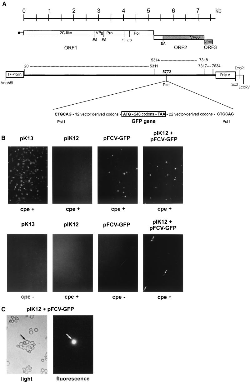
It has been shown recently that FCV with a chimeric major capsid protein containing sequences of two different FCV strains can be recovered from an engineered infectious clone (21). We were interested to find out whether it was also possible to insert a foreign sequence into the FCV genome. As a first test the sequence coding for GFP derived from pEGFP-N3 (Clontech, Palo Alto, Calif.) was inserted into the FCV sequence to obtain pFCV-GFP. We chose the PstI site of pIK12 that corresponds to position 5714 of the FCV sequence for insertion so that the marker gene was located in frame of ORF2 close to the 5′ end of the VP60-encoding region (Fig. 6A ) (further details of the cloning procedure are available from the authors upon request). The inserted sequence in construct pFCV-GFP contained an in-frame translational stop codon at the 3′ end of the foreign gene. The expression of the GFP gene should occur via translation of a precursor composed of the capsid protein leader, 29 amino acids of the capsid protein, 12 vector-derived amino acids, and GFP. By processing at the EA site separating the capsid leader and capsid protein (4), a GFP with 41 foreign residues at the amino terminus should be generated. Because of the translation termination codon at the end of the GFP gene, the genetic manipulation present in pFCV-GFP inhibits expression of VP60, and recovery of infectious recombinant viruses could not be expected after transfection of pFCV-GFP derived RNA. However, since all nonstructural proteins should be expressed from such an RNA, this molecule could represent an autonomous replicon that should be able to express a subgenomic mRNA of about 3 kb containing the capsid protein-encoding region and the GFP gene. As expected as a consequence of the low efficiency of virus recovery after RNA transfection, introduction of RNA transcribed from pFCV-GFP in vitro resulted in detection of only about five single fluorescent cells per 3.5-cm dish 20 h posttransfection (average of six independent experiments). The positive cells had a round shape that was most likely due to CPE (data not shown). Attempts to rescue and amplify the replicon by cotransfection of RNA from pIK12 failed. The same was true for experiments in which transfected cells were superinfected with FCV. Again, these results are most likely due to the low efficiency of the RNA transfection and virus recovery system. In order to overcome this problem we again used the transient expression system based on MVA-T7. Five different transfection reactions were conducted with either no DNA, a GFP control plasmid (pK13 containing the GFP gene inserted via XhoI and KpnI into plasmid pCI [Promega, Heidelberg, Germany; kindly provided by C. Meyer, Federal Research Centre for Virus Diseases of Animals]), pIK12, pFCV-GFP, and a mixture of the latter two constructs. Eighteen hours posttransfection the cells showed MVA-T7-induced CPE and were analyzed for GFP fluorescence. As expected, signals could not be detected for the transfection control or the pIK12-transfected cells. In the dish with the positive control pK13, about 60% of the cells were positive. Also, the two dishes transfected with pFCV-GFP contained fluorescent cells (about 5 to 10% of the cells) (Fig. 5). This result could be due to FCV gene expression based on transcription of subgenomic FCV RNA or on internal initiation of translation, as published before (28). FCV mRNA transcription should in theory be dependent on FCV genome replication to provide a negative-strand template for transcription. However, DNA recombination in vaccinia virus-infected cells is a well known and efficient process which makes it difficult to interpret such results. Indeed, transfection of a pFCV-GFP variant with a translational stop codon introduced into ORF1 (codon 692, upstream of the known VPg-protease-polymerase region) resulted in a considerably lower but detectable number of fluorescent cells (results not shown). We therefore tried to passage virus that might have been generated after the transfection. To do so, supernatant of the transfected cells was harvested, filtered to eliminate MVA-T7, and used for infection of CRFK cells. After 18 h of incubation at 37°C, the dishes were analyzed for fluorescent cells. As expected, the dishes infected with supernatant from the cultures transfected with no DNA (data not shown), the GFP control pK13, or FCV infectious clone pIK12 were all negative with regard to fluorescence. For the reaction with pIK12, CPE could be detected which proved the successful recovery of infectious FCV. The dish with the sample obtained after transfection of FCV-GFP alone was free of fluorescent cells and showed no CPE. This result was expected, since the replicon should not be able to express VP60 and should therefore not generate infectious virus particles. In contrast, positive fluorescence signals and CPE were visible when cells were infected with supernatant derived from cultures that had been transfected with a mixture of pFCV-GFP and pIK12 (Fig. 6). On the average, about 500 single fluorescent cells were found in the dishes infected with the first passage of the mixture of recovered FCV and replicon. Both the detection of the CPE and the fluorescence signal were prevented when the extract of the MVA-T7-infected and DNA-transfected cells was incubated with the anti-FCV serum (1 μl of serum per 0.2 ml of extract; incubation at 37°C for 10 min), whereas treatment with a rabbit preimmune serum had no effect (data not shown). These results represent a further argument for the conclusion that the RNA derived from pFCV-GFP was packaged into FCV particles that were able to infect new cells and could be neutralized by FCV-specific antibodies.
FIG. 6.
(A) Schematic representation of the cDNA construct pFCV-GFP. On top, a scale in kilobases is shown; shown below are schemes of the FCV genome organization and major features of the plasmid pFCV-GFP. The plasmid was obtained from the infectious FCV clone pIK12 by insertion of the GFP-encoding sequence into the PstI site within ORF2. The GFP gene of about 720 nt (box designated GFP gene) was derived from pEGFP-N3 (Clontech) by PCR with primers Ol-GFPup and Ol-GFPdown that also contained the PstI sites (shown in bold face). Note that there is a translational stop codon present at the 3′ end of the GFP gene that prevents expression of the major part of the FCV capsid protein gene located downstream of the inserted foreign sequence. (B) Fluorescence analysis of cells transfected with different cDNA constructs or infected with material obtained after DNA transfection. The upper set of panels shows detection of CRFK cells expressing GFP after MVA-T7-dependent transient expression of the DNA constructs indicated on top of the photos. Construct pK13 represents a positive control with the GFP gene under the control of a T7 promoter (vector also includes a eucaryotic promoter); pIK12 represents the FCV infectious full-length construct from which pFCV-GFP is derived by insertion of the GFP-encoding sequence into ORF2. Below the fluorescence images, the notations cpe + and cpe − indicate whether or not CPE was detectable. The lower set of panels shows detection of the GFP signal in CRFK cells infected with the supernatants from the upper set of cultures. Fluorescent cells are marked by arrows. (C) Higher magnification to demonstrate GFP expression in a cell after infection with the lysate of cells transfected with a mixture of pIK12 and pFCV-GFP. The same field of the dish is shown either with light microscopy (left) or fluorescence microscopy (right). The localization of the fluorescent cell (marked by arrows in both parts of the figure) was done with computer-aided image analysis using a set of three positive cells for positioning of the field. Note that all cells visible in the left part are rounded as a consequence of the CPE resulting from FCV infection.
We tried to demonstrate recombinant FCV genomic and subgenomic RNA by Northern blot hybridization with a GFP probe, but presumably because of the low number of positive cells these attempts did not lead to unequivocal results. Longer incubation times did not result in an increase in the number of fluorescent cells. Later on, the number of cells expressing GFP even decreased due to the pronounced CPE resulting from FCV infection. In a second passage, fluorescent cells could no longer be detected reproducibly. The observation that the replicon was apparently lost during a further passage is most likely due to the fact that the efficiently replicating wt FCV has considerable advantages over the defective virus. However, our results clearly show that foreign sequences can be inserted into the FCV genome without loss of functions necessary for RNA replication. Moreover, such sequences can be expressed by the viral system, and the recombined sequences are packaged by viral proteins provided in trans. The resulting viral particles are able to infect cells and start a new replication cycle. The results presented here provide a basis for a set of new approaches aimed at elucidation of calicivirus replication and transcription mechanisms.
Acknowledgments
We thank Maren Ziegler, Petra Wulle, and Silke Esslinger for excellent technical assistance, B. Moss for MVA-T7, M. Büttner for the anti-FCV antiserum, C. Meyer for the GFP plasmid pK13, and K. Danner for FCV 2024 and the CRFK cells.
This study was supported by grants Me1367/1-3 and Me1367/1-5 from the Deutsche Forschungsgemeinschaft.
REFERENCES
- 1.Boyer, J.-C., and A.-L. Haenni. 1994. Infectious transcripts and cDNA clones of RNA viruses. Virology 198:415-426. [DOI] [PubMed] [Google Scholar]
- 2.Carroll, M. W., and B. Moss. 1997. Host range and cytopathogenicity of the highly attenuated MVA strain of vaccinia virus: propagation and generation of recombinant viruses in a nonhuman mammalian cell line. Virology 238:198-211. [DOI] [PubMed] [Google Scholar]
- 3.Carter, M. J. 1990. Transcription of feline calicivirus RNA. Arch. Virol. 114:143-152. [DOI] [PMC free article] [PubMed] [Google Scholar]
- 4.Carter, M. J., I. D. Milton, P. C. Turner, J. Meanger, M. Bennett, and R. M. Gaskell. 1992. Identification and sequence determination of the capsid protein gene of feline calicivirus. Arch. Virol. 122:233-235. [DOI] [PMC free article] [PubMed] [Google Scholar]
- 5.Chirgwin, J. M., A. E. Przybyla, R. J. MacDonald, and W. J. Rutter. 1979. Isolation of biologically active ribonucleic acid from sources enriched in ribonuclease. Biochemistry 18:5294-5299. [DOI] [PubMed] [Google Scholar]
- 6.Clarke, I. N., and P. R. Lambden. 1997. The molecular biology of caliciviruses. J. Gen. Virol. 78:291-301. [DOI] [PubMed] [Google Scholar]
- 7.Clarke, I. N., and P. R. Lambden. 2000. Organization and expression of calicivirus genes. J. Infect. Dis. 181(Suppl. 2):309-316. [DOI] [PubMed] [Google Scholar]
- 8.Conzelmann, K.-K. 1996. Genetic manipulation of non-segmented negative-strand RNA viruses. J. Gen. Virol. 77:381-389. [DOI] [PubMed] [Google Scholar]
- 9.Ehresmann, D. W., and F. L. Schaffer. 1977. RNA synthesized in calicivirus-infected cells is atypical of picornaviruses. J. Virol. 22:572-583. [DOI] [PMC free article] [PubMed] [Google Scholar]
- 10.Gaskell, R. M. 1985. Feline medicine and therapeutics, p. 257-270. Blackwell, Oxford, United Kingdom.
- 11.Glass, P. J., L. J. White, J. M. Ball, I. Leparc-Goffart, M. E. Hardy, and M. K. Estes. 2000. Norwalk virus open reading frame 3 encodes a minor structural protein. J. Virol. 74:6581-6591. [DOI] [PMC free article] [PubMed] [Google Scholar]
- 12.Green, K. Y., T. Ando, M. S. Balayan, T. Berke, I. N. Clarke, M. K. Estes, D. O. Matson, S. Nakata, J. D. Neill, M. J. Studdert, and H.-J. Thiel. 2000. Family Caliciviridae, p. 725-734. In M. H. V. Regenmortel, C. M. Fauquet, and D. H. L. Bishop. Virus taxonomy. Academic Press, New York, N.Y.
- 13.Herbert, T. P., I. Brierley, and T. D. Brown. 1997. Identification of a protein linked to the genomic and subgenomic mRNA of feline calicivirus and its role in translation. J. Gen. Virol. 78:1033-1040. [DOI] [PubMed] [Google Scholar]
- 14.König, M., H.-J. Thiel, and G. Meyers. 1998. Detection of viral proteins after infection of cultured hepatocytes with rabbit hemorrhagic disease virus. J. Virol. 72:4492-4497. [DOI] [PMC free article] [PubMed] [Google Scholar]
- 15.Kümmerer, B. M., and G. Meyers. 2000. Correlation between point mutations in NS2 and the viability and cytopathogenicity of bovine viral diarrhea virus strain Oregon analyzed with an infectious cDNA clone. J. Virol. 74:390-400. [DOI] [PMC free article] [PubMed] [Google Scholar]
- 16.Machín, A., J. S. Martín Alonso, and F. Parra. 2001. Identification of the amino acid residue involved in rabbit hemorrhagic disease virus VPg uridylylation. J. Biol. Chem. 276:27787-27792. [DOI] [PubMed] [Google Scholar]
- 17.Meyers, G., C. Wirblich, and H.-J. Thiel. 1991. Genomic and subgenomic RNAs of rabbit hemorrhagic disease virus are both protein-linked and packaged into particles. Virology 184:677-686. [DOI] [PMC free article] [PubMed] [Google Scholar]
- 18.Meyers, G., C. Wirblich, H.-J. Thiel, and J. O. Thumfart. 2000. Rabbit hemorrhagic disease virus: genome organization and polyprotein processing of a calicivirus studied after transient expression of cDNA constructs. Virology 276:349-363. [DOI] [PubMed] [Google Scholar]
- 19.Meyers, G., N. Tautz, P. Becher, H.-J. Thiel, and B. M. Kümmerer. 1996. Recovery of cytopathogenic and noncytopathogenic bovine viral diarrhea viruses from cDNA constructs. J. Virol. 70:8606-8613. [DOI] [PMC free article] [PubMed] [Google Scholar]
- 20.Meyers, G., T. Rümenapf, and H.-J. Thiel. 1989. Molecular cloning and nucleotide sequence of the genome of hog cholera virus. Virology 171:555-567. [DOI] [PubMed] [Google Scholar]
- 21.Neill, J. D., S. V. Sosnovtsev, and K. Y. Green. 2000. Recovery and altered neutralization specificities of chimeric viruses containing capsid protein domain exchanges from antigenically distinct strains of feline calicivirus. J. Virol. 74:1079-1084. [DOI] [PMC free article] [PubMed] [Google Scholar]
- 22.Rümenapf, T., G. Meyers, R. Stark, and H.-J. Thiel. 1989. Hog cholera virus—characterization of specific antiserum and identification of cDNA clones. Virology 171:18-27. [DOI] [PubMed] [Google Scholar]
- 23.Sambrook, S., E. F. Fritsch, and T. Maniatis. 1989. Molecular cloning: a laboratory manual, 2nd ed. Cold Spring Harbor Laboratory Press, Cold Spring Harbor, N.Y.
- 24.Schaffer, F. L., D. E. Ehresmann, M. K. Fretz, and M. E. Soergel. 1980. A protein, VPg, covalently linked to 36S calicivirus RNA. J. Gen. Virol. 47:215-220. [DOI] [PubMed] [Google Scholar]
- 25.Schnell, M. J., T. Mebatsion, and K.-K. Conzelmann. 1994. Infectious rabies virus from cloned cDNA. EMBO J. 13:4195-4203. [DOI] [PMC free article] [PubMed] [Google Scholar]
- 26.Sosnovtsev, S. V., and K. Y. Green. 2000. Identification and genomic mapping of the ORF3 and VPg proteins in feline calicivirus virions. Virology 277:193-203. [DOI] [PubMed] [Google Scholar]
- 27.Sosnovtsev, S. V., and K. Y. Green. 1995. RNA transcripts derived from a cloned full-length copy of the feline calicivirus genome do not require VPg for infectivity. Virology 210:383-390. [DOI] [PubMed] [Google Scholar]
- 28.Sosnovtsev, S. V., S. A. Sosnovtseva, and K. Y. Green. 1998. Cleavage of the feline calicivirus capsid precursor is mediated by a virus-encoded proteinase. J. Virol. 72:3051-3059. [DOI] [PMC free article] [PubMed] [Google Scholar]
- 29.Sosnovtseva, S. A., S. V. Sosnovtsev, and K. Y. Green. 1999. Mapping of the feline calicivirus proteinase responsible for autocatalytic processing of the nonstructural polyprotein and identification of a stable proteinase-polymerase precursor protein. J. Virol. 73:6626-6633. [DOI] [PMC free article] [PubMed] [Google Scholar]
- 30.Wei, L., J. S. Huhn, A. Mory, H. B. Pathak, S. V. Sosnovtsev, K. Y. Green, and C. E. Cameron. 2001. Proteinase-polymerase precursor as the active form of feline calicivirus RNA-dependent RNA polymerase. J. Virol. 75:1211-1219. [DOI] [PMC free article] [PubMed] [Google Scholar]
- 31.Wirblich, C., H.-J. Thiel, and G. Meyers. 1996. Genetic map of the calicivirus rabbit hemorrhagic disease virus as deduced from in vitro translation studies. J. Virol. 70:7974-7983. [DOI] [PMC free article] [PubMed] [Google Scholar]
- 32.Wyatt, L. S., B. Moss, and S. Rozenblatt. 1995. Replication-deficient vaccinia virus encoding bacteriophage T7 RNA polymerase for transient gene expression in mammalian cells. Virology 210:202-205. [DOI] [PubMed] [Google Scholar]


