Abstract
The generation of replication-competent measles virus (MV) depends on the incorporation of biologically active, fusogenic glycoprotein complexes, which are required for attachment and penetration into susceptible host cells and for direct virus spread by cell-to-cell fusion. Whereas multiple studies have analyzed the importance of the ectodomains of the MV glycoproteins hemagglutinin (H) and fusion protein (F), we have investigated the role of the cytoplasmic tails of the F and H proteins for the formation of fusogenic complexes. Deletions in the cytoplasmic tails of transiently expressed MV glycoproteins were found to have varying effects on receptor binding, fusion, or fusion promotion activity. F tail truncation to only three amino acids did not affect fusion capacity. In contrast, truncation of the H cytoplasmic tail was limited. H protein mutants with cytoplasmic tails of <14 residues no longer supported F-mediated cell fusion, predominantly due to a decrease in surface expression and receptor binding. This indicates that a minimal length of the H protein tail of 14 amino acids is required to ensure a threshold local density to have sufficient accumulation of fusogenic H-F complexes. By using reverse genetics, a recombinant MV with an F tail of three amino acids (rMV-FcΔ30), as well as an MV with an H tail of 14 residues (rMV-HcΔ20), could be rescued, whereas generation of viruses with shorter H tails failed. Thus, glycoprotein truncation does not interfere with the successful generation of recombinant MV if fusion competence is maintained.
One of the major obstacles in the development of recombinant measles viruses (rMV) carrying either altered MV glycoproteins or foreign glycoproteins is the necessity of preserving the biological activities of the surface proteins required for efficient virus replication (7, 12, 16, 40, 46, 47, 52). Therefore, it is crucial to identify important protein domains that are essential for biological activities. The MV surface glycoprotein complex is composed of two integral membrane proteins, the hemagglutinin (H) and the fusion (F) protein. The H protein is a type II membrane protein which is assumed to exist at the viral envelope or on the surfaces of infected cells as a tetramer of two covalently linked dimers (26). H is responsible for binding to host cells carrying a suitable receptor, such as CD46 or SLAM, and is an essential cofactor for virus-induced membrane fusion (9, 11, 23, 25, 30, 49, 55). The F protein is a type I membrane protein with an N-terminal ectodomain that has to be cleaved into the F1 and F2 subunits to allow pH-independent fusion (22). Cleaved F trimers have to interact with H oligomers to constitute biologically active MV glycoprotein complexes. Membrane-proximal regions in the ectodomains of both proteins appear to be involved in the formation of these fusogenic H-F complexes (15, 56). Whereas the importance of the ectodomains of the glycoproteins for receptor binding activity, fusion activity, and the formation of fusogenic complexes has been intensively studied (2, 3, 14, 15, 20, 26, 36, 40, 41, 53, 54), the importance of the cytoplasmic domains for these biological properties is not well understood.
The cytoplasmic tails of the glycoproteins are clearly involved in virus assembly, since they bind to the matrix protein, which acts as a bridge between the virus envelope and the viral nucleocapsid (5, 29, 34, 47). Subacute sclerosing panencephalitis (SSPE) MV strains, which often have altered glycoprotein tails, and rMV resembling these naturally occurring SSPE strains were shown to be defective in virus assembly (7, 8). Furthermore, tail alterations may affect the fusion competence of the MV glycoproteins. We have reported recently that a tyrosine-dependent sorting signal in the respective cytoplasmic tails directs both the H and the F proteins to the basolateral surfaces of polarized epithelial cells. Only cells expressing both proteins on the basolateral side were able to fuse with neighboring cells. Alteration of the critical tyrosines in either of the two glycoproteins did not affect fusion competence in nonpolarized cells but completely prevented fusion of epithelial cells (24, 27). Cathomen et al. (7) observed positive and negative effects on fusion activity by shortening the cytoplasmic tails of the F or H protein. Viruses having either a truncated F tail (24 of the 33 C-terminal amino acids deleted; designated FcΔ24) or a truncated H tail (14 of the 34 N-terminal amino acids deleted; designated HcΔ14) showed enhanced fusion competence due to a defective glycoprotein M interaction. Unlike HcΔ14, H protein with a cytoplasmic domain of only 10 amino acids (HcΔ24) did not allow rMV rescue. Although surface expression appeared not to be reduced, HcΔ24 did not support fusion (7). From these results, it has been concluded that membrane-proximal sequences (>10 but <20 amino acids) in the MV H cytoplasmic tail are directly involved in the fusion process.
In this study, we define the minimal length of the cytoplasmic domains that still support fusogenic activity of MV glycoprotein complexes and thereby the minimal requirements for the successful generation of recombinant MV. We found that the F tail can be reduced to 3 residues, whereas the H tail must have a minimal length of 14 amino acids. Further truncation of the H tail affected the expression level of the H protein. In contrast to conclusions drawn from earlier data, our results clearly indicate that the membrane-proximal sequences are not directly involved in fusion promotion activity but that (i) the H tail is predominantly required to ensure sufficient protein expression and surface transport of H and (ii) the total length and not the N-terminal sequence determines the expression level.
(This work was done in partial fulfillment by M.M. of the requirements for a Ph.D. degree from Philipps-Universität Marburg.)
MATERIALS AND METHODS
Cell culture.
Vero cells (African green monkey kidney cells) were grown in Dulbecco's modified minimal essential medium (DMEM; Gibco-BRL) supplemented with 10% fetal calf serum (FCS; Gibco-BRL), 100 U of penicillin/ml, and 100 μg of streptomycin/ml. BJAB cells (human B lymphocytes) were grown in RPMI 1640 medium (Gibco-BRL) supplemented with 10% FCS, penicillin, and streptomycin. Helper cell line 293-3-46 (human embryonic kidney cells) was maintained in DMEM containing 1 mg of G418/ml. These cells stably express MV nucleoprotein (NP) and phosphoprotein (P), as well as T7 RNA polymerase (37).
Plasmid construction and site-specific mutagenesis.
Cloning of the viral glycoprotein (H and F protein) genes into the expression vector pCG under the control of the cytomegalovirus early promoter has been described previously (6). Truncated H protein (HcΔ15-HcΔ24) genes were prepared by introduction of site-specific deletions with complementary primers into the double-stranded pCG plasmids using the Quikchange site-directed mutagenesis kit (Stratagene). Mutagenic oligonucleotide primers were arranged to bind with 21 to 24 bases on both sites of the sequence being deleted. Clones encoding HcΔ21-A, HcΔ24-4A, HcΔ26-6A, or HcΔ30-10A were constructed by insertion of nucleotides directly after the initial methionine: GCT into the plasmid pCG-HcΔ21, GCTGCCGCAGCG into pCG-HcΔ24, GCTGCCGCAGCGGCCGCA into pCG-HcΔ26, or GCTGCCGCAGCGGCCGCAGCAGCTGCTGCG into pCG-HcΔ30. The FcΔ30 gene was generated by introducing a premature translational stop codon by mutation at nucleotide position 2900 (T to A).
For the generation of recombinant MV, the plasmid p(+)MVNSe (46) carrying the full-length MV Edmonston B-based genome was used. Plasmids p(+)MV-HcΔ20, p(+)MV-HcΔ22, and p(+)MV-HcΔ24 were constructed by ligation of a PacI-SpeI fragment containing the mutated H gene from the respective pCG plasmid into p(+)MVNSe. To construct a full-length MV genome with the F tail truncation mutant (FcΔ30), the F gene was mutagenized in the shuttle vector peF1 (37). A NarI-PacI fragment containing the mutated F gene was subcloned into p(+)MVNSe. All mutated full-length antigenomes abide by the rule of six. The sequences of all constructs were confirmed by dideoxy sequencing.
Fusion assays.
Vero cells were cotransfected with 2.5 μg of plasmid DNA encoding MV Edmonston H (MV HEdm) or MV H truncation mutants and 2.5 μg of plasmid DNA encoding MV FEdm by using the cationic lipid transfection reagent Lipofectamine 2000 (Gibco-BRL), following the instructions of the supplier. At 24 h posttransfection, the transiently expressing cells were fixed with ethanol and stained with 1:10-diluted Giemsa's staining solution. To quantify the fusion helper activity of the H truncation mutants, four randomly chosen microscopic fields for each sample were photographed. The number of nuclei in single, nonfused cells and the number of nuclei in syncytia were counted and averaged. The relation of the total number of nuclei and the number of nuclei in syncytia per microscopic field indicated the percent fusion.
Surface biotinylation and Western blot analysis.
At 24 h posttransfection, transiently expressing Vero cells were washed three times with cold phosphate-buffered saline (PBS) containing 0.1 mM CaCl2 and 1 mM MgCl2 and incubated twice for 20 min at 4°C with 2 mg of sulfo-N-hydroxysuccinimidobiotin (Pierce)/ml. After biotinylation, the cells were washed once with cold PBS containing 0.1 M glycine and three times with cold PBS. The cells were lysed in 0.5 ml of radioimmunoprecipitation assay buffer (1% Triton X-100, 1% sodium deoxycholate, 0.1% SDS, 0.15 M NaCl, 10 mM EDTA, 10 mM iodoacetamide, 1 mM phenylmethylsulfonyl fluoride, 50 U of aprotinin/ml, 20 mM Tris-HCl, pH 8.5) followed by centrifugation for 20 min at 100,000 × g. The supernatant was immunoprecipitated with the H-specific monoclonal antibody K83 at a final dilution of 1:100. After the addition of 20 μl of a suspension of protein A-Sepharose CL-4B (Sigma, Deisenhofen, Germany), the immunocomplexes were washed three times with radioimmunoprecipitation assay buffer, suspended in reducing sample buffer for SDS-polyacrylamide gel electrophoresis (PAGE), and divided into two aliquots. Following separation on a 10% polyacrylamide gel, the proteins were blotted to nitrocellulose by the semidry-blot technique. Nonspecific binding sites were blocked by 5% nonfat dry milk in PBS. Biotinylated cell surface H proteins were detected by incubation with a streptavidin-biotinylated horseradish peroxidase complex (Amersham Pharmacia Biotech) diluted 1:2,000 in PBS. Total cellular H protein was detected by using a polyclonal MV antiserum (1:1,000) and horseradish peroxidase-conjugated swine anti-rabbit immunoglobulin G (IgG) (1:5,000; DAKO, Roskilde, Denmark). Proteins were visualized with the enhanced chemiluminescence system (Amersham Pharmacia Biotech) by exposure to an autoradiography film (Kodak BIOMAX).
Metabolic labeling.
At 20 h posttransfection, Vero cells grown in six-well tissue culture plates were incubated for 30 min in DMEM lacking cysteine and methionine and then metabolically labeled by incubation with the same medium containing [35S]cysteine and [35S]methionine (Promix; Amersham) at a final concentration of 100 μCi/0.5 ml for 10 to 45 min. Subsequently, labeling medium was replaced by chase medium containing 5% FCS, and the cells were incubated at 37°C for various times. After being radiolabeled, H proteins were immunoprecipitated as described above and subjected to SDS-PAGE. The dried gels were quantified using an imaging system (BioImager; Perkin-Elmer) and then exposed to Kodak BIOMAX films.
Flow cytometry.
Vero cells grown on 60-mm-diameter culture dishes were transfected with plasmids encoding H truncation mutants. At 24 h posttransfection, the cells were detached from the dish by treatment with 0.25% trypsin-1 mM EDTA, washed with PBS, and incubated with the H-specific monoclonal antibody K83 for 1 h at 4°C. Fluorescein-conjugated goat anti-mouse IgG (DAKO) was used as the secondary antibody. After suspension in 1 ml of PBS, the fluorescence intensity of 10,000 cells was measured by a FACScan flow cytometer (Becton Dickinson).
Hemadsorption assay.
At 24 h posttransfection, Vero cells transiently expressing standard or mutant MV H proteins were washed twice with PBS and incubated for 1 h at 37°C with a 1% suspension of African green monkey red blood cells (RBC). After unadsorbed RBC were removed by repeated washings with cold PBS, the cells were observed and photographed under an inverted light microscope. To quantify the hemadsorption, bound erythrocytes were lysed with distilled water, and the adsorbance value of released hemoglobin was measured at 540 nm.
Virus rescue and preparation of recombinant-virus stocks.
Transfection and rescue of MV were performed as described previously (37). Briefly, 293-3-46 helper cells mediating both artificial T7 transcription and NP and P functions were transfected with 5 μg of either p(+)MVNSe, p(+)MV-HcΔ20, p(+)MV-HcΔ22, p(+)MV-HcΔ24, or p(+)MV-FcΔ30 and 10 ng of plasmid encoding the MV polymerase (pEMC-La) using the calcium phosphate transfection procedure. After reaching confluence, the cell monolayers were expanded from 35-mm-diameter wells to 100-mm-diameter dishes and subsequently transferred to Vero cells. The cultures were regularly examined for syncytium formation. The viruses were harvested when they showed 80 to 90% cytopathic effects (CPEs). Virus stocks were produced following plaque purification. The identity of the rescued viruses was confirmed by reverse transcription-PCR followed by dideoxy sequencing.
Virus infection and plaque assay.
Virus growth characteristics were analyzed by infecting confluent Vero cells in 35-mm-diameter dishes with either rMVEdm, rMV-FcΔ30, or rMV-HcΔ20 at a multiplicity of infection (MOI) of 0.001. After incubation with virus for 1.5 h at 37°C, the inocula were removed, the cells were washed three times with PBS, and cultures were grown in DMEM containing 2% FCS at 33°C. At various times postinfection (p.i.), cell-free virus was prepared by clarifying cell supernatants by centrifugation, and cell-associated virus was recovered by scraping the cells into OptiMEM followed by one round of freezing and thawing. Virus titers were determined by plaque assay. Plaque assays were carried out with Vero cells in 35-mm-diameter wells. After 1.5 h of virus adsorption, the inoculum was removed and the cells were overlaid with 3 ml of DMEM containing 2% FCS and 0.8% Bacto Agar (Difco). After 4 days, the agar overlay was removed and the cells were fixed with formalin and stained with crystal violet to determine the number and size of the plaques.
Confocal double immunofluorescence.
Vero cells were grown on coverslips and infected with rMV for 30 h p.i. at 33°C. To prevent disruption of the cell architecture due to extensive syncytium formation, a fusion-inhibitory peptide was added to the infected cells (54). Without prior fixation, the cells were incubated for 1 h at 4°C with PBS containing 0.3% bovine serum albumin and a monoclonal antibody directed against the F protein (A504) or directed against the H protein (K83). After extensive washing with PBS, bound antibodies were detected with rhodamine-labeled anti-mouse IgG (Dianova, Hamburg, Germany). After permeabilization with methanol-acetone (1:1) and blocking with 5% normal mouse serum for 30 min at room temperature, the cells were incubated with a fluorescein isothiocyanate (FITC)-labeled monoclonal antibody directed against the M protein (MAb 8910; Chemicon, Temecula, Calif.) for 1 h at 4°C. Labeling of the antibody with FITC (Sigma) was performed as described by the manufacturer. The samples were mounted in Mowiol (Hoechst, Frankfurt, Germany) and 10% 1,4-diazabicyclo(2,2,2)octane (Sigma) and examined using a confocal laser scanning microscope (Carl Zeiss, Oberkochem, Germany).
RESULTS
Expression of cytoplasmic-tail truncation mutants of MV glycoproteins.
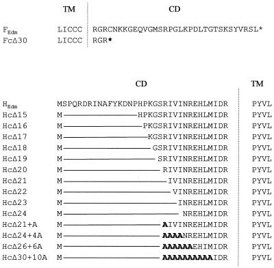
To investigate the impact of F and H cytoplasmic-tail truncations on the generation of replication-competent MV, several truncation mutants were constructed by PCR-based mutagenesis. Figure 1 shows a schematic representation of the various mutants with shortened cytoplasmic tails used in this study. A mutant F protein with a truncation of 24 amino acids identified in an MV SSPE strain (FcΔ24) was described as being fusion competent and allowing the generation of rMV (7). Since the FcΔ24 cytoplasmic tail with nine residual amino acids is almost as long as the natural cytoplasmic tails of other type I membrane proteins, such as the influenza HA protein (45), it might be long enough to bind intracellularly to other cellular or viral components, influencing the biological activity of the MV F protein. To rule out this possibility, we further truncated the protein. Because the transmembrane domain of a type I integral membrane protein is usually followed by two charged residues, we completely deleted the F cytoplasmic tail except for the three membrane-proximal residues, RGR, thereby ensuring stable integration in the membrane. The resulting truncation mutant was designated FcΔ30. To elucidate the importance of the H cytoplasmic tail, a series of truncated H genes was constructed. Cathomen et al. (7) reported that rMV encoding an H protein with a deletion of the 14 N-terminal residues could be rescued whereas a deletion of 24 amino acids did not allow the generation of rMV. Therefore, we constructed genes encoding H proteins with progressive N-terminal deletions from 15 (HcΔ15) up to 24 amino acids (HcΔ24). All of the truncation mutants were cloned into the eucaryotic expression vector pCG and transfected into Vero cells. Surface expression of the proteins was analyzed by immunostaining of living cells using monoclonal antibodies directed against either the F or the H protein. Bright fluorescence signals on the surfaces of the transfected cells were observed for all mutants, indicating that all truncation mutants were expressed on the cell surface (data not shown).
FIG. 1.
Schematic diagram of cytoplasmic domains of standard and mutant F and H proteins of MV (Edmonston strain). The vertical line separates the transmembrane sequences (TM) from the cytoplasmic domains (CD).
FcΔ30 is fusion competent.
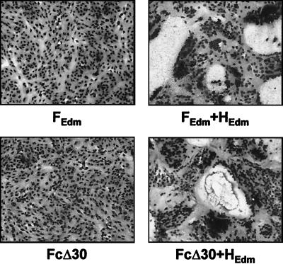
To determine whether FcΔ30 can mediate membrane fusion, Vero cells were transfected with pCG-FEdm or pCG-FcΔ30 either alone or in combination with a standard H protein (pCG-HEdm). The cells were fixed and stained 24 h posttransfection. As shown in Fig. 2, mutant and standard F proteins were not able to mediate fusion in the absence of H, whereas both proteins induced cell-to-cell fusion with similar efficiencies when coexpressed with standard H protein. Thus, F protein can mediate cell fusion regardless of whether it has a full-length or a truncated cytoplasmic domain. This indicates that the F protein needs only its extracellular domain and its transmembrane region for the formation of oligomers, proteolytic cleavage, cell surface transport, and interaction with the H protein.
FIG. 2.
Cell fusion induced by standard and truncated F proteins. Vero cells were transfected with the standard F gene (FEdm) or with FcΔ30 alone or in combination with the standard H gene (HEdm). At 24 h posttransfection, the cells were fixed and stained with Giemsa's staining solution. Magnification, ×100.
The membrane-proximal 14 amino acids of the H tail are required for fusion helper function.
To assess the competence of truncated H proteins to support F-mediated fusion, Vero cells were transfected with the standard F gene (pCG-FEdm) in combination with pCG-HEdm, pCG-HcΔ15, pCG-HcΔ16, pCG-HcΔ17, pCG-HcΔ18, pCG-HcΔ19, pCG-HcΔ20, pCG-HcΔ21, pCG-HcΔ22, pCG-HcΔ23, or pCG-HcΔ24. At 24 h posttransfection, the cells were fixed and stained with Giemsa's staining solution. Figure 3 shows that all H proteins with cytoplasmic tails of 14 or more amino acids (from HcΔ15 to HcΔ20) promoted extensive fusion similarly to standard H protein (HEdm). In contrast, HcΔ21, HcΔ22, HcΔ23, and HcΔ24 did not support prominent syncytium formation. Quantification of the cell fusion (see Material and Methods) showed that HcΔ21, HcΔ22, HcΔ23, and HcΔ24 induced fusion of less than 30% of the cells whereas standard H protein and the other truncation mutants mediated 80% cell fusion (Fig. 3, bottom). This clearly indicates that the fusion helper function of H is severely affected if the H tail contains less than the 14 membrane-proximal amino acids.
FIG. 3.
Fusion of Vero cells transiently expressing standard and truncated H proteins. (A) Cells were transfected with pCG-FEdm in combination with a plasmid containing standard H or truncated H genes (HEdm or HcΔ15 to HcΔ24). At 24 h posttransfection, the cells were fixed and stained with Giemsa's staining solution. Magnification, ×100. (B) Syncytium formation (% cell fusion) was quantified as described in Materials and Methods. The error bars indicate standard deviation.
Cell fusion involves both mixing of the outer leaflet membrane lipids (hemifusion) and mixing of the aqueous contents of donor and recipient cells (17). To analyze whether H tail mutants that are defective in the promotion of complete fusion can support hemifusion, we tested the ability of Vero cells expressing F in combination with H truncation mutants to fuse with target cells (BJAB cells) preloaded with the lipid dye R18. After a 1-h incubation, the dye was still retained in the membranes of target cells when overlaid on cells expressing HcΔ21, HcΔ22, HcΔ23, or HcΔ24, whereas the dye rapidly spread to cells expressing standard H protein or HcΔ20 (not shown). This clearly indicates that shortening of the H tail to <14 amino acids prevents the mixing of the outer membrane leaflets and thus interferes with an early step in the fusion process.
Importance of the sequence of the H cytoplasmic tail for fusion helper function.
We wanted to determine whether specific sequences in the H tail are required for fusion promotion activity. Therefore, we constructed four H genes with a minimal cytoplasmic tail of 14 amino acids in which either 1, 4, 6, or 10 of the N-terminal residues were replaced by alanines. The resulting proteins had the sequence M-AIVI (HcΔ21-A), M-AAAA (HcΔ24-4A), M-AAAAAA (HcΔ26-6A), or M-AAAAAAAAAA (HcΔ30-10A) (Fig. 1). Coexpression of these mutants with standard F protein clearly showed that HcΔ21-A and HcΔ24-4A, but not HcΔ26-6A and HcΔ30-10A, support fusion similarly to HcΔ20 (Fig. 4). This demonstrates that in an H protein with a minimal cytoplasmic tail of 14 residues, only the four N-terminal amino acids can be exchanged without affecting the ability to promote fusion.
FIG. 4.
Cell fusion induced by truncated H proteins with mutations at the N terminus. (A) Syncytium formation in Vero cells transiently expressing standard F protein in combination with truncated H proteins (HcΔ20, HcΔ21, and HcΔ24) or truncated H proteins elongated by the addition of alanines at the N terminus (HcΔ21-A, HcΔ24-4A, HcΔ26-6A, and HcΔ30-10A) was visualized at 24 h posttransfection by Giemsa staining. Magnification, ×100. (B) Quantification was performed as described in Materials and Methods. The error bars indicate standard deviation.
Surface expression of H truncation mutants.
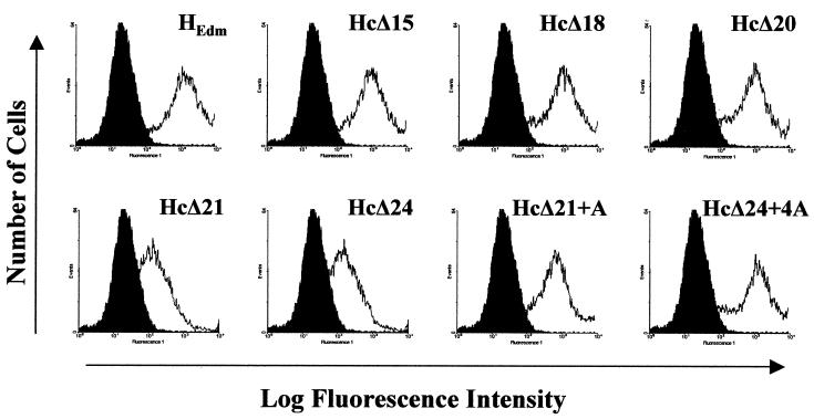
Although all truncation mutants showed bright immunofluorescent surface staining (not shown), the possibility that slight differences in the surface expression level existed which could be responsible for the differences in fusion helper function could not be ruled out. To assess this possibility, flow cytometry was performed (Fig. 5). The results in Table 1 show that the numbers of cells expressing the different truncation mutants were comparable (Table 1, positive cells). In contrast, the geometric mean levels of fluorescence intensity, representing the average amount of surface-expressed H protein, differed noticeably among the mutants (Table 1, cell surface expression). The surface expression levels of HcΔ21 and HcΔ24 (compared with that of the standard HEdm, set as 1.00) were 0.62 and 0.63, respectively.
FIG. 5.
Surface expression of standard and mutant H proteins. Vero cells were transfected with the indicated H genes. At 24 h posttransfection, mock- and H-transfected cells were stained for surface expression by using an H-specific antibody and subsequently analyzed by FACS scanning.
TABLE 1.
Surface expression of truncated H proteins
| Protein | % Positive cellsa | Cell surface expressionb | Cell fusionc | Fusion promotion activityd |
|---|---|---|---|---|
| HEdm | 68.2 | 1.00 | 1.00 | 1.00 |
| HcΔ15 | 64.2 | 0.93 | 1.12 | 1.20 |
| HcΔ18 | 67.0 | 0.88 | 1.11 | 1.26 |
| HcΔ20 | 62.6 | 0.93 | 1.13 | 1.21 |
| HcΔ21 | 59.0 | 0.62 | 0.31 | 0.49 |
| HcΔ24 | 58.7 | 0.63 | 0.09 | 0.14 |
| HcΔ21-A | 65.9 | 0.80 | 1.08 | 1.35 |
| HcΔ24-4A | 66.0 | 0.98 | 1.06 | 1.08 |
To test whether the reduced surface expression was due to an intracellular retention of H or to intracellular degradation of the protein, the amounts of intracellular and surface-expressed proteins were compared. For this purpose, a cell surface biotinylation assay was performed. Transfected cells were labeled by adding the non-membrane-permeating reagent sulfo-N-hydroxysuccinimidobiotin. The cells were lysed, and the H protein mutants were immunoprecipitated by an H-specific monoclonal antibody (K83). The precipitates were divided into two parts, separated on an SDS-10% polyacrylamide gel, and blotted to nitrocellulose. On one blot, biotin-labeled H proteins were detected with streptavidin-peroxidase. As shown in Fig. 6A, comparable amounts of HEdm, HcΔ18, and HcΔ20 were found on the surfaces of transfected cells. In contrast, the surface expression levels of HcΔ21 and HcΔ24 were clearly decreased, confirming the flow cytometric analysis. Parallel staining of the second blot with a polyclonal anti-MV antiserum and peroxidase-conjugated anti-rabbit immunoglobulins showed a similar decrease in the total amount of HcΔ21 and HcΔ24 (Fig. 6B). To evaluate whether the reduced expression level is due to rapid degradation of aberrantly folded protein or to an overall decrease in H translation, cells transfected with HEdm, HcΔ21, and HcΔ24 were metabolically labeled with 35S-labeled Promix for different times (Fig. 6C). After lysis, the H proteins were immunoprecipitated and subjected to SDS-PAGE. Quantification of the radioactivity of the precipitated proteins showed that the total amount of HcΔ21 and HcΔ24 synthesized in 10 and 30 min was clearly reduced (only about 35% of the expression found for standard H protein). As shown in Fig. 6D, the pulse-chase analysis with HEdm and HcΔ24 demonstrates similar stabilities of both proteins (half-life, about 3 h). This strongly suggests that the reduced expression levels of fusion-promotion-defective mutants is due to a decrease in the translation level of H proteins with cytoplasmic tails of <14 residues.
FIG. 6.
Expression levels of standard and truncated H proteins analyzed by cell surface biotinylation assay, Western blot analysis, and metabolic labeling. Vero cells were transfected with plasmids carrying the H genes indicated. At 24 h posttransfection, the cell surfaces were labeled with biotin and lysed. After immunoprecipitation of the H proteins, the samples were divided into two parts. Each was subjected to SDS-PAGE and blotted to nitrocellulose. (A) One blot was incubated with streptavidin-peroxidase to visualize surface-labeled H proteins. (B) The second blot was stained with a polyclonal MV antiserum and peroxidase-conjugated anti-rabbit immunoglobulins to detect the total amount of H proteins. (C) Transfected cells were radiolabeled with [35S]methionine and [35S]cysteine for 10 or 30 min, respectively. After lysis, H was immunoprecipitated and separated on a 10% SDS gel. (D) For pulse-chase analysis, cells were radiolabeled for 45 min and then incubated in chase medium for 0, 90, or 180 min. Quantification of the signals was performed using a BioImager.
To calculate the fusion helper activities of the truncation mutants, we normalized the cell fusion of the mutant H proteins to the amount of surface expression. This calculation (Table 1, cell fusion and fusion promotion activity) shows that even if HcΔ21 and HcΔ24 were expressed at wild-type levels, their fusion helper activities would be markedly reduced (0.49 and 0.14 versus 1.00 for standard H protein). This suggests that besides its impact on the surface transport of the H protein, the cytoplasmic domain also plays a direct role in fusion promotion. Although HcΔ21 and HcΔ24 were expressed at similar reduced levels, the calculated fusion promotion activity of HcΔ24 was clearly lower. This indicates that further shortening of the tail does not further increase impairment of H translation or intracellular degradation but increases the direct deleterious effect on the ability of the truncated H protein to support fusion.
Receptor binding activities of H truncation mutants.
The receptor binding activity of the H protein can be monitored by RBC binding (hemadsorption) to H protein-expressing cells (20). In order to determine whether the defect in fusion helper function is due to impaired receptor binding function, the hemadsorption activities of fusion-promoting and fusion-defective H mutants were analyzed. Figure 7A shows that RBC were efficiently bound to the surfaces of cells transfected with HcΔ20, HcΔ21, and HcΔ21-A. The binding was quantified by lysing bound RBC and measuring the released hemoglobin. The results shown in Fig. 7B (hemadsorption) demonstrate that the decrease in surface expression of the H protein was accompanied by a quantitative decrease in receptor binding of the mutant-expressing cells (0.075 for HcΔ20 versus 0.045 for HcΔ21). But when we normalized hemadsorption to surface expression, we found the receptor binding activities of the mutants to be almost equal (Fig. 7B, receptor binding activity). This indicates that the tail truncations did not significantly influence the receptor binding activities of the mutant H proteins.
FIG. 7.
Hemadsorption of Vero cells transiently expressing truncated H proteins. (A) At 16 h posttransfection, cells were incubated for 1 h with a 1% suspension of RBC. After removal of unbound erythrocytes by extensive washing, the cells were photographed using an inverted light microscope. Magnification, ×250. (B) To quantify hemadsorption, bound RBC were lysed and hemoglobin release was measured at A540. Receptor binding activity was calculated by normalizing hemadsorption to cell surface expression (Table 1).
Generation of infectious glycoprotein tail-truncated MV recombinants from cloned DNA.
Fusion of viral envelopes with target cell membranes and fusion of cells with neighboring cells are distinct processes which may have different requirements concerning the interaction with cellular components or the lateral mobility of the viral glycoproteins, since the surface densities of the viral glycoproteins in the virus envelope and infected or transfected cells are different (13, 42). Therefore, glycoprotein truncation mutants which promote cell-to-cell fusion in the transient-expression assays do not necessarily mediate virus-cell fusion in the background of a standard MV infection. To assess the importance of the H and F truncation mutants for the replication of MV, recombinant viruses were generated. cDNAs encoding the F and H variants were cloned into a full-length MV genome plasmid. Only rMV genomes harboring glycoprotein tail truncations differing in length from standard H and F proteins by 2n amino acids were generated, since they remained even multiples of 6 nucleotides, thus abiding by the so-called rule of six found for MV and many other paramyxoviruses (4, 37). To generate viruses, rMV genome plasmids were transfected into 293-3-46 helper cells in the presence of a plasmid containing the gene for the MV polymerase. Recombinant viruses were rescued with the standard protocol (37). Similar to rMVEdm, rMV-FcΔ30 and rMV-HcΔ20 could be successfully recovered on the first attempt. Reverse transcription-PCR sequencing analysis confirmed that the recombinant viruses encode glycoproteins with the respective deletions in the cytoplasmic tails. Rescue of infectious rMV-HcΔ24 (four attempts), as well as recovery of rMV-HcΔ22 (four attempts), failed. This clearly shows that H truncation mutants that did not support F-mediated cell-to-cell fusion in our transient-expression assay did not allow the generation of infectious MV.
Replication of glycoprotein tail-truncated viruses in cultured cells.
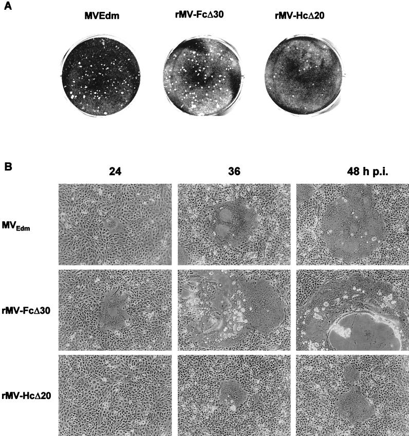
By performing a plaque assay to titrate the virus stocks of the rMV, differences in plaque sizes were noticed. As shown in Fig. 8A, plaques formed at 33°C by rMV-HcΔ20 were noticeably smaller than those found for standard MVEdm or rMV-FcΔ30. This was also observed when the plaque assay was carried out at 37°C (not shown). When we monitored the CPE in cells infected with the three viruses at 24, 36, and 48 h p.i., we clearly observed differences. Syncytia induced by MV-FcΔ30 and MVEdm are visible at 24 h p.i. In accordance with what has been reported for rMV-FcΔ24 (7), syncytia formed in rMV-FcΔ30-infected cells grew much faster than those induced by standard MVEdm and began to detach at 36 h p.i. In contrast to standard MVEdm and rMV-FcΔ30, rMV-HcΔ20 caused no visible CPE during the first 24 h. Then, within the next 12 h small syncytia formed. Syncytia detected at 48 h p.i. were still much smaller than those of rMV-FcΔ30 or MVEdm at 36 h p.i. Thus, the ability of rMV-HcΔ20 to induce syncytium formation is severely reduced. rMV-HcΔ20 also showed differences in multistep virus growth. For the growth curve experiment shown in Fig. 9, Vero cells were infected with the different rMVs at an MOI of 0.001 PFU/cell. At various times p.i., the culture media were harvested and cell-associated viruses were isolated by one freezing-thawing cycle. Virus titers were determined by plaque assay. In comparison to standard MVEdm and rMV-FcΔ30, the growth of rMV-HcΔ20 was delayed. Titers of both released and cell-associated rMV-HcΔ20 at early time points up to 48 h p.i. were detectably lower. However, after multiple replication cycles (72 h p.i.), maximum titers similar to those of standard MV were reached. This indicates that despite the reduced ability to mediate cell-to-cell fusion, rMV-HcΔ20 shows no substantial decrease in infectious-virus production.
FIG. 8.
Syncytium formation in cells infected with MV glycoprotein cytoplasmic-tail-truncated viruses. (A) Plaques in Vero cells infected with MVEdm, rMV-FcΔ30, or rMV-HcΔ20 were stained with crystal violet after 5 days of incubation at 33°C. (B) CPEs were monitored by phase-contrast microscopy at different times p.i. Magnification, ×100.
FIG. 9.
Growth curve analysis of MV glycoprotein cytoplasmic-tail-truncated rMV. Vero cells were infected with an MOI of 0.001 PFU/cell, and the culture medium (dashed lines) and the cells (solid lines) were harvested after incubation at 33°C for the indicated times. To obtain cell-associated viruses, cells were subjected to one freezing-thawing cycle. Virus titers were determined by plaque assay. Growth curves of MVEdm (▪), rMV-FcΔ30 (•), and rMV-HcΔ20 (▴) are shown. The values plotted represent averages of results from two experiments.
Colocalization of tail-truncated glycoproteins and M protein.
Cathomen et al. (5, 7) proposed that M protein downregulates cell-to-cell fusion by binding to the cytoplasmic portion of the H-F complex. In order to examine whether the difference in fusion activity of our tail-truncated viruses is due to a difference in the ability to interact with the M protein, colocalization of M and the truncated glycoproteins on the surfaces of rMV-HcΔ20- and rMV-FcΔ30-infected cells was analyzed. At 30 h p.i., the surfaces of the unfixed cells were labeled with a monoclonal anti-H antibody (Fig. 10A) or with a monoclonal anti-F antibody (Fig. 10B) and a rhodamine-conjugated secondary antibody. To visualize the M protein, the cells were permeabilized and FITC-conjugated monoclonal anti-M antibodies were added. In standard MV infection (MVEdm), H and F clearly colocalized with M, whereas neither H and M in rMV-HcΔ20-infected cells (Fig. 10A) nor F and M in rMV-FcΔ30-infected cells (Fig. 10B) showed marked colocalization. This indicates that the differences in the fusion activities of rMV-FcΔ30 and rMV-HcΔ20 are not due to differences in the ability of the truncated glycoprotein to interact with the M protein.
FIG. 10.
Colocalization of M with the viral glycoproteins in rMV-infected cells. Vero cells were infected in the presence of a fusion-inhibitory peptide with MVEdm, rMV-FcΔ30, or rMV-HcΔ20. At 30 h p.i., double immunostaining of the H and M proteins (A) or F and M proteins (B) was performed using either a monoclonal anti-F antibody or a monoclonal anti-H antibody and a rhodamine-conjugated secondary antibody followed by permeabilization and incubation with a FITC-conjugated anti-M antibody. Magnification, ×1,000.
DISCUSSION
In recent years, much has been learned about the mechanism of paramyxovirus-mediated cell fusion (19, 35, 39). In the fusion process, the glycoproteins operate as a coupled molecular scaffold to release and direct the fusion peptide to the target membrane. Accordingly, binding of the MV H protein to the cellular receptor (CD46 or SLAM) is a prerequisite to draw the membranes into juxtaposition for the fusion process to begin. It has been suggested that a conformational change in the H protein after receptor binding triggers a conformational change in F to release the fusion peptide, which inserts into the target membrane and induces the merging of the membranes. The role of MV H protein is more than a nonspecific attachment function. The sequential conformational changes very likely require precise lateral interactions between H and F oligomers (51). As in other paramyxoviruses, these interaction domains appear to be located in the ectodomains of the proteins. Here, part of the cysteine-rich region (amino acids 337 to 381) in the F protein and amino acid 98 in the stalk region of H are known to be required for the formation of fusion-active H-F complexes (15, 56). The data presented here make it clear that not only are regions in the ectodomain of H required but a minimal length of the H cytoplasmic tail is also crucial for the formation of adequate amounts of fusion-competent glycoprotein complexes. Since fusion competence is essential for MV infectivity, successful generation of recombinant MV requires an H protein with at least 14 cytoplasmic amino acids.
Our results show that the cytoplasmic tail of the F protein is not required for fusion in the transient-expression assay. Oligomerization, proteolytic cleavage, and interaction with the H protein appear not to be affected by the F tail truncation. In this regard, MV F resembles the F protein of human parainfluenza virus type 2 (HPIV-2) but differs from the F protein of HPIV-3, simian virus 5, and Newcastle disease virus (NDV) (1, 43, 58).
The cytoplasmic tails of integral membrane proteins are exposed at the cytoplasmic face of the ER membrane and thus may interact with cellular proteins that promote or delay the intracellular transport process. Deletions in the cytoplasmic portions of viral glycoproteins can have small, but also drastic, effects on intracellular transport and surface expression (1, 28, 32, 38, 45, 48, 57, 58). As we have shown here, deletions in the cytoplasmic tails of the MV glycoproteins can also have different effects on the biological functions of the proteins. F tail deletions did not remarkably affect the biological activity of the protein. In contrast, truncation of the H cytoplasmic tail was only tolerated up to 20 amino acids and further truncation severely affected translation efficiency. De novo synthesis of H truncation mutants with cytoplasmic tails of <14 residues was clearly reduced, and steady-state cell surface expression was only about 60% of the expression observed with standard H protein.
There are major differences among the paramyxoviruses in the relative importance of a proper ratio between F and HN proteins needed for efficient fusion promotion. Simian virus 5 F protein can promote fusion even in the absence of HN protein (1). In contrast, the ratio of the two surface glycoproteins directly affects fusion of HPIV-3. The extent of fusion was found to increase with increasing densities of HPIV-3 HN until F trimers and HN tetramers were present in equimolar ratios similar to that found in infected cells (10). Our data indicate that for MV as well, sufficient quantities of H on the surface are required for F-mediated fusion.
A decrease in surface expression of the H protein was accompanied by a comparable decrease in receptor binding, whereas fusion promotion decreased disproportionately. As there were relatively large amounts of H protein present on the surface at even the lowest surface density observed yet fusion was not observed, we propose that a threshold local density of H protein must be present to allow sufficient receptor interactions and to have sufficient accumulation of H-F complexes in the region of contact. To ensure sufficient local density of H on the cell surface, the H cytoplasmic tail must have at least 14 amino acids. Similar to MV H, substantial truncation of the NDV HN cytoplasmic tail resulted in clearly decreased surface expression (44), but in contrast to what we found when we normalized the fusion promotion activity of the MV H truncation mutants HcΔ21 and HcΔ24 to surface expression (49 and 14% of standard H activity), the NDV HN truncation mutant had a calculated fusion promotion activity of 213% compared with wild-type activity. This indicates that truncation of the MV H tail, but not that of the NDV HN protein, directly affects the fusion helper function of the attachment protein.
MV glycoproteins show lateral mobility on the surfaces of infected cells (21). This mobility is probably required to enable several such proteins to associate into active fusogenic entities. H proteins with <14 cytoplasmic amino acids might have decreased ability to undergo lateral redistribution so that the formation of fusion pore complexes is inhibited. Alternatively, the direct influence of a cytoplasmic-tail truncation on the fusion promotion activity of surface-expressed H protein might be due to a conformational change in the ectodomain or the transmembrane domain, affecting the membrane-proximal region in the ectodomain that is required for efficient H-F interaction and/or tetramerization of H. There is some evidence that the transmembrane domain and/or the cytoplasmic portion of paramyxovirus H/HN proteins is involved in the tetrameric structure, which in turn may be crucial for the biological activity of the proteins (33, 50, 58). If such changes in H-F interaction and/or H tetramerization are induced by H tail truncations, they obviously affect only fusion promotion activity. Receptor binding activities of HcΔ21 and HcΔ24 expressed on the cell surface were clearly not reduced. Whatever this more direct negative effect(s) on fusion promotion is due to, it only enhances the drastic negative effect which the tail truncations have on the surface expression of H.
In MV-infected cells, H and F tails are known to downregulate cell fusion by binding to the viral M protein. Consequently, truncations of the tails increase the fusion competence of the mutated viruses due to an impairment of the glycoprotein M interaction. This model was mainly based on observations made with SSPE strains and the corresponding rMVs (7). Supporting this model, our virus mutant rMV-FcΔ30 showed an enhanced fusion competence. Unexpectedly, rMV-HcΔ20 did not. Here, cell fusion capacity was found to be reduced, although H-M interaction was impaired. It could be speculated that in MV-HcΔ20-infected cells, H truncation not only impaired the interaction with M but also the interaction with cellular components enhancing MV-mediated cell fusion. Molecules promoting or hindering virus cell entry or fusion have been described for other paramyxoviruses, such as Sendai virus and NDV (18, 31). Interaction of the H tail with such a cellular-fusion-enhancing component appeared to be required only for virus-induced cell fusion, since HcΔ20 induced cell fusion in the transient expression assay as efficiently as standard H protein.
Our data emphasize that the influence of cytoplasmic-tail truncations on the biological activities of paramyxovirus proteins cannot be predicted. We have shown that the cytoplasmic tail of one MV envelope protein (F) is not required for surface expression and functionality. On the other hand, one amino acid more or less in the cytoplasmic tail of the second envelope protein (H) can make a decisive difference in the protein expression level, which in turn is crucial for the biological activities. Whereas a slightly reduced surface expression of H proportionally reduced the binding of H to cellular receptors, promotion of F-mediated cell fusion was completely abolished, thereby preventing the generation of infectious rMV.
Acknowledgments
This work was supported by a grant from the Deutsche Forschungsgemeinschaft to A.M.
We thank M. Billeter for kindly providing the MV rescue system; C. Coulibaly, R. Cattaneo, and J. and S. Schneider-Schaulies for African green monkey erythrocytes, monoclonal antibodies, and cloned MV genes; and G. Herrler for critical reading of the manuscript.
REFERENCES
- 1.Bagai, S., and R. A. Lamb. 1996. Truncation of the COOH-terminal region of the paramyxovirus SV5 fusion protein leads to hemifusion but not complete fusion. J. Cell Biol. 135:73-85. [DOI] [PMC free article] [PubMed] [Google Scholar]
- 2.Buchholz, C., D. Koller, P. Devaux, C. Mumenthaler, J. Schneider-Schaulies, W. Braun, D. Gerlier, and R. Cattaneo. 1997. Mapping of the primary binding site of measles virus to its receptor CD46. J. Biol. Chem. 272:22072-22079. [DOI] [PubMed] [Google Scholar]
- 3.Buckland, R., E. Malvoisin, P. Beauverger, and F. Wild. 1992. A leucine zipper structure present in the measles virus fusion protein is not required for its tetramerization but is essential for fusion. J. Gen. Virol. 73:1703-1707. [DOI] [PubMed] [Google Scholar]
- 4.Calain, P., and L. Roux. 1993. The rule of six, a basic feature for efficient replication of Sendai virus defective interfering RNA. J. Virol. 67:4822-4830. [DOI] [PMC free article] [PubMed] [Google Scholar]
- 5.Cathomen, T., B. Mrkic, D. Spehner, R. Drillien, R. Naef, J. Pavlovic, A. Aguzzi, M. A. Billeter, and R. Cattaneo. 1998. A matrix-less measles virus is infectious and elicits extensive cell fusion: consequence for propagation in the brain. EMBO J. 17:3899-3908. [DOI] [PMC free article] [PubMed] [Google Scholar]
- 6.Cathomen, T., C. J. Buchholz, P. Spielhofer, and R. Cattaneo. 1995. Preferential initiation at the second AUG of the measles virus F mRNA: a role for the long untranslated region. Virology 214:628-632. [DOI] [PubMed] [Google Scholar]
- 7.Cathomen, T., H. Y. Naim, and R. Cattaneo. 1998. Measles virus with altered envelope protein cytoplasmic tails gain cell fusion competence. J. Virol. 72:1224-1234. [DOI] [PMC free article] [PubMed] [Google Scholar]
- 8.Cattaneo, R., A. Schmid, D. Eschle, K. Baczko, V. ter Meulen, and M. A. Billeter. 1988. Biased hypermutation and other genetic changes in defective measles viruses in human brain infections. Cell 55:255-265. [DOI] [PMC free article] [PubMed] [Google Scholar]
- 9.Dörig, R. E., A. Marcil, A. Chopra, and C. D. Richardson. 1993. The human CD46 molecule is a receptor for measles virus (Edmonston strain). Cell 75:295-305. [DOI] [PubMed] [Google Scholar]
- 10.Dutch, R. E., S. B. Joshi, and R. A. Lamb. 1998. Membrane fusion promoted by increasing surface densities of the paramyxovirus F and HN proteins: comparison of fusion reactions mediated by simian virus 5 F, human parainfluenza virus type 3 F, and influenza virus HA. J. Virol. 72:7745-7753. [DOI] [PMC free article] [PubMed] [Google Scholar]
- 11.Erlenhoefer, C., W. J. Wurzer, S. Loffler, S. Schneider-Schaulies, V. ter Meulen, and J. Schneider-Schaulies. 2001. CD150 (SLAM) is a receptor for measles virus but is not involved in viral contact-mediated proliferation inhibition. J. Virol. 75:4499-4505. [DOI] [PMC free article] [PubMed] [Google Scholar]
- 12.Hammond, A. L., R. K. Plemper, J. Zhang, U. Schneider, S. J. Russell, and R. Cattaneo. 2001. Single-chain antibody displayed on a recombinant measles virus confers entry through the tumor-associated carcinoembryonic antigen. J. Virol. 75:2087-2096. [DOI] [PMC free article] [PubMed] [Google Scholar]
- 13.Henis, Y. I., Y. Herman-Barhom, B. Aroeti, and O. Gutman. 1989. Lateral mobility of both envelope proteins (F and HN) of Sendai virus in the cell membrane is essential for cell-cell fusion. J. Biol. Chem. 264:17119-17125. [PubMed] [Google Scholar]
- 14.Hsu, E. C., F. Sarangi, C. Iorio, M. S. Sidhu, S. A. Udem, D. L. Dillehay, W. Xu, P. A. Rota, W. J. Bellini, and C. D. Richardson. 1998. A single amino acid change in the hemagglutinin protein of measles virus determines its ability to bind CD46 and reveals another receptor on marmoset B cells. J. Virol. 72:2905-2916. [DOI] [PMC free article] [PubMed] [Google Scholar]
- 15.Hummel, K. B., and W. J. Bellini. 1995. Localization of monoclonal antibody epitopes and functional domains in the hemagglutinin protein of measles virus. J. Virol. 69:1913-1916. [DOI] [PMC free article] [PubMed] [Google Scholar]
- 16.Johnston, I. C., V. ter Meulen, J. Schneider-Schaulies, and S. Schneider-Schaulies. 1999. A recombinant measles vaccine virus expressing wild-type glycoproteins: consequences for viral spread and cell tropism. J. Virol. 73:6903-6915. [DOI] [PMC free article] [PubMed] [Google Scholar]
- 17.Kemble, G. W., T. Danieli, and J. M. White. 1994. Lipid-anchored influenza hemagglutinin promotes hemifusion, not complete fusion. Cell 76:383-391. [DOI] [PubMed] [Google Scholar]
- 18.Kumar, M., M. Q. Hassan, S. K. Tyagi, and S. P. Sarkar. 1997. A 45,000-Mr glycoprotein in the Sendai virus envelope triggers virus-cell fusion. J. Virol. 71:6398-6406. [DOI] [PMC free article] [PubMed] [Google Scholar]
- 19.Lamb, R. A. 1993. Paramyxovirus fusion: a hypothesis for changes. Virology 197:1-11. [DOI] [PubMed] [Google Scholar]
- 20.Lecouturier, V., J. Fayolle, M. Caballero, J. Carabana, M. L. Celma, R. Fernandez-Munoz, T. F. Wild, and R. Buckland. 1996. Identification of two amino acids in the hemagglutinin glycoprotein of measles virus (MV) that govern hemadsorption, HeLa cell fusion, and CD46 downregulation: phenotypic markers that differentiate vaccine and wild-type MV strains. J. Virol. 70:4200-4204. [DOI] [PMC free article] [PubMed] [Google Scholar]
- 21.Lydy, S. L., S. Basak, and R. W. Compans. 1990. Host cell-dependent lateral mobility of viral glycoproteins. Microb. Pathog. 9:375-386. [DOI] [PubMed] [Google Scholar]
- 22.Maisner, A., B. Mrkic, G. Herrler, M. Moll, M. A. Billeter, R. Cattaneo, and H.-D. Klenk. 2000. Recombinant measles virus requiring an exogenous protease for activation of infectivity. J. Gen. Virol. 81:441-449. [DOI] [PubMed] [Google Scholar]
- 23.Maisner, A., and G. Herrler. 1995. Membrane cofactor protein with different types of N-glycans can serve as measles virus receptor. Virology 210:479-481. [DOI] [PubMed] [Google Scholar]
- 24.Maisner, A., H.-D. Klenk, and G. Herrler. 1998. Polarized budding of measles virus is not determined by viral glycoproteins. J. Virol. 72:5276-5278. [DOI] [PMC free article] [PubMed] [Google Scholar]
- 25.Maisner, A., J. Schneider-Schaulies, M. K. Liszewski, J. P. Atkinson, and G. Herrler. 1994. Binding of measles virus to membrane cofactor protein (CD46): importance of disulfide bonds and N-glycans for the receptor function. J. Virol. 68:6299-6304. [DOI] [PMC free article] [PubMed] [Google Scholar]
- 26.Malvoisin, E., and F. Wild. 1993. Measles virus glycoproteins: studies on the structure and interaction of the haemagglutinin and fusion proteins. J. Gen. Virol. 74:2365-2372. [DOI] [PubMed] [Google Scholar]
- 27.Moll, M., H.-D. Klenk, G. Herrler, and A. Maisner. 2001. A single amino acid change in the cytoplasmic domains of measles virus glycoproteins, H, and F alters targeting, endocytosis and fusion in polarized Madin-Darby canine kidney cells. J. Biol. Chem. 276:17887-17894. [DOI] [PubMed] [Google Scholar]
- 28.Mulligan, M. J., G. V. Yamshchikov, G. D. Ritter, Jr., F. Gao, M. J. Jin, C. D. Nail, C. P. Spies, B. H. Hahn, and R. W. Compans. 1992. Cytoplasmic domain truncation enhances fusion activity by the exterior glycoprotein complex of human immunodeficiency virus type 2 in selected cell types. J. Virol. 66:3971-3975. [DOI] [PMC free article] [PubMed] [Google Scholar]
- 29.Naim, H. Y., E. Ehler, and M. A. Billeter. 2000. Measles virus matrix proteins specify apical virus release and glycoprotein sorting in epithelial cells. EMBO J. 19:3576-3585. [DOI] [PMC free article] [PubMed] [Google Scholar]
- 30.Naniche, D., G. Varior-Krishnan, F. Cervoni, T. F. Wild, B. Rossi, C. Rabourdin-Combe, and D. Gerlier. 1993. Human membrane cofactor protein (CD46) acts as a cellular receptor for measles virus. J. Virol. 67:6025-6032. [DOI] [PMC free article] [PubMed] [Google Scholar]
- 31.Okamoto, K., S. Ohgimoto, M. Nishio, M. Tsurudome, M. Kawano, H. Komada, M. Ito, Y. Sakakura, and Y. Ito. 1997. Paramyxovirus-induced syncytium cell formation is suppressed by a dominant negative fusion regulatory protein-1 (FRP-1)/CD98 mutated construct: an important role of FRP-1 in virus-induced cell fusion. J. Gen. Virol. 78:775-783. [DOI] [PubMed] [Google Scholar]
- 32.Parks, G. D., and R. A. Lamb. 1990. Defective assembly and intracellular transport of mutant paramyxovirus hemagglutinin-neuraminidase proteins containing altered cytoplasmic domains. J. Virol. 64:3605-3616. [DOI] [PMC free article] [PubMed] [Google Scholar]
- 33.Parks, G. D., and R. A. Lamb. 1990. Folding and oligomerization properties of a soluble and secreted form of the paramyxovirus hemagglutinin-neuraminidase glycoprotein. Virology 178:498-508. [DOI] [PubMed] [Google Scholar]
- 34.Peeples, M. E. 1991. Paramyxovirus M proteins. Pulling it all together and taking it on the road, p. 427-456. In D.W. Kingsbury (ed.), The paramyxoviruses. Plenum Press, New York, N.Y.
- 35.Peisajovich, S. G., O. Samuel, and Y. Shai. 2000. Paramyxovirus F1 protein has two fusion peptides: implications for the mechanism of membrane fusion. J. Mol. Biol. 296:1353-1365. [DOI] [PMC free article] [PubMed] [Google Scholar]
- 36.Plemper, R. K., A. L. Hammond, and R. Cattaneo. 2000. Characterization of a region of the measles virus hemagglutinin sufficient for its dimerization. J. Virol. 74:6485-6493. [DOI] [PMC free article] [PubMed] [Google Scholar]
- 37.Radecke, F., P. Spielhofer, H. Schneider, K. Kaelin, M. Huber, C. Dötsch, G. Christiansen, and M. A. Billeter. 1995. Rescue of measles viruses from cloned DNA. EMBO J. 14:5773-5784. [DOI] [PMC free article] [PubMed] [Google Scholar]
- 38.Rose, J. K., and J. E. Bergmann. 1983. Altered cytoplasmic domains affect intracellular transport of the vesicular stomatitis virus glycoprotein. Cell 34:513-524. [DOI] [PubMed] [Google Scholar]
- 39.Russell, C. J., T. S. Jardetzky, and R. A. Lamb. 2001. Membrane fusion machines of paramyxoviruses: capture of intermediates of fusion. EMBO J. 20:4024-4034. [DOI] [PMC free article] [PubMed] [Google Scholar]
- 40.Schneider, U., F. Bullough, S. Vongpunsawad, S. J. Russell, and R. Cattaneo. 2000. Recombinant measles viruses efficiently entering cells through targeted receptors. J. Virol. 74:9928-9936. [DOI] [PMC free article] [PubMed] [Google Scholar]
- 41.Schneider-Schaulies, J., J. Schnorr, U. Brinckmann, L. M. Dunster, K. Baczko, U. G. Liebert, S. Schneider-Schaulies, and V. ter Meulen. 1995. Receptor usage and differential downregulation of CD46 by measles virus wild-type and vaccine strains. Proc. Natl. Acad. Sci. USA 92:3943-3947. [DOI] [PMC free article] [PubMed] [Google Scholar]
- 42.Sekiguchi, K., and A. Asano. 1978. Participation of spectrin in Sendai virus-induced fusion of human erythrocyte ghosts. Proc. Natl. Acad. Sci. USA 75:1740-1744. [DOI] [PMC free article] [PubMed] [Google Scholar]
- 43.Sergel, T., and T. G. Morrison. 1995. Mutations in the cytoplasmic domain of the fusion glycoprotein of Newcastle disease virus depress syncytia formation. Virology 210:264-272. [DOI] [PubMed] [Google Scholar]
- 44.Sergel, T., L. W. McGinnes, M. E. Peeples, and T. G. Morrison. 1993. The attachment function of the Newcastle disease virus hemagglutinin-neuraminidase protein can be separated from fusion promotion by mutation. Virology 193:717-726. [DOI] [PubMed] [Google Scholar]
- 45.Simpson, D. A., and R. A. Lamb. 1992. Alterations to influenza virus hemagglutinin cytoplasmic tail modulate virus infectivity. J. Virol. 66:790-803. [DOI] [PMC free article] [PubMed] [Google Scholar]
- 46.Singh, M., R. Cattaneo, and M. A. Billeter. 1999. A recombinant measles virus expressing hepatitis B virus surface antigen induces humoral immune responses in genetically modified mice. J. Virol. 73:4823-4828. [DOI] [PMC free article] [PubMed] [Google Scholar]
- 47.Spielhofer, P., T. Bächi, T. Fehr, G. Christiansen, R. Cattaneo, K. Kaelin, M. A. Billeter, and H. Y. Naim. 1998. Chimeric measles virus with a foreign envelope. J. Virol. 72:2150-2159. [DOI] [PMC free article] [PubMed] [Google Scholar]
- 48.Tanaka, Y., and M. S. Galinski. 1995. Human parainfluenza virus type 3: analysis of the cytoplasmic tail and transmembrane anchor of the hemagglutinin-neuraminidase protein in promoting cell fusion. Virus Res. 36:131-149. [DOI] [PubMed] [Google Scholar]
- 49.Tatsuo, H., N. Ono, K. Tanaka, and Y. Yanagi. 2000. SLAM (CDw150) is a cellular receptor for measles virus. Nature 406:893-897. [DOI] [PubMed] [Google Scholar]
- 50.Thompson, S. D., W. G. Laver, K. G. Murti, and A. Portner. 1988. Isolation of a biologically active soluble form of the hemagglutinin-neuraminidase protein of Sendai virus. J. Virol. 62:4653-4660. [DOI] [PMC free article] [PubMed] [Google Scholar]
- 51.Von Messling, V., G. Zimmer, G. Herrler, L. Haas, and R. Cattaneo. 2001. The hemagglutinin of canine distemper virus determines tropism and cytopathogenicity. J. Virol. 75:6418-6427. [DOI] [PMC free article] [PubMed] [Google Scholar]
- 52.Wang, Z., L. Hangartner, T. I. Cornu, L. R. Martin, A. Zuniga, M. A. Billeter, and H. Y. Naim. 2001. Recombinant measles viruses expressing heterologous antigens of mumps and simian immunodeficiency viruses. Vaccine 19:2329-2336. [DOI] [PubMed] [Google Scholar]
- 53.Weidmann, A., C. Fischer, S. Ohgimoto, C. Ruth, V. ter Meulen, and S. Schneider-Schaulies. 2000. Measles virus-induced immunosuppression in vitro is independent of complex glycosylation of viral glycoproteins and of hemifusion. J. Virol. 74:7548-7553. [DOI] [PMC free article] [PubMed] [Google Scholar]
- 54.Weidmann, A., A. Maisner, W. Garten, M. Seufert, V. ter Meulen, and S. Schneider-Schaulies. 2000. Proteolytic cleavage of the fusion protein but not membrane fusion is required for measles virus-induced immunosuppression in vitro. J. Virol. 74:1985-1993. [DOI] [PMC free article] [PubMed] [Google Scholar]
- 55.Wild, T. F., E. Malvoisin, and R. Buckland. 1991. Measles virus: both the haemagglutinin and fusion glycoproteins are required for fusion. J. Gen. Virol. 72:439-442. [DOI] [PubMed] [Google Scholar]
- 56.Wild, T. F., J. Fayolle, P. Beauverger, and R. Buckland. 1994. Measles virus fusion: role of the cysteine-rich region of the fusion glycoprotein. J. Virol. 68:7546-7548. [DOI] [PMC free article] [PubMed] [Google Scholar]
- 57.Wilson, C., R. Gilmore, and T. Morrison. 1990. Aberrant membrane insertion of a cytoplasmic tail deletion mutant of the hemagglutinin-neuraminidase glycoprotein of Newcastle disease virus. Mol. Cell. Biol. 10:449-457. [DOI] [PMC free article] [PubMed] [Google Scholar]
- 58.Yao, Q., X. Hu, and R. W. Compans. 1997. Association of the parainfluenza virus fusion and hemagglutinin-neuraminidase glycoproteins on cell surfaces. J. Virol. 71:650-656. [DOI] [PMC free article] [PubMed] [Google Scholar]


