

Abstract
1. Blue, green, and red sensitive cone mechanisms have been studied in two types of on-centre ganglion cells in the Rhesus monkey's retina.
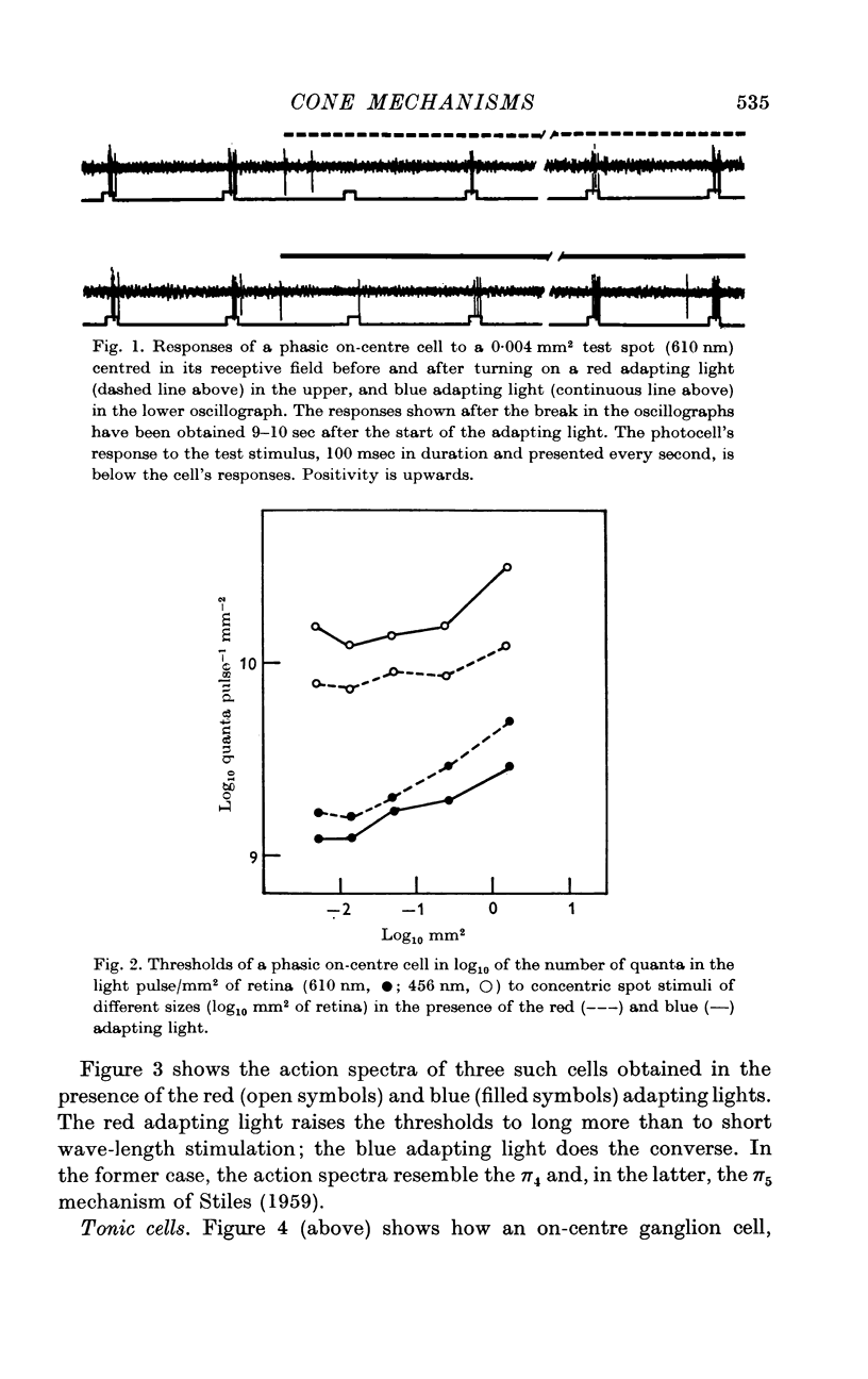
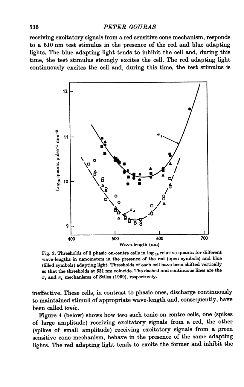
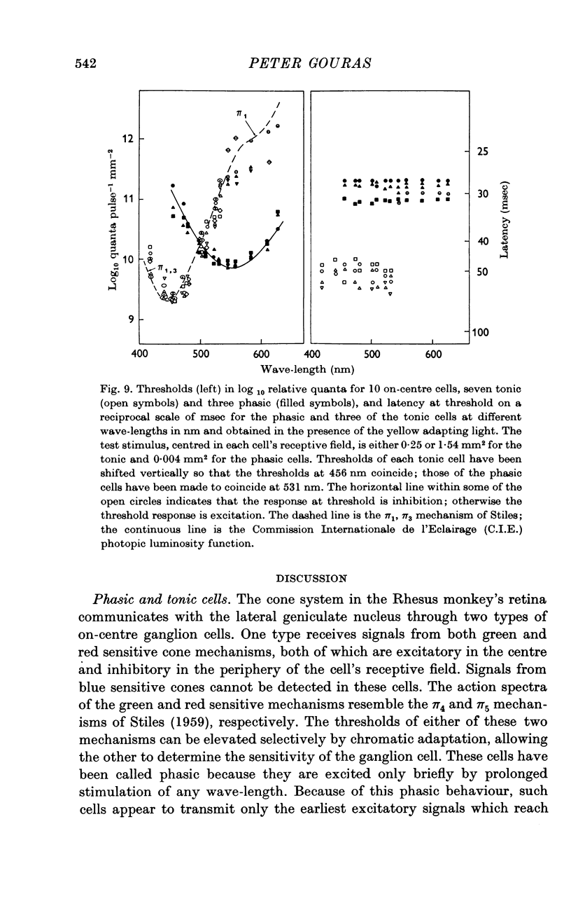
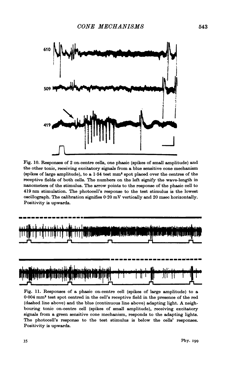
2. One type of cell receives signals from both green and red sensitive cone mechanisms, both of which excite in the centre and inhibit in the periphery of the cell's receptive field. These cells discharge transiently to maintained stimuli of any wave-length and are called phasic.
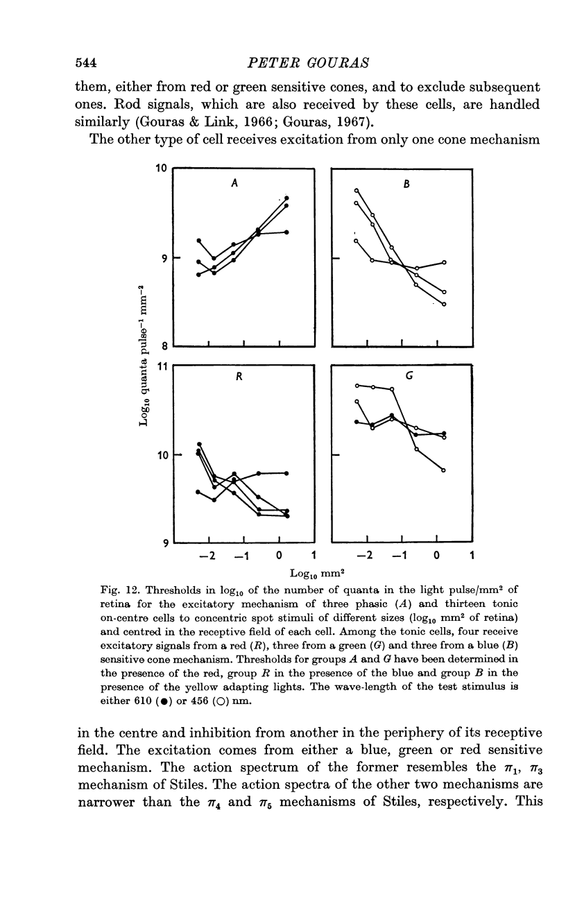
3. The second type of cell receives excitatory signals from only one cone mechanism, either blue, green or red sensitive, in the centre, and inhibition from another cone mechanism in the periphery of its receptive field. These cells discharge continuously to maintained stimuli of appropriate wave-length and are called tonic.
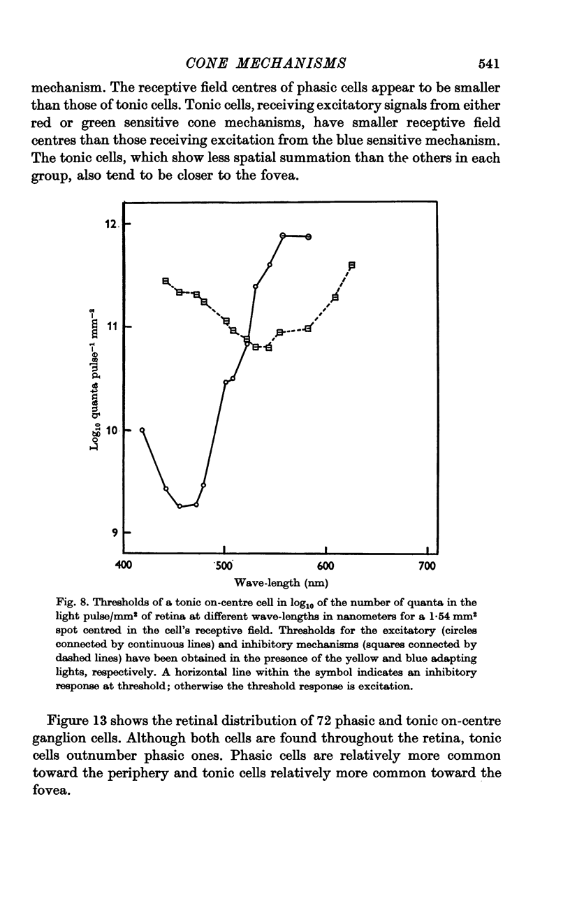
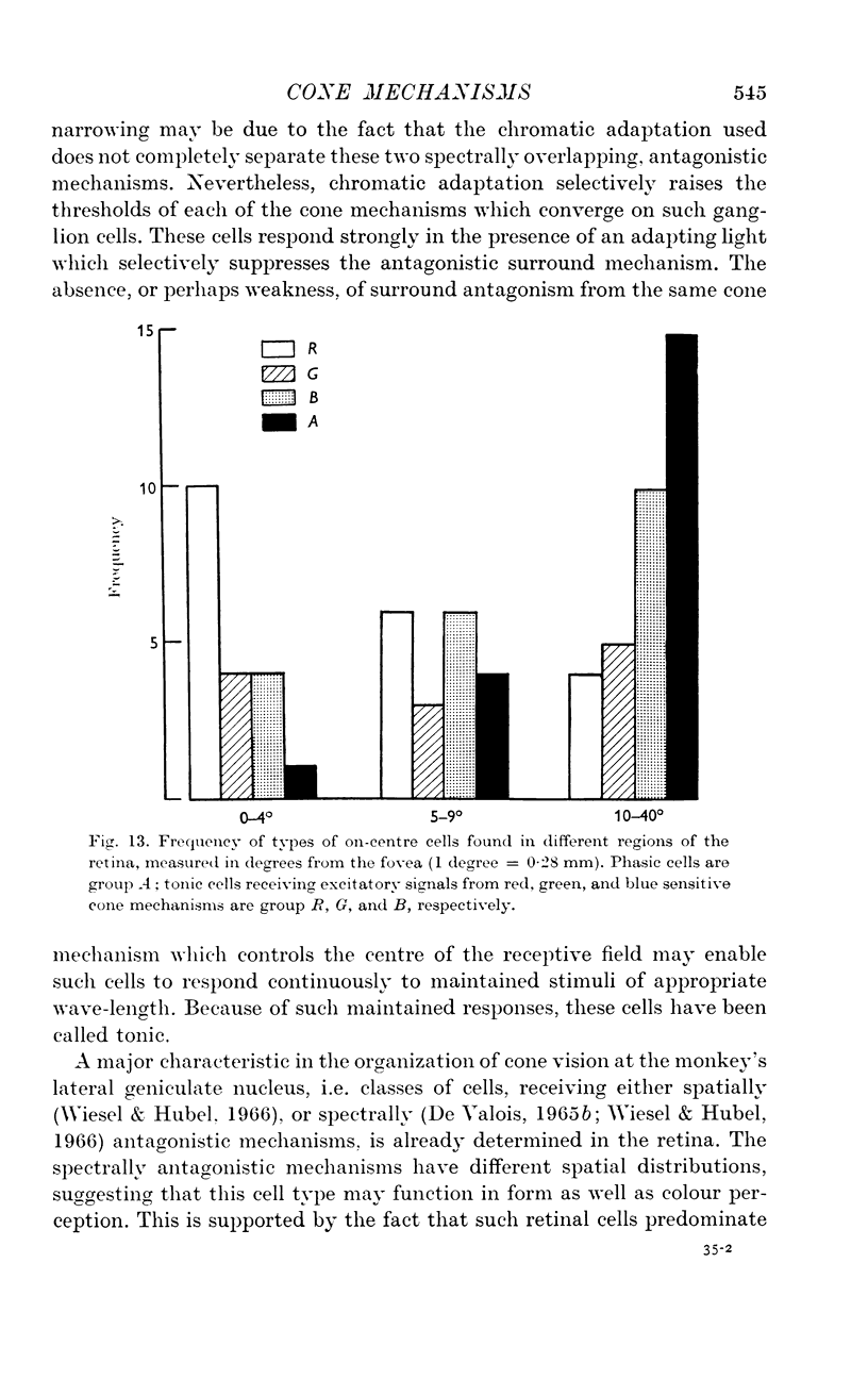
4. Tonic cells outnumber phasic cells although both are found adjacent to one another throughout the retina. Phasic cells are relatively more common toward the periphery and tonic cells relatively more common toward the fovea.
Full text
PDF
Selected References
These references are in PubMed. This may not be the complete list of references from this article.
- BARLOW H. B., FITZHUGH R., KUFFLER S. W. Change of organization in the receptive fields of the cat's retina during dark adaptation. J Physiol. 1957 Aug 6;137(3):338–354. doi: 10.1113/jphysiol.1957.sp005817. [DOI] [PMC free article] [PubMed] [Google Scholar]
- BROWN P. K., WALD G. VISUAL PIGMENTS IN HUMAN AND MONKEY RETINAS. Nature. 1963 Oct 5;200:37–43. doi: 10.1038/200037a0. [DOI] [PubMed] [Google Scholar]
- Brindley G. S., Du Croz J. J., Rushton W. A. The flicker fusion frequency of the blue-sensitive mechanism of colour vision. J Physiol. 1966 Mar;183(2):497–500. doi: 10.1113/jphysiol.1966.sp007879. [DOI] [PMC free article] [PubMed] [Google Scholar]
- DAS S. R. FOVEAL INCREMENT THRESHOLDS IN DARK ADAPTATION. J Opt Soc Am. 1964 Apr;54:541–546. doi: 10.1364/josa.54.000541. [DOI] [PubMed] [Google Scholar]
- De Valois R. L. Analysis and coding of color vision in the primate visual system. Cold Spring Harb Symp Quant Biol. 1965;30:567–579. doi: 10.1101/sqb.1965.030.01.055. [DOI] [PubMed] [Google Scholar]
- Du Croz J. J., Rushton W. A. The separation of cone mechanisms in dark adaptation. J Physiol. 1966 Mar;183(2):481–496. doi: 10.1113/jphysiol.1966.sp007878. [DOI] [PMC free article] [PubMed] [Google Scholar]
- Gouras P., Link K. Rod and cone interaction in dark-adapted monkey ganglion cells. J Physiol. 1966 May;184(2):499–510. doi: 10.1113/jphysiol.1966.sp007928. [DOI] [PMC free article] [PubMed] [Google Scholar]
- Gouras P. The effects of light-adaptation on rod and cone receptive field organization of monkey ganglion cells. J Physiol. 1967 Oct;192(3):747–760. doi: 10.1113/jphysiol.1967.sp008328. [DOI] [PMC free article] [PubMed] [Google Scholar]
- MARKS W. B., DOBELLE W. H., MACNICHOL E. F., Jr VISUAL PIGMENTS OF SINGLE PRIMATE CONES. Science. 1964 Mar 13;143(3611):1181–1183. doi: 10.1126/science.143.3611.1181. [DOI] [PubMed] [Google Scholar]
- Sperling H. G., Sidley N. A., Dockens W. S., Jolliffe C. L. Increment-threshold spectral sensitivity of the rhesus monkey as a function of the spectral composition of the background field. J Opt Soc Am. 1968 Feb;58(2):263–268. doi: 10.1364/josa.58.000263. [DOI] [PubMed] [Google Scholar]
- Wiesel T. N., Hubel D. H. Spatial and chromatic interactions in the lateral geniculate body of the rhesus monkey. J Neurophysiol. 1966 Nov;29(6):1115–1156. doi: 10.1152/jn.1966.29.6.1115. [DOI] [PubMed] [Google Scholar]