Abstract
The nucleoprotein (NP) of Newcastle disease virus (NDV) functions primarily to encapsidate the virus genome for the purpose of RNA transcription, replication, and packaging. This conserved multifunctional protein is also efficient in inducing NDV-specific antibody in chickens. Here, we localized a conserved B-cell immunodominant epitope (IDE) spanning residues 447 to 455 and successfully generated a recombinant NDV lacking the IDE by reverse genetics. Despite deletion of NP residues 443 to 460 encompassing the NP-IDE, the mutant NDV propagated in embryonated specific-pathogen-free chicken eggs to a level comparable to that of the parent virus. In addition, a B-cell epitope of the S2 glycoprotein of murine hepatitis virus (MHV) was inserted in-frame to replace the NP-IDE. Recombinant viruses properly expressing the introduced MHV epitope were successfully generated, demonstrating that the NP-IDE not only is dispensable for virus replication but also can be replaced by foreign sequences. Chickens immunized with the hybrid recombinants produced specific antibodies against the S2 glycoprotein of MHV and completely lacked antibodies directed against the NP-IDE. These marked-NDV recombinants, in conjunction with a diagnostic test, enable serological differentiation of vaccinated animals from infected animals and may be useful tools in ND eradication programs. The identification of a mutation-permissive region on the NP gene allows a rational approach to the insertion of protective epitopes and may be relevant for the design of NDV-based cross-protective marker vaccines.
The negative-strand RNA virus genome of Newcastle disease virus (NDV) contains six genes encoding six major structural proteins: nucleoprotein (NP), phosphoprotein (P), matrix protein (M), fusion protein (F), hemagglutinin-neuraminidase (HN), and RNA-dependent RNA polymerase (L). The RNA together with NP, P, and L proteins forms the ribonucleoprotein complex (RNP), which serves as a template for RNA synthesis (15). The NP together with the polymerase proteins, P and L, plays an eminent role in encapsidating the RNA. Moreover, NP regulates transcription and replication of the viral genome by interacting with P alone, with P and L, or with itself (NP-NP interaction). For Sendai paramyxovirus, it was shown that a conserved N-terminal region of NP was involved in NP-RNA and NP-NP interaction (5), whereas the carboxy-terminal domain was shown to be required for template function (8). Most of the NP is thus absolutely essential for virus replication due to multifold engagement of NP in the assembly and biologic activity of the RNP. In addition, NPs of negative-strand RNA viruses are highly immunogenic in nature and have been used as antigens for diagnostic purposes, including the NP of rabies (12), measles (34), vesicular stomatitis virus (1), and NDV (9). NP-based immunoassays are used mainly to monitor vaccination programs and as a diagnostic test in differentiating between vaccinated and infected animals in conjunction with subunit vaccines (18).
NDV is responsible for one of the most devastating diseases of poultry and has a substantial economic impact on the poultry industry. Vaccination of chickens, particularly those raised for commercial consumption, is carried out throughout the world. Although effective live or inactivated Newcastle disease (ND) vaccines are currently available, the virus remains an ongoing threat to commercial flocks. For continuation of successful international poultry trades, introduction of a systematic ND control measure is desirable. Recently intensive vaccination with marker vaccines and stamping-out strategies have been gaining popularity in veterinary medicine where eradication of specific diseases is of national or international interest (reviewed in reference 2). A marker vaccine is a vaccine that, in conjunction with a diagnostic test, enables serological differentiation of vaccinated animals from infected animals. An animal diagnosed as positive for the presence of a field infection has to be eliminated regardless of prior vaccination with a marker vaccine. A major drawback of all currently used whole-virus-based live and inactivated NDV vaccines is that vaccinated animals cannot be distinguished from infected animals with standard serological tests, such as hemagglutination inhibition (HI) or virus neutralization. Approaches to develop marker vaccines include deletion of one or more nonessential but immunogenic proteins. This is mainly applicable for large DNA viruses containing several dispensable genes (e.g., herpesviruses). An alternative approach for the development of a marker vaccine is the use of “subunit vaccines.” This approach has been implemented for many antigens involved in inducing protective immunity, including the two glycoproteins F and HN of NDV (4, 20, 31). The disadvantage of most subunit vaccines is that they are less effective than whole-virus-based live vaccines, emphasizing the importance of an NDV marker vaccine based on live attenuated virus.
For RNA viruses, in which most of the genes are essential, a deleted immunogenic gene may be complemented in trans. Classical swine fever viruses lacking the Erns or E2 genes were successfully recovered in cells constitutively expressing the glycoprotein Erns or E2, respectively (32, 35). For NDV, most vaccines are derived from low-virulence strains (lentogenic) that can only propagate in embryonated chicken eggs, where in trans complementation is very difficult. Another approach to generate live attenuated marker vaccines is generation of chimeric RNA viruses by replacing a whole immunogenic gene or part of a gene with a corresponding gene from another virus. This approach was employed for NDV by generating a recombinant NDV containing a hybrid HN protein. The chimeric HN is composed of only a one-fourth part of the HN protein from NDV, while the remaining part of the HN protein is derived from avian paramyxovirus type 2 or 4 (23). Since F and HN surface glycoproteins are the major viral proteins responsible for the induction of protective antibodies, replacement of three-fourth of the HN protein might impair the full effectiveness of a live vaccine, particularly for commercial chickens possessing passive neutralizing antibodies.
In this work we focused on the highly conserved nucleoprotein of NDV, which is involved not only in important biologic functions in the virus life cycle but also in inducing a high level of NDV-specific antibody in chickens. Interestingly, the NP-specific immunity alone is not protective against a lethal challenge (25, 33), indicating that the immunogenic NP region(s) is dispensable for protection. To determine whether an immunogenic region of the NP is permissive for modification, we first localized a conserved B-cell immunodominant epitope (IDE) on the NP gene of NDV. A recombinant NDV lacking 18 residues encompassing the IDE was successfully generated by reverse genetics (reviewed in references 7 and 26), and this site was used to insert foreign epitopes from an unrelated virus. Chickens immunized with the hybrid viruses lacked antibodies directed against the IDE and produced antibodies specific for the introduced epitopes. We describe the properties of these recombinant viruses and discuss the potential use of such viruses for displaying foreign immunogenic epitope and development of marker NDV vaccines.
MATERIALS AND METHODS
Transient expression of NP.
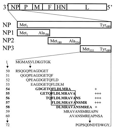
To identify an immunodominant region on the NP of NDV, the authentic open reading frame (ORF) containing a total of 489 amino acids was arbitrarily divided into three ORFs corresponding to amino acids 1 to 186, 180 to 334, and 335 to 489 (Fig. 1). The NP expression plasmid, pCiteNP (27), was digested with PstI, and relegation of an approximately 4.4-kb fragment resulted in an expression plasmid (NP1) coding for the amino-terminal 186 amino acids. To express the NP region corresponding to residues 180 to 334, PCR was performed on pCiteNP template using primers 5′-AAAGCCATGGCTGCGTATGAG-3′ (nucleotide positions 653 to 673; nucleotide numbering is according to that of Römer-Oberdörfer et al. [27]; EMBL accession no. Y18898) and 5′-GATGCCATCTAGATGGCAAAGGAG-3′ (nucleotide positions 1114 to 1137). The PCR product was digested with NcoI and XbaI and ligated into the same site of the pCite 2a vector (Novagen) to generate the expression plasmid NP2. A third plasmid (NP3) coding for the C-terminal amino acids 335 to 489 was obtained by digesting pCiteNP with NcoI and PstI and ligating an approximately 0.5-kb fragment into the same site of the pCite 2a vector. BSR-T7/5 cells stably expressing T7 RNA polymerase (6) were transfected with one of the plasmids by using the mammalian transfection kit (CaPO4 protocol; Stratagene) and processed for immunofluorescence (IF) analysis.
FIG. 1.
Localization of a B-cell epitope on the NP gene of NDV. A schematic representation of the NDV gene order (top) and the ORFs of the authentic NP gene (NP) and three ORFs (NP1 to NP3) prepared for transient expression are shown. The first and last amino acids in three-letter code and their corresponding positions on the authentic NP ORF are indicated. The peptide library containing 72 overlapping peptides corresponds to C-terminal NP residues 335 to 489. Pepscan analysis revealed very strong reactions (+++) with peptides 55 to 57 and weaker reactions (+) with peptides 54 and 58 (all shown in bold). Amino acids that are in common in the three strongly reacting peptides (representing the sequence of the epitope) are boxed.
Pepscan analysis.
After localizing the IF-reactive NP region, 72 overlapping 13-mer peptides were synthesized (shifted by two amino acids) and bound to a cellulose membrane for detailed mapping. The peptide library corresponded to C-terminal residues 335 to 489 (nucleotide positions 1124 to 1591) (Fig. 1). The membrane was incubated with chicken anti-NDV sera raised against live or inactivated NDV vaccines followed by incubation with goat anti-chicken immunoglobulin G conjugated with horseradish peroxidase. The membrane was washed at each incubation step with Tris-buffered saline (pH 8.0) containing 0.05% Tween 20. Proteins bound to the membrane were detected by chemoluminescent substrates as recommended by the supplier (Boehringer Mannheim, Mannheim, Germany).
Introduction of deletions into the full-length NDV cDNA.
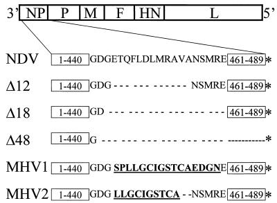
In order to generate mutant NDVs, the plasmid pflNDV expressing the full-length antigenome RNA of the lentogenic ND vaccine strain Clone-30 (27) was used to introduce mutations. After localizing an IDE, the region containing the epitope was deleted using the “quick change site-directed mutagenesis kit” according to the supplier's instructions (Stratagene). First, a DNA fragment encompassing nucleotides 77 to 2289 of the NDV genome was prepared by digestion of pflNDV with MluI, filling with Klenow, and treatment with ApaI. The ∼2.2-kb DNA fragment was ligated into the pBluescript SK vector (Stratagene) digested with ApaI and EcoRV. PCR mutagenesis was then performed on the above-described template using the following sets of primers: #P1A (5′-CCAGAAGCCGGGGATGGGAATAGCAT GAGGGAG-3′) and #P1B (5′-CTCCCTCATGCTATTCCCATCCCCGGCTTCTGG-3′) to delete nucleotides 1451 to 1486; #P2A (5′-CCAGAAGCCGGGGATGCGCCAAACTCTGCACAGG-3′) and #P2B (5′-CCTGTGCAGAGTTTGGCGCATCCCCGGCTTCTGG-3′) to delete nucleotides 1448 to 1501; and #P3A (5′-GGCAACCAGAAGCCGGGTGATGGACAAAACCCAGC-3′) and #P3B (5′-GCTGGGTTTTGTCCATCACCCGGCTTCTGGTTGCC-3′) to delete nucleotides 1445 to 1588. Mutagenized clones were digested with AatII/ApaI and cloned into the same sites of pflNDV. The presence of the introduced mutations was confirmed by sequencing the respective clones. The resultant full-length clones, with deletions on the NP gene corresponding to amino acid positions 444 to 455, 443 to 460, or 442 to 489 were named NDV-Δ12, NDV-Δ18, and NDV-Δ48, respectively (Fig. 2).
FIG. 2.
Recombinant NDV constructs. A schematic representation of the NDV gene order in the negative-strand genomic RNA is shown. Sequences around the IDE spanning amino acid residues 441 to 460 on the NP gene are shown in one-letter code. Broken lines show internal deletions of 12 or 18 amino acids in NDV-Δ12 (Δ12) and NDV-Δ18 (Δ18), respectively. NDV-Δ48 (Δ48) possesses a C-terminal truncated NP (deletion of 48 residues). Amino acid replacements in the region around the NP-IDE with MHV sequence are shown in bold and underlined. In NDV-MHV1 (MHV1), 16 amino acids of the NP sequence are replaced with an equivalent number from the MHV sequence, whereas in NDV-MHV2 (MHV2), 12 amino acids of the NP are replaced with only 10 amino acids derived from MHV. Therefore, the entire genome length of NDV-MHV2 is 2 amino acids shorter than the parent virus (NDV). An asterisk indicates a stop codon.
Replacing an immunodominant epitope with a foreign epitope.
In order to determine the possibility of replacing the identified NP-IDE by a foreign epitope, a well-characterized B-cell epitope of the S2 glycoprotein of murine hepatitis virus (MHV) was chosen (14, 16, 17, 30). PCR mutagenesis was performed on the 2.2-kb MluI/ApaI subclone described above to replace (in-frame) NP amino acids 444 to 459 (nucleotide positions 1451 to 1499) with the MHV-A59 S2 glycoprotein sequence 846SPLLGCIGSTCAEDGN861 (MHV-1) or NP amino acids 444 to 455 (nucleotide positions 1451 to 1486) with the sequence 848LLGCIGSTCA857 (MHV-2) (Fig. 2). The primer pairs used for the PCR mutagenesis are the following: #138 (5′-GAAGCCGGGGATGGGAGTCCTCTACTTGGATGCATAGGTTCAACATGTGCTGAAGACGGCAATGAGGCGCCAAACTCTGC-3′) and #139 (5′-GCAGAGTTTGGCGCCTCA TTGCCGTCTTCAGCACATGTTGAACCTATGCATCCAAGTAGAGGACTCCCATCCCCGGCTTC-3′) to replace nucleotides 1451 to 1499 with MHV-1 sequence and #140 (5′-GAAGCCGGGGATGGGCTACTTGGATGCATAGGTTCAACATGTGCTAATAGCATGAGGGAGGCG-3′) and #141 (5′-CGCCTCCCTCATGCTATTAGCACATGTTGAACCTAT GCATCCAAGTAGCCCATCCCCGGCTTC-3′) to replace nucleotides 1451 to 1486 with MHV-2 sequence. Mutagenized clones were digested by AatII/ApaI and cloned into the same sites of pflNDV. The presence of the introduced mutations was confirmed by sequencing the respective clones. The resultant full-length clones possessing MHV-1 sequence or MHV-2 sequence were named NDV-MHV1 and NDV-MHV2, respectively.
Recovery of recombinant viruses.
Transfection and virus recovery were done essentially as described previously (19). Approximately 1.5 × 10⁶ BSR-T7/5 cells stably expressing phage T7 RNA polymerase (6) were grown overnight to 90% confluency in 3.2-cm-diameter culture dishes. Cells were transfected with plasmid mixtures containing 5 μg of pCite-NP, 2.5 μg of pCite-P, 2.5 μg of pCite-L, and 10 μg of one of the full-length clones using a mammalian transfection kit (CaPO4 transfection protocol; Stratagene). Three days after transfection, supernatants were harvested and inoculated into the allantoic cavity of 9- to 11-day-old embryonated specific-pathogen-free (SPF) chicken eggs. After 3 to 4 days of incubation, the presence of virus in the allantoic fluid was determined by rapid plate hemagglutination (HA) test using chicken erythrocytes (3). The infectious titer, expressed as 50% embryo-infectious dose (EID50), was calculated using the method of Reed and Muench (24).
RT-PCR and sequence analysis.
The recombinant viruses were serially passaged five to eight times in embryonated SPF eggs to determine the stability of the introduced modifications. In addition, virus reisolation experiments were performed with the tracheas of SPF chickens vaccinated at 1 day of age. Tracheal homogenates were first inoculated into 9- to 11-day-old embryonated SPF chicken eggs to grow the virus to adequate titer for RNA isolation. BSR-T7/5 cells were infected with the viruses, and total RNA was prepared 24 to 36 h after infection using the Rneasy kit (Qiagen). Reverse transcription (RT) was performed by avian myeloblastosis virus reverse transcriptase on 1 μg of total RNA. The PCR products were analyzed on 1% agarose gel and used directly for sequencing.
Replication of recombinant viruses in embryonated eggs.
To determine the effect of the introduced mutations on the efficiency of virus propagation, 9- to 11-day-old embryonated SPF chicken eggs were inoculated with 6.0 log10 EID50/egg and incubated at 37°C. Allantoic fluid from each group of 10 eggs was pooled at 12, 24, 48, and 72 h after infection, and serial 10-fold dilutions were prepared and inoculated into the allantoic cavity of 9- to 11-day-old embryonated SPF chicken eggs. After 3 to 4 days of incubation, the titer was determined by a rapid-plate HA test (3).
Immunization of chickens with the recombinant viruses.
To determine the absence of antibodies directed against the NP-IDE or the presence of antibodies specific for the introduced MHV epitopes, three groups of each 10 SPF chickens at the age of 3 weeks were immunized with NDV-Δ18, NDV-MHV1, or NDV-MHV2. Animals were vaccinated by the oculo-nasal route with a dose of 6.0 log10 EID50 per 0.2 ml and boosted 5 weeks after the first immunization with the same dose. Serum samples were collected just before vaccination and 5 and 7 weeks after the first immunization. For comparison, two groups of each 10 SPF chickens at the age of 3 weeks were vaccinated with commercial live or inactivated Clone-30 vaccines. Serum samples were collected just before vaccination and 5 weeks after immunization. Serum samples were tested by HI test (3) and enzyme-linked immunosorbent assays (ELISAs) to determine the level of seroconversion.
To determine the safety and efficacy of the recombinant viruses, 1-day-old SPF chickens were vaccinated by eye-drop route at a dose of 6 log10 EID50/chick. At 3 weeks, blood samples were taken and all animals were challenged with the virulent NDV Herts strain, intramuscularly administered. Sera were assayed for NDV antibodies in the HI test and ELISAs. Animals were observed for a period of 2 weeks after challenge for the occurrence of clinical signs of NDV or mortality.
ELISA.
An 18-mer synthetic peptide comprising the NP amino acid sequence 443 to 460 was synthesized and coupled to bovine serum albumin (BSA). Briefly, synthetic peptide (0.5 mg/ml) was coupled to BSA (0.4 mg/ml) in phosphate-buffered saline (PBS) by adding a 2.5% glutaraldehyde solution (final concentration, 0.067 mM). Following overnight incubation at ambient temperature in darkness, the coupling reaction was stopped by the addition of glycine at a final concentration of 100 mM. Peptide-BSA complexes were stored frozen at −20°C. As a control, BSA alone was treated in a similar way with glutaraldehyde. Microtiter plates were coated overnight at 2 to 8°C with 0.5-μg synthetic peptide coupled to BSA in coating buffer (carbonate-buffer, pH 9.6). Unbound antigen was removed by washing with PBST solution (PBS, Tween 20). Serum samples diluted 50-fold were added to the coated wells and incubated for 90 min at 37°C in a humidified atmosphere. Wells were washed with PBST solution and incubated with 100 μl of horseradish peroxidase-conjugated rabbit anti-chicken immunoglobulins. After 45 min of incubation at 37°C, wells were washed and the antigen-antibody complexes were detected by the addition of 100 μl of 3,3′,5,5′-tetramethylbenzidine substrate. After 15 min of incubation at ambient temperature, enzymatic reaction was stopped by adding 50 μl of 2 M sulfuric acid to each well.
To determine whether chickens immunized with NDV-MHV1 and NDV-MHV2 viruses had developed antibodies specific for the expressed MHV epitopes, ELISA plates were coated using a 16-mer synthetic peptide encompassing the MHV 5B19 epitope (846SPLLGCIGSTCAEDGN861). The peptide was coupled to BSA by means of glutaraldehyde as described above. Removal of unbound antigen, blocking, and the remaining incubation steps of the ELISA procedure were identical to the NP 18-mer peptide ELISA. The entire NDV-specific antibody response was measured in ELISA plates coated with sucrose gradient-purified NDV Clone 30 antigens. Wells of microtiter plates were coated overnight with 1% Triton X-100-treated viral antigen in 40 mM PBS (pH 7.2). Optical densities (OD) were measured at 450 nm. The cutoff value was set three standard deviations above the average P/N ratios of negative control sample from SPF chickens (where P is the OD of samples from peptide coated wells, and N is the OD of samples from BSA-coated wells).
Immunofluorescence analysis.
In order to confirm the absence of the NP-IDE and determine the expression of the introduced MHV epitope, BSR-T7 cells were infected at a multiplicity of infection of 0.1 with the parent recombinant Clone-30, NDV-Δ18, NDV-MHV1, or NDV-MHV2. After 18 h of incubation, infected cells were fixed with cold ethanol (96%) for 1 h at room temperature. After three washes with PBS, cells were incubated with monoclonal antibodies (MAbs) directed against the F protein, NDV-185 (19), the immunodominant epitope of the NP protein, NDV-36 (19), or the 5B19 MHV epitope (13, 14). Cells were washed and stained with fluorescein isothiocyanate-conjugated anti-mouse antibody and examined by fluorescence microscopy.
Immunoblotting.
For virus purification, 9- to 11-day-old embryonated SPF chicken eggs were infected and allantoic fluid was collected 3 to 4 days postinfection. Virus in the allantoic fluid was then purified and concentrated by centrifugation through a 20% sucrose cushion in a Beckman SW28 rotor at 21,000 rpm for 90 min. The pellet was resuspended and mixed with protein sample buffer to disrupt the virions. Viral proteins from purified virions were then resolved by sodium dodecyl sulfate-polyacrylamide gel electrophoresis, transferred to polyvinylidene difluoride membranes (Millipore), and incubated with anti-NP, anti-MHV MAbs, or a polyclonal chicken serum raised against NDV Clone-30 vaccine. Membranes were then incubated with peroxidase-conjugated goat anti-chicken or anti-mouse immunoglobulin G. Proteins were visualized after incubation with peroxidase substrate (Vector).
Immunization and challenge of mice.
To determine the protective ability of the MHV epitope expressed by NDV, groups of 4-week-old BALB/c female MHV-seronegative mice were used for immunization and challenge experiments. Two groups of 10 mice each were immunized with NDV-MHV1 or NDV-MHV2 at a dose of 6.0 log10 EID50/mice and boosted with a similar dose 4 weeks later. Immunized animals, as well as 10 age-matched controls, were challenged 2 weeks after the booster immunization with the wild-type MHV A59 strain at a dose of 10 50% lethal doses/mice by the intraperitoneal route. Animals were observed for clinical signs of MHV and mortality for 2 weeks after challenge.
RESULTS
Identification of an immunodominant epitope on the NP of NDV.
In addition to the surface glycoproteins F and HN that induce protective immunity, the NP of NDV was shown to be highly immunogenic. However, chickens immunized with the NP alone, though demonstrating very high NP-specific antibody responses, were not protected against a lethal challenge (33). This finding suggests that immunogenic region(s) on the NP gene may be dispensable for protection. In order to localize a conserved immunodominant epitope on the NP gene, the NP was expressed in three small ORFs: NP1, NP2, and NP3 (Fig. 1). IF analysis of the three transiently expressed NP fragments using NDV-specific chicken sera revealed a strong positive IF reaction with NP3, suggesting localization of relevant epitopes in the region encompassing residues 335 to 489. To precisely localize the epitope(s), a pepscan analysis was performed using 72 overlapping peptides covering the entire region coded by NP3. The analysis showed a very strong positive reaction with peptides #55, #56 and #57 and a reduced reactivity with two flanking peptides, #54 and #58 (Fig. 1). The strong positive reaction with the three peptides, which have the sequence 447FLDLMRAVA455 in common, indicates that residues 447 to 455 of the NP protein represent an IDE. A comparison of the deduced amino acid sequences of the entire NP of Clone-30 with the corresponding sequences of the velogenic Texas GB and the mesogenic Beaudette C strains of NDV reveals 100 and 99.3% identity, respectively. The amino acid sequence in the above IDE is identical in these strains.
Generation of recombinant viruses.
In order to delete the IDE on the NP gene or block expression of the C-terminal part of NP, the modifications described under Materials and Methods were carried out (Fig. 2). Each modified full-length cDNA clone, together with three support plasmids expressing NDV NP, P, and L proteins, was transfected into BSR-T7/5 cells as described previously (19, 27). After 3 to 5 days of incubation, supernatants were harvested and inoculated into 9- to 11-day-old embryonated SPF chicken eggs. After 3 to 4 days of incubation, allantoic fluid samples were harvested and subjected to an HA test. HA was detected in allantoic fluid from eggs inoculated with the supernatant from cells transfected with NDV-Δ18, indicating that the region encompassing the identified epitope is dispensable. However, despite repeated transfection experiments and successive egg passages, we were unable to recover virus from NDV-Δ12 and NDV-Δ48 constructs. To determine whether the deleted site in NDV-Δ18 could be used to insert foreign epitopes, constructs possessing a long or short MHV epitope were prepared (Fig. 2) and transfected into BSR-T7/5 cells. After passaging into 9- to 11-day-old embryonated SPF chicken eggs, HA was detected in the allantoic fluid of eggs inoculated with the supernatant from cells transfected with NDV-MHV1 and NDV-MHV2 constructs. The recombinant viruses were then serially passaged five to eight times in embryonated SPF eggs or at least once in day-old SPF chickens. RNAs isolated from cells infected with the viruses serially passaged in eggs or with the viruses reisolated from chickens were subjected to RT-PCR. For all three viruses the obtained sequences at all the passage levels or after a chicken passage corresponded exactly to the alterations introduced into the respective cDNA clones, demonstrating the stability of the introduced modifications.
Characterization of recombinant NDVs.
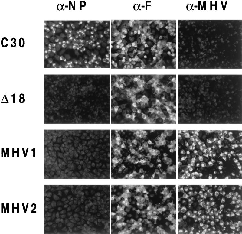
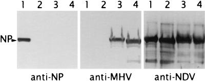
To determine whether the recombinant viruses lack the deleted NP-IDE and also express the newly introduced epitope (NDV-MHV1 and NDV-MHV2), BSR cells were infected with the respective recombinant viruses and subjected to IF (Fig. 3) and Western blot analysis (Fig. 4). Using the anti-F MAb, the expression of F protein was indistinguishable in all viruses (Fig. 3). In contrast, a MAb directed against the NP-IDE reacted only with the parent virus Clone-30. As expected, the MAb directed against the MHV epitope reacted only with cells infected with NDV-MHV1 or NDV-MHV2 (Fig. 3 and 4), demonstrating that the epitope is properly expressed after in-frame insertion into the ORF of the NP gene. The slight differences in mobility of the NP proteins (Fig. 4) most likely result from the deletion of 18 amino acids (NDV-Δ18) or the presence or absence of specific residues of the S2 epitope sequence (NDV-MHV1 and NDV-MHV2).
FIG. 3.
Expression of foreign epitopes by recombinant NDVs. BSR cells were infected with the parent recombinant Clone-30 (C30), NDV-Δ18 (Δ18), NDV-MHV1 (MHV1), or NDV-MHV2 (MHV2). Approximately 18 h after infection, cells were processed for indirect immunofluorescence after incubation with MAbs specific for NP protein (α-NP), for F protein (α-F), or for MHV epitope (α-MHV). As expected, the MAb specific for the NP-IDE reacted only with the parent virus, and the anti-MHV MAb recognized the MHV epitope inserted in-frame into the NP gene.
FIG. 4.
Engineered nucleoproteins of sucrose-purified recombinant NDVs. Virions in the allantoic fluid of infected embryonated eggs were purified by centrifugation through 20% sucrose. Samples were loaded in triplicate (lane 1, parent recombinant Clone-30; lane 2, NDV-Δ18; lane 3, NDV-MHV1; lane 4, NDV-MHV2), and blots were incubated with MAbs specific for the NP (anti-NP) or specific for the MHV 5B19 epitope (anti-MHV). Polyclonal chicken serum raised against NDV was used as a control (anti-NDV).
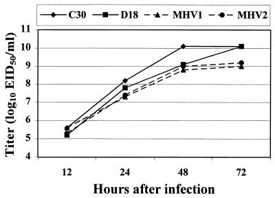
To determine if the deletion of NP-IDE and/or insertions of heterologous sequences into the NP of NDV affected replication, the growth kinetics of the recombinants was analyzed in 9- to 11-day-old embryonated SPF chicken eggs. The titer of the parent virus peaked 2 days after infection and remained the same on further incubation. The deletion mutant NDV-Δ18 also reached a similar peak titer of 10 log10 EID50/ml, albeit 1 day later (Fig. 5). A maximum difference of ∼10-fold in the kinetics of replication between the deletion mutant and the parent virus, which was compensated upon further incubation, was observed at 2 days of infection. Taking the involvement of the NP in many viral functions into consideration, this slight delay in replication kinetics is not surprising. The two recombinants expressing the MHV epitope also had replication kinetics similar to that of NDV-Δ18 for the first 2 days after infection. At 3 days of infection, both recombinants reached a maximum titer of only ∼9 log10 EID50/ml, which is ∼10-fold lower than that of the parent virus or that of NDV-Δ18 mutant (Fig. 5). To determine the significance of the differences in growth kinetics, Friedman two-way analysis of variance was used for statistical analysis. No significant difference was found between the growth kinetics of the parent Clone-30 and the mutant NDV-Δ18, whereas the differences between the parent virus and the two hybrid viruses expressing the MHV epitopes were significant (P < 0.05). For the two hybrid viruses, no further increase in titer was observed even 4 days after infection (data not shown). The specific cause of this defect remains to be elucidated.
FIG. 5.
Replication kinetics of recombinant NDVs. Ten embryonated SPF chicken eggs per group were inoculated with the parent recombinant Clone-30 (C30), NDV-Δ18 (D18), NDV-MHV1 (MHV1), or NDV-MHV2 (MHV2) at a dose of 6 log10 EID50/egg and incubated at 37°C. Allantoic fluid from each 10 eggs was harvested at 12, 24, 48, and 72 h after infection. The virus titers were determined by inoculation of serial 10-fold dilutions of the harvested material into the allantoic cavity of 9- to 11-day-old embryonated SPF chicken eggs and conducting a rapid-plate HA test after 3 to 4 days of incubation.
Immunization, serology, and challenge of chickens.
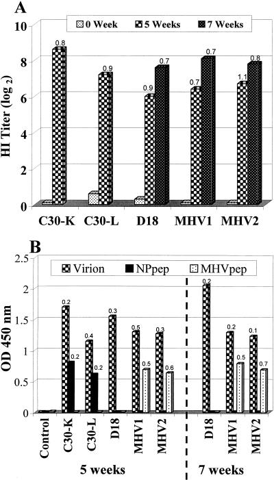
To determine the NDV-specific serological response, sera collected from immunized chickens were first subjected to HI test and whole-virus-based ELISA (Fig. 6). The immune responses measured by these two tests were comparable in the group of animals immunized with the recombinants and the live conventional vaccine. The NDV-specific response against the killed vaccine containing an adjuvant was in general superior to the response in the group of animals immunized with live viruses. Animals immunized with the recombinant viruses were given a booster dose at 5 weeks after the first vaccination to determine the absence of antibody response against the deleted NP-IDE even after repeated vaccinations. As shown in Fig. 6, the HI titers for all chickens at 2 weeks after booster vaccination were above 7 log2 HI units, and the reaction measured in the whole NDV based ELISA was of similar magnitude.
FIG. 6.
Serological response of immunized SPF chickens. Ten SPF chickens per group were vaccinated with commercial Clone-30 killed (C30-K) or live (C30-L) vaccines or with live recombinant virus NDV-Δ18, NDV-MHV1, or NDV-MHV2. Chickens were immunized at 3 weeks of age, and serum samples collected just before vaccination (0 weeks) or 5 weeks after the first vaccination (5 weeks) were analyzed for all the groups. The groups of animals immunized with NDV-Δ18, NDV-MHV1, and NDV-MHV2 were boosted 5 weeks after the first immunization. Results for the sera collected 2 weeks after the booster vaccination were shown to demonstrate the absence of antibodies against the deleted NP-IDE even after revaccination. Each bar shows the mean values for 10 samples, and the numbers at the tops of the bars indicate the respective standard deviations. (A) Hemagglutination-inhibition titer (log2) against NDV. (B) ELISA assays based on whole NDV antigen (Virion), on plates coated with the 18-mer NP peptide (NPpep), or on plates coated with 16-mer MHV peptide (MHVpep). In contrast to chickens vaccinated with conventional live or inactivated vaccines, none of the chickens immunized with the three recombinant viruses showed reactivity in the NP-peptide based ELISA. Up to 90% of the chickens immunized with NDV-MHV1 or NDV-MHV2 showed an antibody response specific for the introduced MHV epitope.
To determine whether animals vaccinated with the recombinant viruses could be serologically distinguished from NDV-infected animals or from animals vaccinated with conventional NDV vaccines, sera were subjected to ELISAs based on 18-mer NP peptide or 16-mer MHV peptide (Fig. 6B). Analysis of sera obtained from animals vaccinated with conventional inactivated or live NDV vaccines showed reactivity against the native 18-mer NP peptide by ELISA. In contrast, none of the sera obtained from animals vaccinated with NDV-Δ18, NDV-MHV1, or NDV-MHV2 reacted in the ELISA, demonstrating that chickens immunized with these recombinants completely lack antibodies directed against the NP-IDE even after a repeated vaccination. Interestingly, as many as 90% of chickens immunized with NDV-MHV1 or NDV-MHV2 reacted positively in the 16-mer MHV-peptide-based ELISA, demonstrating that chickens produce specific antibodies against the MHV epitope expressed by recombinant NDVs (Fig. 6B). The simultaneous presence of anti-MHV antibodies and complete absence of antibodies directed against NP-IDE make these recombinant viruses attractive dual marker candidates and provide extra security in the diagnostic test.
Since routine vaccination against ND is carried out at day 1, day-old SPF chickens were vaccinated with NDV-Δ18 and challenged 3 weeks later. During the time before challenge, no vaccination-related adverse signs were detected. Animals developed NDV-specific antibodies and none of the sera reacted with the ELISA based on the 18-mer NP peptide. Eleven out of twelve animals survived the challenge (92% protection), whereas all control chickens died within 3 days of challenge. For comparison, the commercial live Clone-30 vaccine, which complies with the European Pharmacopoeia requirements, provides complete protection to at least 90% of vaccinated SPF chickens. Taken together, these data demonstrate that the recombinant viruses lacking the NP-IDE are safe and efficacious marker vaccines.
Immunization and challenge of mice.
To further demonstrate the ability of the introduced epitopes to protect against a lethal MHV challenge, mice were immunized with the recombinant viruses and challenged 2 weeks after the booster immunization. The animals that survived the challenge were 70 and 60% in the groups immunized with NDV-MHV1 and NDV-MHV2, respectively. In nonvaccinated control groups only 20% of the challenged animal survived. Although mice are not natural hosts for NDV, the level of protection was surprisingly high. It is conceivable that in the natural host, chickens, such recombinant viruses may induce adequate levels of protective antibodies against a foreign epitope from relevant avian pathogens. Therefore, these recombinant viruses may be attractive not only as marker vaccines but also as vectors to express foreign epitopes.
DISCUSSION
Viruses with negative-strand RNA genomes encode an RNA-binding nucleoprotein, which primarily functions to encapsidate the virus genome for the purpose of RNA transcription, replication, and packaging. The NP also has the ability to interact with itself and other viral proteins, primarily with two polymerase proteins, P and L, to form the RNP complex. Here, we demonstrate that the NP of NDV, which is already engaged in performing multiple essential functions throughout the virus cycle, can be engineered to fulfill additional functions. We first localized an IDE on the NP gene of NDV and successfully generated a recombinant virus lacking this region. In addition, by inserting heterologous epitopes into the NP-IDE region of NDV, we generated viruses that express foreign epitopes and efficiently induced antibodies against these epitopes. Characterization of the recombinant viruses demonstrated the viability of this strategy for designing marker vaccines and for expressing foreign epitopes that are important in inducing protective immunity.
The NP gene of NDV is 489 amino acids long and is highly conserved within lentogenic (low-virulence), mesogenic (moderate-virulence), and velogenic (high-virulence) strains. A comparison of the deduced amino acid sequences of the NP of Clone-30 (27) with the corresponding sequences of the velogenic Texas GB (33) and the mesogenic Beaudette C strains of NDV reveals 100 and 99.3% identity. The NP of Texas GB expressed by recombinant vaccinia viruses could induce higher anti-NDV titers than birds inoculated with the live NDV LaSota strain (33). However, the strong anti-NDV response did not protect birds against lethal challenge with NDV Texas GB, demonstrating that anti-NP immunity is insufficient to provide protection (25, 33). The amino acid sequence in the region comprising the identified IDE (amino acids 447 to 455) is 100% identical among so-far-sequenced NP genes of velogenic, mesogenic, and lentogenic strains of NDV, reinforcing the significance of this region as a robust diagnostic tool. Interestingly, it has been found that not all deletions in the region of the NP gene encompassing the IDE are permissible. Although a deletion of residues 443 to 460 of the NP gene led to the recovery of an infectious virus (NDV-Δ18), we failed to recover infectious viruses from constructs possessing deletions of amino acids 444 to 455 (NDV-Δ12) or 442 to 489 (NDV-Δ48). Analysis of the protein secondary structure showed that amino acid positions 444 to 459 of the NP form an alpha helix, indicating that the amino acids constituting the IDE are incorporated into an alpha-helix peak. Removal of the complete helix in NDV-Δ18 did not prevent the generation of infectious recombinant NDV, whereas removal of the partial helix structure in NDV-Δ12 did not lead to the recovery of infectious NDV. This suggests that secondary structural constraints on the NP may influence virus viability. Failure to recover infectious virus from the construct with extensive deletion of 48 residues in NDV-Δ48 might be due to the loss of important functional domains in addition to structural constraints. Indeed, the carboxy-terminal region of the NP of Sendai paramyxovirus was shown to be required for template function (8). Whether the corresponding region of the NP of NDV possesses similar function remains to be elucidated.
The successful recovery of NDV-MHV1 and NDV-MHV2 recombinants further demonstrated that the NP-IDE is not only dispensable for virus replication but also can be replaced by entirely foreign sequences or epitopes. A well-characterized B-cell epitope of the S2 glycoprotein of MHV (17, 30) was used to replace the NP-IDE of NDV. The in-frame replacement of NP sequence (amino acids 444 to 459) with an equivalent number of MHV amino acids or replacement of 12 amino acids at positions 444 to 455 of the NP with 10 amino acids comprising the 5B19 MHV epitope resulted in genetically stable viruses. Analysis by a Western blot showed the presence of the 5B19 peptide fused within the NP protein. Moreover, as much as 70% of the mice immunized with the NDV expressing the MHV epitope were protected against MHV challenge. This change in function provides additional evidence that amino acids around 444 to 459 of the NP gene contribute to an immunodominant surface structure. Taken together, these results demonstrate that the antigenic sequence of the 5B19 MHV epitope is displayed on the NP and induces protective antibodies in mice against MHV. In a study using tobacco mosaic virus carrying similar MHV epitopes (13), 83% of the mice immunized by three subcutaneous injections or a repetitive intranasal immunization protocol were protected. Several viral systems, including hepatitis B virus, poliovirus, and influenza virus (reviewed in references 10, 22, and 28) have also been used as vectors to express foreign epitopes and induce protective immunity against unrelated pathogens. To our knowledge, this is the first example of a nonsegmented negative-strand RNA virus in which the NP is used to display a foreign peptide and may be an attractive strategy for generation of novel NDV-based vaccine vectors.
In the past, the employment of marker vaccines was proved to be effective for eradication of herpesviruses of pigs and cattle (2). For NDV, legislation to control outbreaks already exists in many countries. In some countries, eradication policies with compulsory elimination of infected birds are practiced. Due to the success in eradicating diseases like pseudorabies in many countries (2), there is a growing interest in the introduction of marker NDV vaccines in conjunction with robust diagnostic tests. In this study all chickens vaccinated with NDV-Δ18, NDV-MHV1, or NDV-MHV2 completely lacked antibodies directed against the deleted IDE as determined by 18-mer NP-peptide-based ELISA. In contrast, all sera from animals immunized with conventional live or inactivated vaccines were positive with the NP peptide ELISA, indicating 100% sensitivity and specificity of the test at least under laboratory conditions. The robustness of the diagnostic ELISA should be further confirmed with large-scale field trials, particularly in naturally infected flocks. Interestingly, as many as 90% of the chickens immunized with the recombinants displaying MHV epitope produced specific antibodies against the introduced 5B19 epitope. This property provides extra security in the diagnostic test and makes NDV-MHV1 and NDV-MHV2 viruses attractive dual marker candidates. An additional advantageous property of the whole-virus-based NP-marker vaccines is that they can be combined with other non-NDV vector vaccines expressing the F and/or HN proteins of NDV without affecting the marker diagnostic test based on the 18-mer NP-peptide ELISA. Recombinant vectors, such as herpesvirus of turkey (HVT) and fowlpox virus expressing NDV F and/or HN, have been successfully constructed and their safety and efficacy have been studied (4, 20, 31). Although these vector vaccines are less effective in inducing local NDV-specific immunity and/or provide a delayed onset of protective immunity compared to that of live NDV vaccines (11, 21), vectors like HVT are efficient in providing long-lasting immunity (29). Administration of a combined NP-based live marker NDV vaccine with a vector vaccine, for instance HVT expressing F and/or HN, into day-old chickens will have an advantage in providing long-lasting local and systemic immunity and avoids the necessity of revaccination and repeated handling of chickens. At the same time, the marker aspect would remain unaffected.
The identification and subsequent removal of the NP-IDE of NDV allowed us to generate recombinant viruses that could not induce antibodies specific for the NP-IDE in chickens. The possibility of grafting protective epitopes of choice into the mutation-permissive region of the NP gene may facilitate the design and development of NDV-based cross-protective marker/vector vaccines.
Acknowledgments
We thank Michael J. Buchmeier (Scripps Research Institute, La Jolla, Calif.) for kindly providing anti-MHV MAb, colleagues from the animal services department of Intervet for assistance, E. Schuurmans for digitizing Fig. 3 and 4, Sjo Koumans for help with statistical analysis, and Ian Tarpey for valuable suggestions on the manuscript.
REFERENCES
- 1.Ahmad, S., M. Bassiri, A. K. Banerjee, and T. Yilma. 1993. Immunological characterization of the VSV nucleocapsid (N) protein expressed by recombinant baculovirus in Spodoptera exigua larva: use in differential diagnosis between vaccinated and infected animals. Virology 192:207-216. [DOI] [PubMed] [Google Scholar]
- 2.Babiuk, L. A. 1999. Broadening the approaches to developing more effective vaccines. Vaccine 17:1587-1595. [DOI] [PubMed] [Google Scholar]
- 3.Beard, C. W., and R. P. Hanson. 1984. Newcastle disease, p. 452-470. In M. S. Hofstad, H. J. Barnes, B. W. Calnek, W. M. Reid, and H. W. Yoder (ed.), Diseases of poultry. Iowa State University Press, Ames, Iowa.
- 4.Boursnell, M. E., P. F. Green, A. C. Samson, J. I. Campbell, A. Deuter, R. W. Peters, N. S. Millar, P. T. Emmerson, and M. M. Binns. 1990. A recombinant fowlpox virus expressing the hemagglutinin-neuraminidase gene of Newcastle disease virus (NDV) protects chickens against challenge by NDV. Virology 178:297-300. [DOI] [PubMed] [Google Scholar]
- 5.Buchholz, C. J., D. Spehner, R. Drillien, W. J. Neubert, and H. E. Homann. 1993. The conserved N-terminal region of Sendai virus nucleocapsid protein NP is required for nucleocapsid assembly. J. Virol. 67:5803-5812. [DOI] [PMC free article] [PubMed] [Google Scholar]
- 6.Buchholz, U. J., S. Finke, and K.-K. Conzelmann. 1999. Generation of bovine respiratory syncytial virus (BRSV) from cDNA: BRSV NS2 is not essential for virus replication in tissue culture, and the human RSV leader region acts as a functional BRSV genome promoter. J. Virol. 73:251-259. [DOI] [PMC free article] [PubMed] [Google Scholar]
- 7.Conzelmann, K.-K. 1998. Nonsegmented negative-strand RNA viruses: genetics and manipulation of viral genomes. Annu. Rev. Genet. 32:113-162. [DOI] [PubMed] [Google Scholar]
- 8.Curran, J., H. Homann, C. Buchholz, S. Rochat, W. Neubert, and D. Kolakofsky. 1993. The hypervariable C-terminal tail of the Sendai paramyxovirus nucleocapsid protein is required for template function but not for RNA encapsidation. J. Virol. 67:4358-4364. [DOI] [PMC free article] [PubMed] [Google Scholar]
- 9.Errington, W., M. Steward, and P. T. Emmerson. 1995. A diagnostic immunoassay for Newcastle disease virus based on the nucleocapsid protein expressed by a recombinant baculovirus. J. Virol. Methods 55:357-365. [DOI] [PubMed] [Google Scholar]
- 10.Girard, M., A. Martin, and S. van der Werf. 1993. Potential use of poliovirus as a vector. Biologicals 21:371-377. [DOI] [PubMed] [Google Scholar]
- 11.Heckert, R. A., J. Riva, S. Cook, J. McMillen, and R. D. Schwartz. 1996. Onset of protective immunity in chicks after vaccination with a recombinant herpesvirus of turkeys vaccine expressing Newcastle disease virus fusion and hemagglutinin-neuraminidase antigens. Avian Dis. 40:770-777. [PubMed] [Google Scholar]
- 12.Katayama, S., M. Yamanaka, S. Ota, and Y. Shimizu. 1999. A new quantitative method for rabies virus by detection of nucleoprotein in virion using ELISA. J. Vet. Med. Sci. 61:411-416. [DOI] [PubMed] [Google Scholar]
- 13.Koo, M., M. Bendahmane, G. A. Lettieri, A. D. Paoletti, T. E. Lane, J. H. Fitchen, M. J. Buchmeier, and R. N. Beachy. 1999. Protective immunity against murine hepatitis virus (MHV) induced by intranasal or subcutaneous administration of hybrids of tobacco mosaic virus that carries an MHV epitope. Proc. Natl. Acad. Sci. USA 96:7774-7779. [DOI] [PMC free article] [PubMed] [Google Scholar]
- 14.Koolen, M. J. M., M. A. J. Borst, M. C. Horzinek, and W. J. M. Spaan. 1990. Immunogenic peptide comprising a mouse hepatitis virus A59 B-cell epitope and an influenza virus T-cell epitope protects against lethal infection. J. Virol. 64:6270-6273. [DOI] [PMC free article] [PubMed] [Google Scholar]
- 15.Lamb, R. A., and D. Kolakofsky. 1996. Paramyxoviridae: the viruses and their replication. In B. N. Fields et al. (ed.), Fields virology, 3rd ed. Lippincott-Raven Press, Philadelphia, Pa.
- 16.Luytjes, W., L. S. Sturman, P. J. Bredenbeek, J. Charite, B. A. van der Zeijst, M. C. Horzinek, and W. J. Spaan. 1987. Primary structure of the glycoprotein E2 of coronavirus MHV-A59 and identification of the trypsin cleavage site. Virology 161:479-487. [DOI] [PMC free article] [PubMed] [Google Scholar]
- 17.Luytjes, W., D. Geerts, W. Posthumus, R. Meloen, and W. Spaan. 1989. Amino acid sequence of a conserved neutralizing epitope of murine coronaviruses. J. Virol. 63:1408-1412. [DOI] [PMC free article] [PubMed] [Google Scholar]
- 18.Makkay, A. M., P. J. Krell, and E. Nagy. 1999. Antibody detection-based differential ELISA for NDV-infected or vaccinated chickens versus NDV HN-subunit vaccinated chickens. Vet. Microbiol. 66:209-222. [DOI] [PubMed] [Google Scholar]
- 19.Mebatsion, T., S. Verstegen, L. T. C. de Vaan, A. Römer-Oberdörfer, and C. Schrier. 2001. A recombinant Newcastle disease virus with low-level V protein expression is immunogenic and lacks pathogenicity for chicken embryos. J. Virol. 75:420-428. [DOI] [PMC free article] [PubMed] [Google Scholar]
- 20.Morgan, R. W., J. Gelb, Jr., C. S. Schreurs, D. Lütticken, J. K. Rosenberger, and P. J. Sondermeijer. 1992. Protection of chickens from Newcastle and Marek's diseases with a recombinant herpesvirus of turkeys vaccine expressing the Newcastle disease virus fusion protein. Avian Dis. 36:858-870. [PubMed] [Google Scholar]
- 21.Morgan, R. W., J. Gelb, Jr., C. R. Pope, and P. J. Sondermeijer. 1993. Efficacy in chickens of a herpesvirus of turkeys recombinant vaccine containing the fusion gene of Newcastle disease virus: onset of protection and effect of maternal antibodies. Avian Dis. 37:1032-1040. [PubMed] [Google Scholar]
- 22.Palese, P., F. Zavala, T. Muster, R. S. Nussenzweig, and A. Garcia-Sastre. 1997. Development of novel influenza virus vaccines and vectors. J. Infect. Dis. 176:45-49. [DOI] [PubMed] [Google Scholar]
- 23.Peeters, B. P. H., O. S. De Leeuw, I. Verstegen, G. Koch, and A. L. J. Gielkens. 2001. Generation of a recombinant chimeric Newcastle disease virus vaccine that allows serological differentiation between vaccinated and infected animals. Vaccine 19:1616-1627. [DOI] [PubMed] [Google Scholar]
- 24.Reed, L. J., and H. Muench. 1938. A simple method of estimating fifty percent end point. Am. J. Hyg. 27:493-497. [Google Scholar]
- 25.Reynolds, D. L., and A. D. Maraqa. 2000. Protective immunity against Newcastle disease: the role of cell-mediated immunity. Avian Dis. 44:145-154. [PubMed] [Google Scholar]
- 26.Roberts, A., and J. K. Rose. 1998. Recovery of negative-strand RNA viruses from plasmid DNAs: a positive approach revitalizes a negative field. Virology 247:1-6. [DOI] [PubMed] [Google Scholar]
- 27.Römer-Oberdörfer, A., E. Mundt, T. Mebatsion, U. Buchholz, and T. Mettenleiter. 1999. Generation of recombinant lentogenic Newcastle disease virus from cDNA. J. Gen. Virol. 80:2987-2995. [DOI] [PubMed] [Google Scholar]
- 28.Schodel, F., D. Peterson, J. Hughes, R. Wirtz, and D. Milich. 1996. Hybrid hepatitis B virus core antigen as a vaccine carrier moiety. I. Presentation of foreign epitopes. J. Biotechnol. 44:91-96. [DOI] [PubMed] [Google Scholar]
- 29.Sondermeijer, P. J., J. A. Claessens, P. E. Jenniskens, A. P. Mockett, R. A. Thijssen, M. J. Willemse, and R. W. Morgan. 1993. Avian herpesvirus as a live viral vector for the expression of heterologous antigens. Vaccine 11:349-358. [DOI] [PubMed] [Google Scholar]
- 30.Talbot, P. J., A. A. Salmi, R. L. Knobler, and M. J. Buchmeier. 1984. Topographical mapping of epitopes on the glycoproteins of murine hepatitis virus-4 (strain JHM): correlation with biological activities. Virology 132:250-260. [DOI] [PMC free article] [PubMed] [Google Scholar]
- 31.Taylor, J., C. Edbauer, A. Rey-Senelonge, J. F. Bouquet, E. Norton, S. Goebel, P. Desmettre, and E. Paoletti. 1990. Newcastle disease virus fusion protein expressed in a fowlpox virus recombinant confers protection in chickens. J. Virol. 64:1441-1450. [DOI] [PMC free article] [PubMed] [Google Scholar]
- 32.van Gennip, H. G., A. Bouma, P. A. van Rijn, M. N. Widjojoatmodjo, and R. J. Moormann. 2002. Experimental non-transmissible marker vaccines for classical swine fever (CSF) by trans-complementation of E(rns) or E2 of CSFV. Vaccine 22:1544-1556. [DOI] [PubMed] [Google Scholar]
- 33.Ward, M. D. W., J. Frederick, Y. Mehrotra, and E. V. De Buysscher. 2000. Nucleotide sequence and vaccinia expression of the nucleoprotein of a highly virulent, neurotropic strain of Newcastle disease virus. Avian Dis. 44:34-44. [PubMed] [Google Scholar]
- 34.Warnes, A., A. R. Fooks, and J. R. Stephenson. 1994. Production of measles nucleoprotein in different expression systems and its use as a diagnostic reagent. J. Virol. Methods 49:257-268. [DOI] [PubMed] [Google Scholar]
- 35.Widjojoatmodjo, M. N., H. G. P. van Gennip, A. Bouma, P. A. van Rijn, and R. J. M. Moormann. 2000. Classical swine fever virus Erns deletion mutants: trans-complementation and potential use as non-transmissible, modified, live-attenuated marker vaccines. J. Virol. 74:2973-2980. [DOI] [PMC free article] [PubMed] [Google Scholar]


