Abstract
Replication-competent propagation-deficient virus vectors based on the transmissible gastroenteritis coronavirus (TGEV) genome that are deficient in the essential E gene have been developed by complementation within E+ packaging cell lines. Cell lines expressing the TGEV E protein were established using the noncytopathic Sindbis virus replicon pSINrep21. In addition, cell lines stably expressing the E gene under the CMV promoter have been developed. The Sindbis replicon vector and the ectopic TGEV E protein did not interfere with the rescue of infectious TGEV from full-length cDNA. Recombinant TGEV deficient in the nonessential 3a and 3b genes and the essential E gene (rTGEV-Δ3abΔE) was successfully rescued in these cell lines. rTGEV-Δ3abΔE reached high titers (107 PFU/ml) in baby hamster kidney cells expressing porcine aminopeptidase N (BHK-pAPN), the cellular receptor for TGEV, using Sindbis replicon and reached titers up to 5 × 10⁵ PFU/ml in cells stably expressing E protein under the control of the CMV promoter. The virus titers were proportional to the E protein expression level. The rTGEV-Δ3abΔE virions produced in the packaging cell line showed the same morphology and stability under different pHs and temperatures as virus derived from the full-length rTGEV genome, although a delay in virus assembly was observed by electron microscopy and virus titration in the complementation system in relation to the wild-type virus. These viruses were stably grown for >10 passages in the E+ packaging cell lines. The availability of packaging cell lines will significantly facilitate the production of safe TGEV-derived vectors for vaccination and possibly gene therapy.
Transmissible gastroenteritis coronavirus (TGEV) is a member of the family Coronaviridae of the order Nidovirales (10). TGEV is an enveloped virus with a single-stranded, positive-sense ∼28.5-kb RNA genome (38) for which an infectious cDNA clone has been engineered (1). The 5′ two-thirds of a coronavirus genome comprises open reading frames 1a and 1b, containing the replicase gene. The 3′ one-third of the genome contains the structural and other nonstructural genes (9). TGEV replicates in both the villous epithelial cells of the small intestine and the lung cells of newborn piglets, resulting in a mortality of almost 100% (11, 43). TGEV replicates in the cell cytoplasm and encodes a set of eight or nine nested mRNA molecules of different sizes (depending on the TGEV strain). The largest mRNA is the genomic RNA, which also serves as the mRNA for the rep1a and rep1b genes. The others are subgenomic mRNAs (sgmRNAs) that are produced by a discontinuous transcription process during the synthesis of the negative strand (29, 44, 45, 48).
The TGEV virion presents three structural levels: the envelope, in which the spike (S), envelope (E), and membrane (M) proteins are embedded; the internal core, made up of the nucleocapsid and the C terminus of the M protein; and the nucleocapsid, consisting of the RNA genome and the nucleoprotein (N) (5, 12, 13). The M and E proteins are key factors for virus assembly and release. Ectopic expression of both proteins in cell lines results in the production of virus-like particles (31, 49). The product of the E gene is a transmembrane protein that is present in small amounts in the virus envelope, affecting coronavirus morphogenesis (28, 32, 41, 47). The role of the E protein in viral assembly is probably essential, although it has not been completely elucidated. It has been proposed to be the factor that induces the envelope curvature in pre-Golgi membranes during mouse hepatitis virus (MHV) infection (41), possibly by closing the nascent particle in the final stages of budding (14).
Complementation of deficient viruses is frequently used to generate safe vectors for vaccination and gene therapy. Flavivirus (24, 30), adenovirus (27, 50), lentivirus (23, 37), and recently coronavirus (6) have been successfully complemented in packaging cell lines. This type of packaging cell has been developed using noncytopathic Sindbis virus replicon systems (17, 24, 25, 30), Semliki Forest virus-based vectors (39), Venezuelan equine encephalitis virus replicon particles (6), and stably transformed cell lines by placing the genes under noninducible (50) or inducible tetracycline or ecdysone promoters (23, 37).
In this article, we report the efficient trans-complementation of a recombinant TGEV (rTGEV) deficient in the essential E gene by packaging cell lines expressing the E protein (E+ cells). Packaging cell lines were established by using the noncytopathic Sindbis virus replicon or by stable expression of the E gene under the cytomegalovirus (CMV) promoter. These systems allowed the generation of an efficient replication-competent, propagation-deficient vector based on the coronavirus genome. Virus titers reached up to 5 × 10⁵ and 1 × 10⁷ PFU/ml in stable expression systems and using the Sindbis replicon, respectively. This article also describes a direct correlation between protein E expression levels and virus titers. This is the first description of a system allowing the production of high-titer viral preparations of stable and deficient TGEV virions that can be grown indefinitely in packaging cell lines.
MATERIALS AND METHODS
Virus and cells.
TGEV strain PUR46-MAD was grown and titrated as described previously (22). Baby hamster kidney cells (BHK-21) stably transformed with the gene coding for the porcine aminopeptidase N (BHK-pAPN) (7) were grown in Dulbecco's modified Eagle's medium (DMEM) supplemented with 2% fetal calf serum and containing Geneticin G418 (1.5 mg/ml) as a selection agent. Porcine kidney cells, LLC-PK1 (European Collection of Cell Cultures no. 86121112), were grown in medium 199 supplemented with 2 mM glutamine and 10% fetal bovine serum (FBS). Standard virus titrations were performed in porcine swine testis (ST) cells. Titrations of virus with the E gene deleted were performed in LLC-PK1 cells expressing the E protein.
Construction of plasmids.
The Sindbis virus replicon vector pSINrep21-E was constructed from the pSINrep21 vector (kindly provided by C. M. Rice) (15). The E gene was amplified by PCR with Pwo DNA polymerase (Roche Molecular Biochemicals, Mannheim, Germany) from the plasmid pSL-SC11-3EMN7C8-BGH (21) using a forward 24-mer oligonucleotide (5′-CCTCTAGATGACGTTTCCTAGGGC-3′) that included an XbaI restriction endonuclease site (boldface nucleotides). The E gene initiation codon is underlined. The reverse primer was a 25-mer oligonucleotide (5′-GTACGCGTCAAGCAAGGAGTGCTCC-3′) that included an MluI restriction site (boldface nucleotides) and the stop codon (underlined). The resulting PCR fragment was sequenced, digested, and cloned into the pSINrep21 vector digested with XbaI and MluI.
The plasmid pcDNA3.1(+)E containing the TGEV E gene under the control of the CMV promoter was constructed by PCR amplification of the E gene as described above, using a forward 24-mer oligonucleotide (5′-CGCGGATCCTATGACGTTTCCTAG-3′) that included a BamHI restriction endonuclease site (boldface nucleotides). The E gene initiation codon is underlined. The reverse primer was a 25-mer oligonucleotide (5′-CGCGAATTCTTAGTTCAAGCAAGGA-3′) that included an EcoRI restriction site (boldface nucleotides) and the stop codon (underlined). The resulting PCR fragment was sequenced, digested, and cloned into the pcDNA3.1(+) vector (Invitrogene) digested with BamHI and EcoRI.
Construction of the pBAC-TGEV(MluI-FseI)FL and pBAC-TGEV-Δ3abΔE cDNAs.
To generate the plasmids pBAC-TGEV(MluI-FseI)FL and pBAC-TGEV-Δ3abΔE, the unique restriction sites MluI and FseI and 24 nucleotides from the transcription regulatory sequence (TRS) of gene M downstream of the restriction site FseI were introduced into the genome. MluI was generated by introducing a point mutation (A 24809→C) into the 3′ TRS of gene 3a by overlap extension PCR using the plasmid pBAC-TGEVΔCla (1). The primers S-2450vs (5′-GGTGCTTTTGTTTTTATTAA-3′) and 3′-MluI-rs (5′-CAGGACCTGTAATGACGCGTAAG-3′), including the restriction site MluI (boldface nucleotides), were used to generate a PCR product from nucleotides (nt) 22795 to 24826 of the TGEV genome. The primers 5′-MluI-vs (5′-CTTACGCGTCATTACAGGTCCTG-3′), including an MluI site (boldface nucleotides), and X1-34 (5′-TTAATGACCATTCCATTGTG-3′) were used to generate a PCR product from nt 24803 to 25910 of the TGEV genome. Both overlapping PCR products were used as templates for PCR amplification using the primers S-2450vs and X1-34. The amplified DNA was digested with AvrII and cloned into the AvrII-digested pBAC-TGEVΔCla plasmid to obtain plasmid pBAC-TGEV(MluI)ΔCla. An FseI restriction site was generated by overlapping PCR amplification from the plasmid pSL-SC11-3EMN7C8-BGH. The primers S-24175vs (5′-CAGCCTAGAGTTGCAACTAGTTC-3′) and 3′E-FseI-rs (5′-CAAGCAAGGAGTGGCCGGCCTTCAAGCAAG-3′), which included the restriction site FseI (boldface nucleotides), were used to generate a PCR product from nt 24175 to 26904 of the TGEV genome. The oligonucleotide primers 5′E-FseI-vs (5′-TTGCTTGAAGGCCGGCCACTCCTTGCTTGAAC-3′), including the FseI site (boldface nucleotides) and 16 nucleotides of the TRS of the M gene (underlined), and N-13rs (5′-CCCTGGTTGGCCATTTAGAAGTTTAG-3′) were used to generate a PCR product from nt 26092 to 26930 of the TGEV genome. Both overlapping PCR products were used as templates for a PCR using primers S-24175vs and N-13rs. The final PCR product was digested with SpeI and MscI and cloned into the SpeI-MscI-digested pSL-SC11-3EMN7C8-BGH plasmid to obtain the pSL-SC11-3E (FseI)MN7C8-BGH plasmid. The assembly of pBAC-TGEV(MluI-FseI)-FL from the plasmids pSL-SC11-3E(FseI)MN7C8-BGH and pBAC-TGEV(MluI)ΔCla was performed as described previously (21). To generate the deletion Δ3abΔE, the pBAC-TGEV-(MluI-FseI)ΔCla plasmid was digested with MluI and FseI. The protruding 3′ tails were filled using T4 DNA polymerase (New England Biolabs) and ligated to obtain plasmid pBAC-TGEV-Δ3abΔEΔCla. The assembly of plasmid pBAC-TGEV-Δ3abΔE, encoding the full-length genome with genes 3a, 3b, and E deleted, has been described (21).
Generation of BHK-pAPN cell lines expressing TGEV E protein.
BHK-pAPN cells were transfected with pSINrep21-E plasmid using 15 μg of Lipofectin in OPTIMEN medium (GIBCO Life Technologies) according to the manufacturer's specifications. The transfected cells were grown at 37°C for 6 h, and the medium was replaced by DMEM containing 5% FBS. Three days after transfection, medium with 5 μg of puromycin/ml was added. The cells were maintained in the medium with puromycin during further passages to select cells expressing the TGEV E protein. TGEV E protein expression was monitored by immunofluorescence microscopy and Western blot analysis using the E protein-specific monoclonal antibody (MAb) V27 (20).
Generation of LLC-PK1 cell lines stably expressing TGEV E protein.
LLC-PK1 cells were transfected with pcDNA3.1(+) carrying the gene coding for the TGEV E protein [pcDNA3.1(+)E] using Lipofectin. The transfected cells were grown in DMEM with 1.5 mg of G418/ml to select for Geneticin resistance. The cells were cloned three times in the presence of the antibiotic. Individual colonies were screened for expression of the TGEV E protein by indirect immunofluorescence, reverse transcription (RT)-PCR, and Western blotting.
Indirect immunofluorescence microscopy.
Cells were grown on 12-mm-diameter glass coverslips to 60% confluence. For immunodetection, the cells were washed with phosphate-buffered saline (pH 7.4) containing 1% bovine serum albumin (PBS-BSA), fixed with 4% paraformaldehyde for 30 min at room temperature, and incubated for 90 min at room temperature with the E protein-specific MAb V27 (1:250 dilution in PBS-BSA containing 0.1% Saponin [Superfos Biosector, Vedback, Denmark]). The cells were washed three times with PBS-BSA and incubated with rhodamine-conjugated anti-mouse immunoglobulin G (1:500 dilution; Cappel) for 30 min at room temperature. The coverslips were washed five times with PBS-BSA, mounted on glass slides, and analyzed with a Zeiss Axiophot fluorescence microscope. Representative images were collected with a MicroMax digital camera system (Princeton Instruments, Inc.).
Western blotting.
Cell lysates were analyzed by gradient sodium dodecyl sulfate-polyacrylamide gel electrophoresis (5 to 20% polyacrylamide) (12). The proteins were transferred to a nitrocellulose membrane with a Bio-Rad Mini Protean II electroblotting apparatus at 150 mA for 2 h in 25 mM Tris-192 mM glycine buffer (pH 8.3) containing 20% methanol. Membrane binding sites were blocked for 1 h with 5% dried skim milk in TBS (20 mM Tris-HCl [pH 7.5], 150 mM NaCl). The membranes were then incubated with a MAb specific for protein S (5B.H1), N (3D.C10), M (9D.B4), or E (V27). Bound antibody was detected with horseradish peroxidase-conjugated rabbit anti-mouse antibody and the ECL detection system (Amersham Pharmacia Biotech.).
Transfection and recovery of infectious TGEV from cDNA clones.
BHK-pAPN cells expressing the TGEV E protein and nonexpressing controls cells were grown to 60% confluence in 35-mm-diameter plates and transfected with 10 μg of either pBAC-TGEV-(MluI-FseI)-FL or pBAC-TGEV-Δ3abΔE plasmid with 15 μg of Lipofectin (GIBCO Life Technologies) according to the manufacturer's specifications. The cells were incubated at 37°C for 6 h, after which the transfection medium was replaced with fresh DMEM containing 10% (vol/vol) FBS. Two days later (referred to as passage 0), the cell supernatants were harvested and passaged four times to amplify TGEV on fresh BHK-pAPN cells expressing the TGEV E protein or on control cells. Virus titers were determined by plaque titration and standard immunofluorescence techniques with TGEV-specific MAbs.
RNA analysis by Northern blotting and RT-PCR.
Total RNA was extracted using the Ultraspec RNA isolation system (Biotecx) according to the manufacturer's instructions. For Northern blot analysis, RNA was separated in denaturing 1% agarose-2.2 M formaldehyde gels. Following electrophoresis, the RNAs were irradiated for 12 s using a UVP cross-linker (CL-1000) and were blotted onto nylon membranes (Duralon-UV; Stratagene) by using a Vacugene pump (Pharmacia). The nylon membranes were irradiated with two pulses (70 mJ/cm2) and soaked in hybridization buffer (ultrasensitive hybridization solution [ULTRAHyb]; Ambion). Northern hybridizations were performed in hybridization buffer containing [α-32P]dATP-labeled probe synthesized using a random-priming procedure (Strip-Ezpec DNA; Ambion) according to the manufacturer's instructions. The 3′-untranslated-region-specific single-stranded DNA probe was complementary to nt 28300 to 28544 of the TGEV strain PUR46-MAD genome (2). After hybridization, the RNA was analyzed in a Personal FX Molecular Imager (Bio-Rad).
For RT-PCR analysis, cDNAs were synthesized at 42°C for 1 h using Moloney murine leukemia virus reverse transcriptase (Ambion) and the antisense primer S-449rs (5′-TAACCTGCACTCACTACCCC-3′), complementary to nt 429 to 449 of the S gene, or primer E-249rs (5′-TCAAGCAAGGAGTGCTCC), complementary to nt 321 to 249 of the E gene. The cDNAs generated were used as templates for a specific PCR to amplify the sgmRNA. For sgmRNA amplification, the virus sense primers, leader 15+ (5′-GTGAGTGTAGCGTGGCTATATCTCTTC-3′), complementary to the virus genome from nt 15 to 41 of the TGEV leader sequence, and primer E-1vs (5′-ATGACGTTTCCTAGGGC-3′), complementary to the TGEV E gene (nt 1 to 17), and the reverse sense primers described for the RT reactions were used for the PCR. DNA amplifications were performed with a GeneAmp PCR system 9600 thermocycler (Perkin-Elmer) for 30 cycles. Each cycle comprised 30 s of denaturation at 94°C, 45 s of annealing at 57°C, and 1.5 min of extension at 72°C. The RT-PCR products were separated by electrophoresis in 0.8% agarose gels, purified, and sequenced. Optimization of RT-PCRs for semiquantitative analysis was carried out after normalization using the β-actin mRNA as an internal control. RT-PCR was performed with serial dilutions of the RNAs. Two sets of oligonucleotide primers were used: virus sense primer (5′-CGGGAGATCGTGCGGGACAT-3′), complementary to nt 250 to 270, and antisense primer (5′-AGCACCGTGTTGGCGTAGAG-3′), complementary to nt 491 to 511 of the porcine β-actin gene (U07786). PCR amplification was performed for 25 cycles as described above. The intensities of the PCR products were proportional to the amount of template (data not shown).
Electron microscopy.
Monolayers of TGEV E-expressing BHK-pAPN cells and control cells were infected with rTGEV-(MluI-FseI)FL or rTGEV-Δ3abΔE virus. The cells were fixed in situ at 14 or 24 h postinfection with a mixture of 2% glutaraldehyde and 1% tannic acid in 0.4 M HEPES buffer (pH 7.2) for 2 h at room temperature. The fixed monolayers were removed from the dishes in the fixative and transferred to Eppendorf tubes. After centrifugation in a microcentrifuge, the cells were washed with HEPES buffer and the pellets were processed for embedding in EML-812 (Taab Laboratories, Berkshire, United Kingdom) as described previously (42). The cells were postfixed with a mixture of 1% osmium tetroxide and 0.8% potassium ferricyanide in distilled water for 1 h at 4°C. After four washes with HEPES buffer, samples were incubated with 2% uranyl acetate, washed again, and dehydrated in increasing concentrations of acetone (50, 70, 90, and 100%) for 15 min each at 4°C. Infiltration in the resin EML-812 was done at room temperature for 1 day. Polymerization of infiltrated samples was done at 60°C for 2 days. Ultrathin (50- to 60-nm-thick) sections of the samples were stained with saturated uranyl acetate and lead citrate by standard procedures.
RESULTS
Characterization of a BHK cell line expressing TGEV E protein using Sindbis replicon.
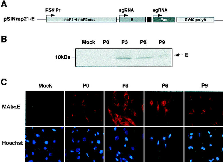
A BHK-pAPN cell line expressing the TGEV E protein was established using the Sindbis virus replicon pSINrep21-E derived from the vector pSINrep21 (Fig. 1A) (15). Following transfection, replicon-containing cell populations were selected with puromycin (5 μg/ml), and the expression of the TGEV E protein was analyzed by Western blotting with MAb V27 specific for E protein (Fig. 1B) (20). MAb V27 recognized the E protein as a single band of 12 kDa. The sequence of TGEV E protein mRNA transcribed in this cell line at passage 5 was identical to that of the wild-type TGEV (strain TGEV-PUR46-MAD) E gene sequence (data not shown). High levels of E protein expression (around 6 μg/106 cells), estimated by Western blot analysis using purified TGEV as a standard, were detected at passage 3. E protein expression was maintained for at least nine additional passages. All cells expressed the E protein in passage 3 as determined by immunofluorescence microscopy (Fig. 1C).
FIG. 1.
Expression of the E protein by a Sindbis virus replicon vector in BHK-pAPN cell lines. (A) Scheme of the Sindbis virus replicon construct encoding the TGEV E gene. RSV Pr, Rous sarcoma virus promoter; sgRNA, subgenomic RNA; Pac, puromycin resistance gene; SV40 poly A, transcription-termination polyadenylation signal from simian virus 40. nsP1-4, nonstructural proteins 1 to 4; nsP2mut, mutant nonstructural protein 2. (B) Western blot analysis of E protein expression at different cell passages (P) of BHK-pAPN transformed with the Sindbis virus replicon expressing the E protein. The position of the E protein is indicated by an arrow. Mock, untransfected. (C) Immunofluorescence microscopy using an E protein-specific MAb at different cell passages of BHK-pAPN transformed with the Sindbis virus replicon expressing the E protein.
Effect of Sindbis replicon and ectopic TGEV E protein expression on TGEV replication and on recovery of infectious TGEV from cDNA.
To determine whether the Sindbis replicon vector or the ectopic expression of the TGEV E protein would interfere with TGEV replication, the growth of TGEV was analyzed in BHK-pAPN cells transfected with either the pSINrep21 or pSINrep21-E vector. No differences in virus production were detected between cells harboring these plasmids and untransfected cells (Fig. 2A), indicating that neither the Sindbis virus replicon nor the expression of the E protein interfered with the replication of TGEV. Similarly, rescue of TGEV from a full-length infectious cDNA was not affected by the previous transformation of BHK-pAPN cells with an empty pSINrep21 or with this replicon expressing the E gene (Fig. 2B).
FIG. 2.
TGEV growth kinetics and rescue from cDNA in BHK-pAPN cells expressing E protein using Sindbis replicon. (A) Growth kinetics of TGEV in BHK-pAPN cells expressing TGEV E protein. Virus titers were determined by plaque assay. (B) Titration of infectious TGEV recovered from a transfected cDNA in BHK-pAPN cells expressing TGEV E protein by Sindbis virus replicon. E, BHK-pAPN cells expressing the TGEV E protein. SIN, cells transfected with pSINrep21 plasmid. C, mock-transfected cells. Mean values from four experiments are represented, and standard deviations are shown as error bars.
Complementation of rTGEV-Δ3abΔE in BHK-pAPN cells expressing TGEV E protein using Sindbis replicon.
rTGEV-Δ3abΔE was constructed using the strategy described to generate a cDNA encoding a full-length TGEV RNA (1). Plasmid pBAC-TGEV-Δ3abΔE was derived from plasmid pBAC-TGEV-(MluI-FseI)-FL by deleting genes 3a, 3b, and E by removing the sequences between the MluI and FseI restriction endonuclease sites (Fig. 3A). Both plasmids were transfected into control cells that did not express the E protein (E− cells) or into cells expressing the E protein (E+ cells) (Fig. 3B). Infectious rTGEV-(MluI-FseI)-FL was rescued from both E+ and E− cells with virus titers around 5 × 10⁸ PFU/ml at passage 2. In contrast, infectious rTGEV-Δ3abΔE was recovered in E+ cells but not in control E− cells, as expected. These results indicated that the complementation was successful. Titers of rTGEV-Δ3abΔE virus increased with passage (Fig. 3B). The mRNA E without the TGEV leader was transcribed from the Sindbis virus replicon as determined by RT-PCR (Fig. 3C). To amplify viral mRNAs, a forward primer complementary to the leader sequence and reverse primers specific for the E or S gene from TGEV were used. In that case, DNA fragments of 550 and 348 bp, corresponding to RT-PCR products derived from the mRNA encoding the S and E proteins, respectively, were identified in passages 1 to 3 in cells transfected with pBAC-TGEV-(MluI-FseI)-FL. In contrast, only the mRNA from the S gene was detected in the cells transfected with pBAC-TGEV-Δ3abΔE, confirming that the E gene was deleted in the TGEV genome.
FIG. 3.
Rescue of rTGEV-Δ3abΔE from cDNA in cells expressing E protein using Sindbis replicon. (A) Scheme of the pBAC-TGEV-(MluI-FseI)-FL plasmid, containing MluI and FseI restriction sites as indicated above the bar, and the pBAC-TGEV-Δ3abΔE plasmid, with 3ab and E genes deleted as indicated above the bar. CMV, CMV promoter; HDV, hepatitis delta virus ribozyme; BGH, bovine growth hormone termination and polyadenylation (An) sequence. (B) Rescue of recombinant TGEV containing the MluI and FseI restriction sites (Vwt) by transfecting BHK-pAPN cells (CE−) and BHK-pAPN cells expressing the TGEV E protein (CE+). VΔE, rTGEV-Δ3abΔE. (C) Detection of virus mRNAs for the E and S genes by RT-PCR analysis in BHK-pAPN cells expressing the TGEV E protein and infected with recombinant viruses rTGEV-(MluI-FseI)-FL (Vwt) and rTGEV-Δ3abΔE (VΔE). V−, uninfected cells; P1, P2, and P3, passages 1, 2, and 3, respectively. (D) Growth kinetics of recombinant viruses rTGEV-(MluI-FseI)-FL (Vwt) and rTGEV-Δ3abΔE (VΔE) in BHK-pAPN cells expressing TGEV E protein (CE+). Mean values from four experiments are represented, and standard deviations are shown as error bars.
The rescue of rTGEV-Δ3abΔE virus from the infectious cDNA throughout passages was slower than that of rTGEV-(MluI-FseI)-FL virus (Fig. 3B). In agreement with these results, the growth kinetics of rTGEV-(MluI-FseI)-FL and rTGEV-Δ3abΔE in E+ cells infected at a multiplicity of infection of 1 showed (Fig. 3D) that the full-length virus generated the highest virus titer (around 7 × 10⁸ PFU/ml) 1 day postinfection, while rTGEV-Δ3abΔE reached the highest titer (around 1 × 10⁷ PFU/ml) 3 days postinfection.
The synthesis of genomic RNA and mRNAs of rTGEV-Δ3abΔE virus was characterized by Northern blotting. BHK-pAPN E+ cells and control E− cells were infected with rTGEV-(MluI-FseI)-FL or rTGEV-Δ3abΔE virus. Total RNA was extracted and evaluated by Northern blotting with a probe complementary to the 3′ end of the TGEV genome. The RNA genome and sgmRNAs were detected in both cell lines infected with the two viruses, indicating that rTGEV-Δ3abΔE virus was replication competent. The smaller amounts of sgmRNAs detected in cells infected with rTGEV-Δ3abΔE virus (Fig. 4A) were consistent with the slower replication kinetics of this virus. The mobilities and relative amounts of the sgmRNAs M, N, and 7 were indistinguishable from those of rTGEV-(MluI-FseI)-FL (Fig. 4A). As expected, sgmRNAs 3 and 4 were not transcribed in cells infected with rTGEV-Δ3abΔE and were not observed even when the autoradiography exposure time was increased 10-fold (Fig. 4A). The sgmRNA 2 from rTGEV-Δ3abΔE migrated faster than that from rTGEV-(MluI-FseI)-FL, due to the deletion of genes 3a, 3b, and E.
FIG. 4.
Characterization of virus mRNAs and structural proteins from recombinant viruses rTGEV-(MluI-FseI)-FL and rTGEV-Δ3abΔE. (A) Northern blot analysis of cytoplasmic RNAs from BHK-pAPN cells (BHK-pAPN-E−) and BHK-pAPN cells expressing the E protein (BHK-pAPN-E+) infected with rTGEV-(MluI-FseI)-FL (wt) or with rTGEV-Δ3abΔE (Δ3abΔE). Δ3abΔE × 10 was exposed 10-fold longer than Δ3abΔE. C−, uninfected BHK-pAPN cells. The subgenomic RNAs are indicated by arrows and named according to the coded gene. (B) Western blot analysis using S, N, M, or E protein-specific MAb (see Materials and Methods) of cell lysates from BHK-pAPN cells (E−) and BHK-pAPN-E+ cells (E+) infected with rTGEV-(MluI-FseI)-FL (wt) or rTGEV- Δ3abΔE (ΔE). The analyses were carried out at 12 and 24 h postinfection, as indicated. V−, uninfected cells.
The production of viral proteins at 12 and 24 h postinfection was studied by Western blotting using MAbs specific for these proteins (Fig. 4B). The levels of the viral proteins S, M, and N were lower in E+ cells infected with rTGEV-Δ3abΔE than in the same cells infected with rTGEV-(MluI-FseI)-FL at 12 h postinfection, consistent with the lower mRNA levels. Nevertheless, the amounts of the three proteins were similar in E+ cells infected with the two viruses at 24 h postinfection. Expression of S, N, and M proteins was also observed in BHK-pAPN cells infected with rTGEV-Δ3abΔE, although the levels of these proteins did not increase with time. This observation was in agreement with the lack of virus propagation in E− cells.
Generation of cell lines constitutively expressing TGEV E protein.
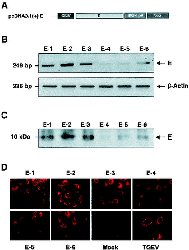
Cell lines constitutively expressing the TGEV E protein were generated to increase the stability of the packaging cell line and to test whether the level of E protein expression influences the titer of the rTGEV-Δ3abΔE virus recovered. The TGEV E gene was expressed under the control of the CMV promoter (Fig. 5A). Swine LLC-PK1 cells were transfected with pcDNA3.1(+)E, and clones resistant to the antibiotic G418 were selected. LLC-PK1 cells, with a higher transfection efficiency (10 to 15%), were used instead of ST cells, which produce high titers of TGEV virus, because of the low transformation efficiency of the ST cells (<4%). Six stably transformed cell lines (named E-1 to E-6) were selected from 52 clones, based on the mRNA-E expression levels determined by RT-PCR (Fig. 5B). These cells were cloned 3 times in the presence of the antibiotic and passed 10 times in culture before their evaluation by Western blotting (Fig. 5C) and immunofluorescence microscopy (Fig. 5D). The selected cell lines were classified as high (E-1, E-2, and E-3), intermediate (E-5 and E-6), and low (E-4) expressers according to the amount of E protein produced (around 1, 0.2, and 0.02 μg of E protein per million cells, respectively). E protein expression levels were homogeneous within each cell line, as determined by immunofluorescence (Fig. 5.D). The sequence of the E gene mRNA transcribed in these cell lines after 20 passages in culture was identical to that of the parental virus (data not shown).
FIG. 5.
Expression of the TGEV E protein in LLC-PK1 cell lines stably transformed with E protein expression plasmids. (A) Scheme of the E gene expression cassette in the pcDNA3.1(+) plasmid. CMV, CMV promoter; BGH pA, bovine growth hormone transcription stop signal and polyadenylation sequence; Neo, gene coding for neomycin resistance. (B) Detection of the mRNA for the E protein and β-actin by semiquantitative RT-PCR analysis in six cell lines (E-1, E-2, E-3, E-4, E-5, and E-6) stably transformed with the E gene. DNA amplification fragments were resolved by agarose gel electrophoresis and stained with ethidium bromide. (C) Western blot analysis of cell lysates from the six LLC-PK1 cell lines stably expressing the E protein, using an E-protein specific MAb. The position of the E protein band is indicated by an arrow. (D) Detection of E protein expression by immunofluorescence microscopy, using an E protein-specific MAb, in the six LLC-PK1 cell lines stably expressing the TGEV E protein. Mock, untransfected LLC-PK1 cells; TGEV, TGEV-infected LLC-PK1 cells.
Effects of E protein expression levels in rTGEV-Δ3abΔE virus complementation.
The titers of the parental E+ virus rTGEV-(MluI-FseI)-FL in the six cell lines ranged between 5 × 10⁵ and 1 × 10⁷ PFU/ml on day 1 postinfection, although the differences were not statistically significant (Fig. 6A). These results indicate that the level of E protein expression did not affect, or affected very little, the replication of the parental virus containing all the TGEV genes. Similar results were obtained in all cell lines expressing E protein by the Sindbis virus replicon. In contrast, the rTGEV-Δ3abΔE virus growth kinetics was dependent on the E protein level (Fig. 6B). Virus levels were undetectable in the cell line E-4, showing the lowest E protein expression level. The other five clones supported virus assembly, and virus titers correlated with E protein expression levels and were highest (5 × 10⁵ PFU/ml) in clones E-1, E-2, and E-3. These data suggest that a minimum level of E protein expression is needed for the successful complementation of the rTGEV-Δ3abΔE genome. Interestingly, the titer of the ΔE virus rescued was directly related to the amount of E protein expressed by each cell line (Fig. 6C).
FIG. 6.
Effects of E protein expression levels in rescue of recombinant viruses rTGEV-(MluI-FseI)-FL and rTGEV- Δ3abΔE. (A and B) Growth kinetics of rTGEV-(MluI-FseI)-FL (A) and rTGEV-Δ3abΔE (B) virus in LLC-PK1 cell lines expressing different levels of E protein (Fig. 5). C, untransformed LLC-PK1 cells; E-1 to E-6, cell lines expressing different amounts of E protein. (C) Relation between virus (rTGEV-Δ3abΔE) titers and E protein expression levels in transformed LLC-PK1 cells. Mean values from four experiments are represented, with standard deviations shown as error bars.
Stability of rTGEV-Δ3abΔE virus at different temperatures and pHs.
The recombinant virus rTGEV-(MluI-FseI)-FL grown in E− cells or rTGEV-Δ3abΔE grown in E+ cells (106 PFU/ml) was incubated at temperatures ranging from 4 to 80°C for 30 min and titrated. Both viruses [rTGEV-(MluI-FseI)-FL and rTGEV-Δ3abΔE] showed the same inactivation profile with heating (Fig. 7A). Interestingly, both viruses showed high inactivation rates at temperatures just above 40°C, a temperature very close to the body temperature of the natural virus host, suggesting that an increase in an infected animal's body temperature could be of great help in eliminating the virus. In both cases, the infectivity was reduced by >105-fold after incubation at 60°C. Heating at 80°C or higher temperatures led to residual virus infectivity. Furthermore, the effect of a range of pH (5 to 9) on the stability of rTGEV-(MluI-FseI)-FL and rTGEV-Δ3abΔE viruses showed a similar profile. Both viruses were stable from pH 6 to 8. Incubation at higher or lower pHs (9 or 5, respectively) produced a decrease of >200-fold in virus infectivity in both recombinants (Fig. 7B). These results indicated that rTGEV-Δ3abΔE virus complemented in packaging cell lines showed the same stability as wild-type virus under different pH and temperature treatments.
FIG. 7.
Effects of temperature and pH changes on rTGEV-(MluI-FseI)-FL and rTGEV-Δ3abΔE virus stabilities. Titrations of virus supernatants from cells infected with recombinant viruses rTGEV-(MluI-FseI)-FL and rTGEV-Δ3abΔE after incubation for 30 min at the indicated temperature (A) or pH (B) are shown. Mean values from four experiments are represented, with standard deviations shown as error bars. The abbreviations are defined in the legend to Fig. 3B.
Intracellular assembly of rTGEV-Δ3abΔE virus in E+ cells.
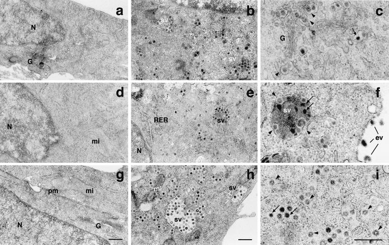
The assembly of rTGEV-Δ3abΔE and rTGEV-(MluI-FseI)-FL in BHK-pAPN E− and E+ cells, respectively, was analyzed by electron microscopy. E+ cells showed no abnormal structures or appreciable changes in the organization of the organelles compared to E− cells (Fig. 8a, d, and g). Similarly, protein E expression showed no effect on the structure of the virions with a full-length genome grown in E+ cells. No aberrant virion or structure due to the cellular expression of E protein was observed. Virions with two different sizes and morphologies were observed in both E− and E+ cells. Large virions exhibited an electron-dense periphery and a less dense central zone (68- to 80-nm diameter) that corresponded to immature virions previously described (42). The smaller virions showed a dense core (60-nm diameter) and corresponded to mature forms (Fig. 8b, c, e, and f). Large virions were more abundant in the perinuclear area of the cell but were also found in smaller amounts in other areas of the cytoplasm. The mature forms were preferentially located close to the plasma membrane and inside secretory vesicles.
FIG. 8.
Characterization of the assembly of recombinant viruses rTGEV-(MluI-FseI)-FL and rTGEV-Δ3abΔE in BHK-pAPN cells by electron microscopy. Electron microscopy sections of BHK-pAPN cells that do not express (a to c) or express (d to i) the TGEV E protein, infected with rTGEV-(MluI-FseI)-FL (b, c, e, and f) or rTGEV-Δ3abΔE (h and i). Large annular viruses are indicated with arrowheads, and small and dense viruses are indicated with arrows. sv, secretory vesicle; ev, extracellular virus; N, nucleus; G, Golgi complex; mi, mitochondria; RER, rough endoplasmic reticulum; pm, plasma membrane. Bars, 400 nm.
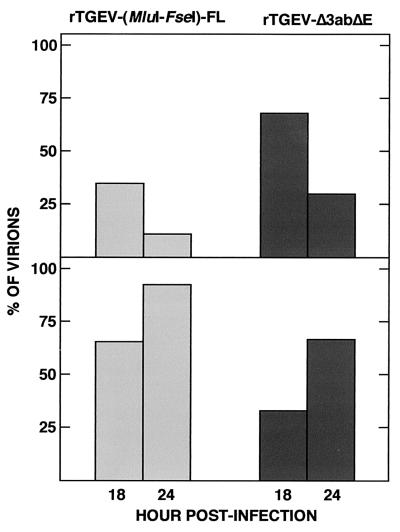
The rTGEV-Δ3abΔE virus growing in the E+ cells showed the same morphology types described for the full-length virus (Fig. 8h and i), but the proportion of large vesicles was higher than in the infection with a full-length virus. The distribution of large and small virions in cells expressing the TGEV E protein infected with rTGEV-Δ3abΔE virus or of E− cells infected with rTGEV-(MluI-FseI)-FL virus was analyzed (Fig. 9). At identical time points after infection, the ratio of mature versus immature virions was higher in the E− cells infected with the full-length genome than in the E+ cells infected with the ΔE virus, consistent with a lower replication rate of the defective rTGEV-Δ3abΔE virus.
FIG. 9.
Quantification of large and small virions in BHK-pAPN cells expressing the TGEV E protein and infected with recombinant virus rTGEV-(MluI-FseI)-FL or rTGEV-Δ3abΔE. Shown are the percentages of intracellular virions with a large (68- to 80-nm diameter) (top) or small (50- to 65-nm) (bottom) core present at 18 and 24 h postinfection. A total of 500 virions were included in the measurements.
DISCUSSION
The construction of a virus vector based on a replication-competent, propagation-deficient coronavirus genome has been described. The TGEV genome deficient in the nonessential genes 3a and 3b and in the essential gene E was complemented in trans by packaging cell lines expressing E protein. Two types of packaging cell lines have been established, one using the noncytopathic Sindbis virus replicon and a collection of cell lines constitutively expressing different levels of E protein. High titers of infectious virus particles with a morphology and stability to temperature and pH identical to that of the wild type virus were generated. Interestingly, the amount of E protein was a limiting factor in the production of infectious TGEV.
To generate packaging cell lines expressing the TGEV E protein, we used two expression systems. One of them was based on the noncytopathic Sindbis virus replicon (17). This system was chosen because of its high expression levels, rapid puromycin selection, and stability for at least 10 passages (24, 30). Previous reports showed that MHV E protein expression by recombinant vaccinia viruses induced apoptosis in DBT cells (3). Interestingly, using the Sindbis virus replicon, we found no evidence of toxicity or apoptosis due to the constitutive expression of the TGEV E protein. The lack of apoptosis induction was not due to the selection of a clone with a mutated E gene, since the mRNA encoding E protein was sequenced at passage 5 and no nucleotide substitution was found in relation to the original gene present in the virus. Therefore, coronavirus E protein, in general, does not induce apoptosis; in fact, this process is cell specific (3). Since it has been reported that TGEV is apoptotic in three porcine cell lines, ST, PDH, and RPTG, and in MDCK-APN, a clone of a canine kidney cell line expressing porcine APN (8), either the protein responsible was other than the E protein or this effect does not take place in the cell lines used in our study.
One concern, in terms of biosafety, is that the packaging cell lines established using RNA replicon systems may produce fully infectious viruses by recombination between the replicon and the viral genome (6, 16, 19, 40). The use of a viral replicon unrelated to the complemented virus reduces the possibility of recombination events. To further reduce the risk of generating a propagation-competent virus by recombination, we generated packaging cell lines stably expressing the TGEV E protein under the control of the CMV promoter. These packaging cell lines also produced high levels of TGEV E protein and practically eliminated the problem of the higher mutation rates in expression systems generated using RNA replicons. In fact, the mRNA encoding E protein in the established cell lines was sequenced, and no mutation was found after 20 passages in culture. These cell lines stably expressing the TGEV E protein represent an advance over previously reported systems based on transient expression (6).
Coronavirus undergoes homologous recombination at high frequency during infections with more than one virus (4, 18). Recombination with sgmRNA may also be possible, although sgmRNAs rarely, if ever, serve as receptor templates for recombination (36). In our system, the rTGEV-Δ3abΔE virus did not recover the E gene in stably transformed E+ cells, at least during 20 passages (data not shown), strongly suggesting that the propagation-deficient virus obtained was stable and functional. To improve the safety of TGEV as a virus vector, viruses deficient in two essential genes separated in the genome are being generated, considerably reducing the possibility of recovering a propagation-competent virus by two potential recombination events.
In the rTGEV-Δ3abΔE virus, genes 3a, 3b, and E were deleted. In this recombinant, the 3′ end of the E gene partially overlaps with the regulatory sequences flanking the M gene 3′ end. To avoid deletion of the E gene affecting transcription levels of the M gene, a TRS of 24 nt derived from the M gene, including the canonical core sequence (5′-CUAAAC-3′) plus 18 nt flanking its 5′ end, was conserved in the engineered genome. The length of this TRS was sufficient to produce standard levels of M protein mRNA, as previously shown (2). In fact, the relative abundances of the viral mRNAs encoded by the engineered virus genome were indistinguishable from those of the wild-type virus, indicating that the TGEV genome is flexible, because neither the insertion of restriction sites nor the duplication of the TRS significantly affected virus replication and transcription levels in cell cultures.
The assembly and morphology of the virus with a deleted genome were apparently unaffected by the acquisition of the E protein from the cell and by the fact that the genome of the virus is 1.2 kb smaller than that of the wild-type virus, due to deletion of genes 3a, 3b, and E. The rTGEV-Δ3abΔE virus was as stable as the wild-type virus under temperature and pH changes, indicating that the supply of E protein in trans had no adverse effect on virus morphogenesis.
The major characteristic of a successful packaging cell system is the production of high-titer virus. The TGEV titers generated in the cell lines stably expressing the E protein under the control of the CMV promoter or expressing the E protein using the Sindbis replicon were 5 × 10⁵ and 1 × 10⁷ PFU/ml, respectively. This system can be used in most research experiments, and the efficacy is fairly close to that required for cost-effective vaccine development. The virus titers obtained with the cells stably expressing the E protein were 20-fold lower than those obtained in the cells expressing the E protein using the Sindbis replicon, and the titers correlated with the E protein expression levels. Since the expression level of the E protein in Sindbis replicon-transformed cells was 6 μg/106 cells and in the stably transformed cells maximum levels were around 1 μg per 106 cells, the increase of stable E protein expression will most likely help to increase virus production by these cells.
Although the virus titers generated by complementation do not generally reach the titers provided by wild-type viruses (23, 30, 50), the virus titers described in this paper are closer to those of the retroviral packaging cell lines, one of the most developed systems, producing virus titers in the range of 105 to 108 PFU/ml (26, 33, 34, 35, 46, 51). The system reported in this paper represents highly significant progress in relation to previously reported coronavirus packaging cell systems owing to (i) its higher expression levels (1,000-fold), (ii) the introduction of stably transformed packaging cell lines that provide better safety and realistic application in vaccine development, (iii) quantification of E protein and virus production levels, and (iv) characterization of the replication-competent, propagation-deficient virus in terms of physical and chemical stability and morphogenesis analyzed by electron microscopy. The most important characteristics of the replication-competent, propagation-deficient virus vector developed are its safety and expression levels that are sufficiently high to permit the realistic testing of coronavirus vector efficacy.
Acknowledgments
This work has been supported by grants from the Comisión Interministerial de Ciencia y Tecnología (CICYT), La Consejería de Educación y Cultura de la Comunidad de Madrid, Fort Dodge Veterinaria, and the European Communities (Frame V, Key Action 2, Control of Infectious Disease Projects QLRT-1999-00002, QLRT-1999-30739, and QLRT-2000-00874). J.O. and D.E. received a contract from the Spanish Department of Education and Culture and from the European Communities (Frame V, Key Action 2, Control of Infectious Diseases).
REFERENCES
- 1.Almazán, F., J. M. González, Z. Pénzes, A. Izeta, E. Calvo, J. Plana-Durán, and L. Enjuanes. 2000. Engineering the largest RNA virus genome as an infectious bacterial artificial chromosome. Proc. Natl. Acad. Sci. USA 97:5516-5521. [DOI] [PMC free article] [PubMed] [Google Scholar]
- 2.Alonso, S., A. Izeta, I. Sola, and L. Enjuanes. 2002. Transcription regulatory sequences and mRNA expression levels in the coronavirus transmissible gastroenteritis virus. J. Virol. 76:1293-1308. [DOI] [PMC free article] [PubMed] [Google Scholar]
- 3.An, S., C. J. Chen, X. Yu, J. L. Leibowitz, and S. Makino. 1999. Induction of apoptosis in murine coronavirus-infected cultured cells and demonstration of E protein as an apoptosis inducer. J. Virol. 73:7853-7859. [DOI] [PMC free article] [PubMed] [Google Scholar]
- 4.Baric, R. S., K. S. Fu, M. C. Schaad, and S. A. Stohlman. 1990. Establishing a genetic recombination map for MHV-A59 complementation groups. Virology 177:646. [DOI] [PMC free article] [PubMed] [Google Scholar]
- 5.Baudoux, P., C. Carrat, L. Besnardeau, B. Charley, and H. Laude. 1998. Coronavirus pseudoparticles formed with recombinant M and E proteins induce alpha interferon synthesis by leukocytes. J. Virol. 72:8636-8643. [DOI] [PMC free article] [PubMed] [Google Scholar]
- 6.Curtis, K. M., B. Yount, and R. S. Baric. 2002. Heterologous gene expression from transmissible gastroenteritis virus replicon particles. J. Virol. 76:1422-1434. [DOI] [PMC free article] [PubMed] [Google Scholar]
- 7.Delmas, B., J. Gelfi, H. Sjöström, O. Noren, and H. Laude. 1994. Further characterization of aminopeptidase-N as a receptor for coronaviruses. Adv. Exp. Med. Biol. 342:293-298. [DOI] [PubMed] [Google Scholar]
- 8.Eleouet, J. F., S. Chilmonczyk, L. Besnardeau, and H. Laude. 1998. Transmissible gastroenteritis coronavirus induces programmed cell death in infected cells through a caspase-dependent pathway. J. Virol. 72:4918-4924. [DOI] [PMC free article] [PubMed] [Google Scholar]
- 9.Enjuanes, L., D. Brian, D. Cavanagh, K. Holmes, M. M. C. Lai, H. Laude, P. Masters, P. Rottier, S. G. Siddell, W. J. M. Spaan, F. Taguchi, and P. Talbot. 2000. Coronaviridae, p. 835-849. In M. H. V. van Regenmortel, C. M. Fauquet, D. H. L. Bishop, E. B. Carsten, M. K. Estes, S. M. Lemon, D. J. McGeoch, J. Maniloff, M. A. Mayo, C. R. Pringle, and R. B. Wickner (ed.), Virus taxonomy. Classification and nomenclature of viruses. Academic Press, New York, N.Y.
- 10.Enjuanes, L., W. Spaan, E. Snijder, and D. Cavanagh. 2000. Nidovirales, p. 827-834. In M. H. V. van Regenmortel, C. M. Fauquet, D. H. L. Bishop, E. B. Carsten, M. K. Estes, S. M. Lemon, D. J. McGeoch, J. Maniloff, M. A. Mayo, C. R. Pringle, and R. B. Wickner (ed.), Virus taxonomy. Classification and nomenclature of viruses. Academic Press, New York, N.Y.
- 11.Enjuanes, L., and B. A. M. Van der Zeijst. 1995. Molecular basis of transmissible gastroenteritis coronavirus epidemiology, p. 337-376. In S. G. Siddell (ed.), The coronaviridae. Plenum Press, New York, N.Y.
- 12.Escors, D., E. Camafeita, J. Ortego, H. Laude, and L. Enjuanes. 2001. Organization of two transmissible gastroenteritis coronavirus membrane protein topologies within the virion and core. J. Virol. 75:12228-12240. [DOI] [PMC free article] [PubMed] [Google Scholar]
- 13.Escors, D., J. Ortego, H. Laude, and L. Enjuanes. 2001. The membrane M protein carboxy terminus binds to transmissible gastroenteritis coronavirus core and contributes to core stability. J. Virol. 75:1312-1324. [DOI] [PMC free article] [PubMed] [Google Scholar]
- 14.Fischer, F., C. F. Stegen, P. S. Masters, and W. A. Samsonoff. 1998. Analysis of constructed E gene mutants of mouse hepatitis virus confirms a pivotal role for E protein in coronavirus assembly. J. Virol. 72:7885-7894. [DOI] [PMC free article] [PubMed] [Google Scholar]
- 15.Frolov, I., E. Agapov, T. A. Hoffman, B. M. Prágai, M. Lippa, S. Schlesinger, and C. M. Rice. 1999. Selection of RNA replicons capable of persistent noncytopathic replication in mammalian cells. J. Virol. 73:3854-3865. [DOI] [PMC free article] [PubMed] [Google Scholar]
- 16.Frolov, I., E. Frolova, and S. Schlesinger. 1997. Sindbis virus replicons and Sindbis virus: assembly of chimeras and of particles deficient in virus RNA. J. Virol. 71:2819-2829. [DOI] [PMC free article] [PubMed] [Google Scholar]
- 17.Frolov, I., T. A. Hoffman, B. M. Prágai, S. A. Dryga, H. V. Huang, S. Schlesinger, and C. M. Rice. 1996. Alphavirus-based expression vectors: strategies and applications. Proc. Natl. Acad. Sci. USA 93:11371-11377. [DOI] [PMC free article] [PubMed] [Google Scholar]
- 18.Fu, K. S., and R. S. Baric. 1992. Evidence for variable rates of recombination in the MHV genome. Virology 189:88-102. [DOI] [PMC free article] [PubMed] [Google Scholar]
- 19.Geigenmuller-Gnirke, U. B. W., R. Wright, and S. Schlesinger. 1991. Complementation between Sindbis viral RNAs produces infectious particles with a bipartite genome. Proc. Natl. Acad. Sci. USA 88:3253-3257. [DOI] [PMC free article] [PubMed] [Google Scholar]
- 20.Godet, M., R. L'Haridon, J. F. Vautherot, and H. Laude. 1992. TGEV coronavirus ORF4 encodes a membrane protein that is incorporated into virions. Virology 188:666-675. [DOI] [PMC free article] [PubMed] [Google Scholar]
- 21.González, J. M., Z. Penzes, F. Almazán, E. Calvo, and L. Enjuanes. 2002. Stabilization of a full-length infectious cDNA clone of transmissible gastroenteritis coronavirus by the insertion of an intron. J. Virol. 76:4655-4661. [DOI] [PMC free article] [PubMed] [Google Scholar]
- 22.Jiménez, G., I. Correa, M. P. Melgosa, M. J. Bullido, and L. Enjuanes. 1986. Critical epitopes in transmissible gastroenteritis virus neutralization. J. Virol. 60:131-139. [DOI] [PMC free article] [PubMed] [Google Scholar]
- 23.Kafri, T., H. van Praag, L. Ouyang, F. H. Gage, and I. M. Verma. 1999. A packaging cell line for lentivirus vectors. J. Virol. 73:576-584. [DOI] [PMC free article] [PubMed] [Google Scholar]
- 24.Khromykh, A. A., P. L. Sedlak, K. J. Guyatt, R. A. Hall, and E. G. Westaway. 1999. Efficient trans-complementation of the flavivirus Kunjin NS5 protein but not of the NS1 protein requires its coexpression with other components of the viral replicase. J. Virol. 73:10272-10280. [DOI] [PMC free article] [PubMed] [Google Scholar]
- 25.Khromykh, A. A., P. L. Sedlak, and E. G. Westaway. 1999. trans-complementation analysis of the flavivirus Kunjin ns5 gene reveals an essential role for translation of its N-terminal half in RNA replication. J. Virol. 73:9247-9255. [DOI] [PMC free article] [PubMed] [Google Scholar]
- 26.Konetschny, C., G. W. Holzer, and F. G. Falkner. 2002. Retroviral vectors produced in the cytoplasmic vaccinia virus system transduce intron-containing genes. J. Virol. 76:1236-1243. [DOI] [PMC free article] [PubMed] [Google Scholar]
- 27.Kozarsky, K. F., and J. M. Wilson. 1993. Gene therapy: adenovirus vectors. Curr. Opin. Genet. Dev. 3:499-503. [DOI] [PubMed] [Google Scholar]
- 28.Lai, M. M. C., R. S. Baric, P. R. Brayton, and S. A. Stohlman. 1984. Characterization of leader RNA sequences on the virion and mRNAs of mouse hepatitis virus, a cytoplasmic RNA virus. Proc. Natl. Acad. Sci. USA 81:3626-3630. [DOI] [PMC free article] [PubMed] [Google Scholar]
- 29.Lai, M. M. C., and D. Cavanagh. 1997. The molecular biology of coronaviruses. Adv. Virus Res. 48:1-100. [DOI] [PMC free article] [PubMed] [Google Scholar]
- 30.Lindenbach, B. D., and C. M. Rice. 1997. trans-complementation of yellow fever virus NS1 reveals a role in early RNA replication. J. Virol. 71:9608-9617. [DOI] [PMC free article] [PubMed] [Google Scholar]
- 31.Maeda, J., A. Maeda, and S. Makino. 1999. Release of coronavirus E protein membrane vesicles from virus-infected cells and E protein-expressing cells. Virology 263:265-272. [DOI] [PMC free article] [PubMed] [Google Scholar]
- 32.Maeda, J., J. F. Repass, A. Maeda, and S. Makino. 2001. Membrane topology of coronavirus E protein. Virology 281:163-169. [DOI] [PMC free article] [PubMed] [Google Scholar]
- 33.Martínez, I., and R. Dornburg. 1995. Improved retroviral packaging lines derived from spleen necrosis virus. Virology 208:234-241. [DOI] [PubMed] [Google Scholar]
- 34.Miller, A. D., and C. Buttimore. 1986. Redesign of retrovirus packaging cell lines to avoid recombination leading to helper virus production. Mol. Cell. Biol. 6:2895-2902. [DOI] [PMC free article] [PubMed] [Google Scholar]
- 35.Miller, A. D., and F. Chen. 1996. Retrovirus packaging cells based on 10A1 murine leukemia virus for production of vectors that use multiple receptors for cell entry. J. Virol. 70:5564-5571. [DOI] [PMC free article] [PubMed] [Google Scholar]
- 36.Ozdarendeli, A., S. Ku, S. Rochat, S. D. Senanayake, and D. A. Brian. 2001. Downstream sequences influence the choice between a naturally occurring noncanonical and closely positioned upstream canonical heptameric fusion motif during bovine coronavirus subgenomic mRNA synthesis. J. Virol. 75:7362-7374. [DOI] [PMC free article] [PubMed] [Google Scholar]
- 37.Pacchia, A. L., M. E. Adelson, M. Kaul, Y. Ron, and J. P. Dougherty. 2001. An inducible packaging cell system for safe, efficient lentiviral vector production in the absence of HIV-1 accessory proteins. Virology 280:77-86. [DOI] [PubMed] [Google Scholar]
- 38.Penzes, Z., J. M. González, E. Calvo, A. Izeta, C. Smerdou, A. Mendez, C. M. Sánchez, I. Sola, F. Almazán, and L. Enjuanes. 2001. Complete genome sequence of transmissible gastroenteritis coronavirus PUR46-MAD clone and evolution of the Purdue virus cluster. Virus Genes 23:105-118. [DOI] [PMC free article] [PubMed] [Google Scholar]
- 39.Polo, J. M., B. A. Belli, D. A. Driver, I. Frolov, S. Sherrill, M. J. Hariharan, K. Townsend, S. Perri, S. J. Mento, D. J. Jolly, S. M. W. Chang, S. Schlesinger, and J. T. W. Dubensky. 1999. Stable alphavirus packaging cell lines for Sindbis virus- and Semliki Forest virus-derived vectors. Proc. Natl. Acad. Sci. USA 96:4598-4603. [DOI] [PMC free article] [PubMed] [Google Scholar]
- 40.Pushko, P., M. Parker, G. V. Ludwing, N. L. Davis, R. E. Johnston, and J. F. Smith. 1997. Replication-helper systems from attenuated Venezuelan equine encephalitis virus: expression of heterologous genes in vitro and immunization against heterologous pathogens in vivo. Virology 239:389-401. [DOI] [PubMed] [Google Scholar]
- 41.Raamsman, M. J. B., J. K. Locker, A. de Hooge, A. A. F. de Vries, G. Griffiths, H. Vennema, and P. J. M. Rottier. 2000. Characterization of the coronavirus mouse hepatitis virus strain A59 small membrane protein E. J. Virol. 74:2333-2342. [DOI] [PMC free article] [PubMed] [Google Scholar]
- 42.Risco, C., M. Muntión, L. Enjuanes, and J. L. Carrascosa. 1998. Two types of virus-related particles are found during transmissible gastroenteritis virus morphogenesis. J. Virol. 72:4022-4031. [DOI] [PMC free article] [PubMed] [Google Scholar]
- 43.Saif, L. J., and R. D. Wesley. 1992. Transmissible gastroenteritis, p. 362-386. In A. D. Leman, B. E. Straw, W. L. Mengeling, S. D'Allaire, and D. J. Taylor (ed.), Diseases of swine, 7th ed. Wolfe Publishing Ltd., Ames, Iowa.
- 44.Sawicki, D. L., T. Wang, and S. G. Sawicki. 2001. The RNA structures engaged in replication and transcription of the A59 strain of mouse hepatitis virus. J. Gen. Virol. 82:386-396. [DOI] [PubMed] [Google Scholar]
- 45.Sawicki, S. G., and D. L. Sawicki. 1998. A new model for coronavirus transcription. Adv. Exp. Med. Biol. 440:215-220. [DOI] [PubMed] [Google Scholar]
- 46.Thaler, S., and B. S. Schnierle. 2001. A packaging cell line generating CD4-specific retroviral vectors for efficient gene transfer into primary human T-helper lymphocytes. Mol. Ther. 4:273-279. [DOI] [PubMed] [Google Scholar]
- 47.Tung, F. Y. T., S. Abraham, M. Sethna, S. L. Hung, P. Sethna, B. G. Hogue, and D. A. Brian. 1992. The 9-kDa hydrophobic protein encoded at the 3′ end of the porcine transmissible gastroenteritis coronavirus genome is membrane-associated. Virology 186:676-683. [DOI] [PMC free article] [PubMed] [Google Scholar]
- 48.van Marle, G., J. C. Dobbe, A. P. Gultyaev, W. Luytjes, W. J. M. Spaan, and E. J. Snijder. 1999. Arterivirus discontinuous mRNA transcription is guided by base pairing between sense and antisense transcription-regulating sequences. Proc. Natl. Acad. Sci. USA 96:12056-12061. [DOI] [PMC free article] [PubMed] [Google Scholar]
- 49.Vennema, H., G. J. Godeke, J. W. A. Rossen, W. F. Voorhout, M. C. Horzinek, D. J. Opstelten, and P. J. M. Rottier. 1996. Nucleocapsid-independent assembly of coronavirus-like particles by co-expression of viral envelope protein genes. EMBO J. 15:2020-2028. [DOI] [PMC free article] [PubMed] [Google Scholar]
- 50.Von Seggern, D. J., J. Kehler, R. I. Endo, and G. R. Nemorow. 1998. Complementation of a fibre mutant adenovirus by packaging cell lines stably expressing the adenovirus type 5 fibre protein. J. Gen. Virol. 79:1461-1468. [DOI] [PubMed] [Google Scholar]
- 51.Wu, N., and M. M. Ataai. 2000. Production of viral vectors for gene therapy applications. Curr. Opin. Biotechnol. 11:205-208. [DOI] [PubMed] [Google Scholar]


