

Abstract
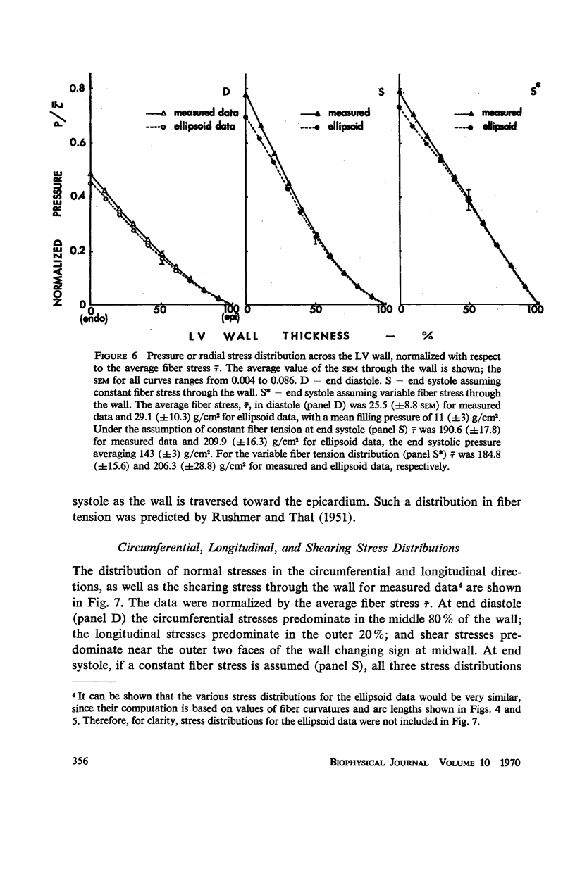
A model is proposed for stress analysis of the left ventricular wall (LV wall) based on the realistic assumption that the myocardium is essentially composed of fiber elements which carry only axial tension and vary in orientation through the wall. Stress analysis based on such a model requires an extensive study of muscle fiber orientation and curvature through the myocardium. Accordingly, the principal curvatures were studied at a local site near the equator in ten dog hearts rapidly fixed in situ at end diastole and end systole; the fiber orientation for these hearts had already been established in a previous study. The principal radii of curvature were (a) measured by fitting templates to the endocardial and epicardial wall surfaces in the circumferential and longitudinal directions and (b) computed from measured lengths of semiaxes of ellipsoids of revolution representing the LV wall (“ellipsoid” data). The wall was regarded as a tethered set of nested shells, each having a unique fiber orientation. Results indicate the following. (a) Fiber curvature, k, is maximum at midwall at end systole; this peak shifts towards endocardium at end diastole. (b) The pressure or radial stress through the wall decreases more rapidly near the endocardium than near the epicardium at end diastole and at end systole when a constant tension is assumed for each fiber through the wall. (c) At end diastole the curve for the circumferential stress vs. wall thickness is convex with a maximum at midwall. In the longitudinal direction the stress distribution curve is concave with a minimum at midwall. Similar distributions are obtained at end systole when a constant tension is assumed for each fiber through the wall. (d) The curvature and stress distributions obtained by direct measurements at a selected local site agree well with those computed from “ellipsoid” data.
Full text
PDF


Selected References
These references are in PubMed. This may not be the complete list of references from this article.
- Eber L. M., Greenberg H. M., Cooke J. M., Gorlin R. Dynamic changes in left ventricular free wall thickness in the human heart. Circulation. 1969 Apr;39(4):455–464. doi: 10.1161/01.cir.39.4.455. [DOI] [PubMed] [Google Scholar]
- FRY D. L. Implications of muscle mechanics in the heart. Discussion. Fed Proc. 1962 Nov-Dec;21:991–993. [PubMed] [Google Scholar]
- Feigl E. O., Simon G. A., Fry D. L. Auxotonic and isometric cardiac force transducers. J Appl Physiol. 1967 Oct;23(4):597–600. doi: 10.1152/jappl.1967.23.4.597. [DOI] [PubMed] [Google Scholar]
- HORT W. [Macroscopic and micrometric research on the myocardium of the left ventricle filled to varying degrees]. Virchows Arch Pathol Anat Physiol Klin Med. 1960;333:523–564. [PubMed] [Google Scholar]
- Hood W. P., Jr, Thomson W. J., Rackley C. E., Rolett E. L. Comparison of calculations of left ventricular wall stress in man from thin-walled and thick-walled ellipsoidal models. Circ Res. 1969 Apr;24(4):575–582. doi: 10.1161/01.res.24.4.575. [DOI] [PubMed] [Google Scholar]
- KIRK E. S., HONIG C. R. AN EXPERIMENTAL AND THEORETICAL ANALYSIS OF MYOCARDIAL TISSUE PRESSURE. Am J Physiol. 1964 Aug;207:361–367. doi: 10.1152/ajplegacy.1964.207.2.361. [DOI] [PubMed] [Google Scholar]
- KREUZER H., SCHOEPPE W. DAS VERHALTEN DES DRUCKES IN DER HERZWAND. Pflugers Arch Gesamte Physiol Menschen Tiere. 1963 Oct 25;278:181–198. [PubMed] [Google Scholar]
- RUSHMER R. F., THAL N. The mechanics of ventricular contraction; a cinefluorographic study. Circulation. 1951 Aug;4(2):219–228. doi: 10.1161/01.cir.4.2.219. [DOI] [PubMed] [Google Scholar]
- Ross J., Jr, Sonnenblick E. H., Covell J. W., Kaiser G., Spiro D. The architecture of the heart in systole and diastole. Technique of rapid fixation and analysis of left ventricular geometry. Circ Res. 1967 Oct;21(4):409–421. doi: 10.1161/01.res.21.4.409. [DOI] [PubMed] [Google Scholar]
- SALISBURY P. F., CROSS C. E., RIEBEN P. A. Intramyocardial pressure and strength of left ventricular contraction. Circ Res. 1962 Apr;10:608–623. doi: 10.1161/01.res.10.4.608. [DOI] [PubMed] [Google Scholar]
- SANDLER H., DODGE H. T. LEFT VENTRICULAR TENSION AND STRESS IN MAN. Circ Res. 1963 Aug;13:91–104. doi: 10.1161/01.res.13.2.91. [DOI] [PubMed] [Google Scholar]
- SONNENBLICK E. H. Implications of muscle mechanics in the heart. Fed Proc. 1962 Nov-Dec;21:975–990. [PubMed] [Google Scholar]
- Sonnenblick E. H., Ross J., Jr, Covell J. W., Spotnitz H. M., Spiro D. The ultrastructure of the heart in systole and diastole. Chantes in sarcomere length. Circ Res. 1967 Oct;21(4):423–431. doi: 10.1161/01.res.21.4.423. [DOI] [PubMed] [Google Scholar]
- Spotnitz H. M., Sonnenblick E. H., Spiro D. Relation of ultrastructure to function in the intact heart: sarcomere structure relative to pressure volume curves of intact left ventricles of dog and cat. Circ Res. 1966 Jan;18(1):49–66. doi: 10.1161/01.res.18.1.49. [DOI] [PubMed] [Google Scholar]
- Streeter D. D., Jr, Spotnitz H. M., Patel D. P., Ross J., Jr, Sonnenblick E. H. Fiber orientation in the canine left ventricle during diastole and systole. Circ Res. 1969 Mar;24(3):339–347. doi: 10.1161/01.res.24.3.339. [DOI] [PubMed] [Google Scholar]
- Taylor R. R., Covell J. W., Ross J., Jr Volume-tension diagrams of ejecting and isovolumic contractions in left ventricle. Am J Physiol. 1969 May;216(5):1097–1102. doi: 10.1152/ajplegacy.1969.216.5.1097. [DOI] [PubMed] [Google Scholar]
- Wong A. Y., Rautaharju P. M. Stress distribution within the left ventricular wall approximated as a thick ellipsoidal shell. Am Heart J. 1968 May;75(5):649–662. doi: 10.1016/0002-8703(68)90325-6. [DOI] [PubMed] [Google Scholar]