Abstract
Rinderpest is an acute and highly contagious viral disease of ruminants, often resulting in greater than 90% mortality. We have constructed a recombinant vaccinia virus vaccine (v2RVFH) that expresses both the fusion (F) and hemagglutinin (H) genes of rinderpest virus (RPV) under strong synthetic vaccinia virus promoters. v2RVFH-infected cells express high levels of the F and H glycoproteins and show extensive syncytium formation. Cattle vaccinated intramuscularly with as little as 103 PFU of v2RVFH and challenged 1 month later with a lethal dose of RPV were completely protected from clinical disease; the 50% protective dose was determined to be 102 PFU. Animals vaccinated with v2RVFH did not develop pock lesions and did not transmit the recombinant vaccinia virus to contact animals. Intramuscular vaccination of cattle with 108 PFU of v2RVFH provided long-term sterilizing immunity against rinderpest. In addition to being highly safe and efficacious, v2RVFH is a heat-stable, inexpensive, and easily administered vaccine that allows the serological differentiation between vaccinated and naturally infected animals. Consequently, mass vaccination of cattle with v2RVFH could eradicate rinderpest.
Rinderpest is an acute, febrile, and highly contagious viral disease of ruminants (particularly cattle and buffalo), with a mortality rate approaching 95% (28). The disease is characterized by inflammation, hemorrhaging, necrosis, and erosion of the gastrointestinal tract accompanied by bloody diarrhea, wasting, and death (2). Rinderpest virus (RPV), along with Measles virus (MV), Canine distemper virus, Peste-des-petits-ruminants virus of goats and sheep, and Phocine distemper virus of seals, is a member of the family Paramyxoviridae, genus Morbillivirus (35). RPV is a pleomorphic, enveloped virus with a helical nucleocapsid. Its negative-sense RNA genome is associated with the large (L), phosphoprotein (P), and nucleocapsid (N) proteins. The fusion (F), hemagglutinin (H), and matrix (M) proteins make up the envelope of the virus (11). Both the F and H glycoproteins of RPV are essential immunogens for inducing protective immunity (42).
The Plowright tissue culture vaccine (PTCV) was developed in Kenya by serial passage of the pathogenic Kabete ‘O’ strain of RPV in primary calf kidney cells (29). PTCV does not induce clinical disease and provides lifelong immunity (27, 29). However, like all classical vaccines for rinderpest, PTCV is heat labile (requiring maintenance of a cold chain), and the lyophilized vaccine must be used within 30 min of reconstitution to ensure potency. Recently, the heat stability of PTCV has been improved by propagation in Vero cells and an extended lyophilization process (19, 20), but the reconstituted vaccine still must be used within 30 min. The logistical problems associated with its delivery in the hot, arid, and often inaccessible regions of Africa and Asia have led to repeated failures of rinderpest eradication programs. In addition, PTCV production requires skilled personnel and well-equipped laboratories, making it economically taxing for many developing countries.
Vaccinia virus (VV), the prototype member of the genus Orthopoxvirus, family Poxviridae, has been used extensively as a vector for the development of recombinant live vaccines (21). We have developed recombinant VV (rVV) vaccines that are heat stable, inexpensive to produce, and easy to administer under the adverse conditions found in countries where rinderpest is prevalent. Initially, we developed single rVV vaccines expressing either the F or H gene of RPV and found that either one was adequate to provide complete protection against challenge. However, only a mixture of the two rVVs provided sterilizing immunity, as manifested by lack of anamnestic responses after challenge with the pathogenic virus (42). To simplify the use of the vaccine in the field, we developed a double rVV vaccine expressing both the F and H genes of RPV (vRVFH). Cattle vaccinated with vRVFH exhibited sterilizing immunity when challenged 1 month postvaccination with pathogenic RPV (8).
In order to ensure long-term protection with a single immunization, we have now developed a second-generation rVV vaccine for rinderpest (v2RVFH), which expresses both the F and H genes of RPV under the control of strong synthetic VV promoters at the thymidine kinase (TK) site of the Copenhagen strain of VV. These promoters have been shown to induce higher levels of expression than the natural P7.5 VV promoter (6) used in vRVFH (8). In addition, we used the same stock of VV that was used to develop V-RG, the rVV vaccine expressing the glycoprotein of Rabies virus. Extensive use of V-RG in Europe and North America to eradicate sylvatic rabies has generated an outstanding safety and efficacy record (18, 25, 26).
Here we report on the generation and characterization of v2RVFH as well as on long-term efficacy and safety studies in cattle carried out at the National Veterinary Institute (NVI), Debre-Zeit, Ethiopia, and at the Kenya Agricultural Research Institute (KARI), Kikuyu, Kenya.
MATERIALS AND METHODS
Cells and viruses.
African green monkey kidney cells (Vero and BS-C-1) and primary calf cells (kidney and testis) were grown at 37°C under 5% CO2 in Dulbecco’s modified Eagle’s medium (DMEM) supplemented with 10% fetal bovine serum (FBS). The Copenhagen strain of VV used for the generation of v2RVFH, obtained from R. Drillien (Etablissement de Transfusion Sanguine de Strasbourg, Strasbourg, France), came from a stock used for generating V-RG (15, 39). The Wyeth strain of VV and its derived single (vRVF and vRVH) and double (vRVFH) recombinants have been described previously (8). All VVs were propagated in Vero cells and titered on BS-C-1 cells.
Generation of v2RVFH and analysis of its genomic DNA, stability, and purity.
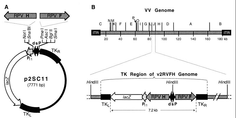
The VV transfer vector p2SC11RVFH was used for the generation of v2RVFH. This plasmid contains cDNA from the F and H genes of the highly virulent Kabete ‘O’ strain of RPV. Briefly, the 273-bp XbaI-SmaI fragment of pSC11 (5) (obtained by partial XbaI digestion and containing the VV P7.5 promoter) was replaced with the 161-bp NheI-SmaI fragment of pJS5 (6), generating p2SC11, which contains two back-to-back strong synthetic VV promoters (dsP) (Fig. 1). Next, the 1,954-bp BamHI-EcoRI fragment of plasmid pvRVH (42), containing the H gene of RPV, was filled in with T4 DNA polymerase and inserted into the SnaBI site of plasmid p2SC11, generating p2SC11RVH. Finally, the 1,813-bp EcoRI fragment of pRVFΔ6 (42), containing the F gene of RPV, was filled in with T4 DNA polymerase and inserted into the PmeI site of p2SC11RVH, generating p2SC11RVFH.
FIG. 1.
Construction of transfer vector p2SC11 and generation of v2RVFH. (A) p2SC11 directs the insertion of genes at the TK site of VV by homologous recombination. It contains the lacZ gene under the control of the VV P11 late promoter for screening of rVVs, and two back-to-back strong synthetic VV promoters (dsP) that are active in both early and late stages of infection. There are multiple cloning sites adjacent to each side of the dsP to facilitate cloning of heterologous genes (only unique sites are shown). The H and F genes of RPV were inserted into the SnaBI and PmeI sites of p2SC11, respectively, and the resulting plasmid was used for the generation of v2RVFH. (B) Genome of the Copenhagen strain of VV, showing HindIII restriction fragments A through P (top). The HindIII J fragment, which contains the TK gene, is shown in detail as it is present in v2RVFH (bottom). ITR, inverted terminal repeat.
v2RVFH was generated by standard homologous recombination using cationic liposome-mediated transfection of plasmid p2SC11RVFH into BS-C-1 cells infected with VV at 0.05 PFU/cell. v2RVFH was plaque purified from transfection supernatants on BS-C-1 cell monolayers, using 5-bromo-4-chloro-3-indolyl-β-d-galactopyranoside (X-Gal) to detect the expression of the lacZ marker gene (coding for β-galactosidase) (7). The expression of the lacZ gene by the final plaque-purified rVV was confirmed by cytochemical staining of infected cell monolayers as described previously (37). Restriction analysis of VV DNA samples was performed with DNA purified by a small-scale method employing micrococcal nuclease (17).
Radioimmunoprecipitation.
The expression of F and H polypeptides of RPV by the rVVs was characterized by radioimmunoprecipitation. Briefly, monolayers of BS-C-1 cells were infected with rVVs at 10 PFU/cell for 1 h at room temperature. Cells were then incubated at 37°C for 30 min in DMEM and for 90 min in methionine- and cysteine-free medium. Then, 100 μCi of l-[35S]methionine/cysteine (Pro-Mix l-[35S] in vitro cell labeling mix; Amersham Pharmacia Biotech, Piscataway, N.J.) was added, and cells were incubated for an additional 24 h. Finally, the medium was changed to DMEM, and the cells were incubated for 12 h before harvesting. The recombinant F and H polypeptides were immunoprecipitated with a mixture of rabbit anti-F of MV, rabbit anti-RPV, and monkey anti-MV sera. Preparation of cell lysates, immunoprecipitation, polyacrylamide gel electrophoresis, and autoradiography were performed as previously described (36). Band intensities were quantified with the Quantity One software package, version 4.2.1 (Bio-Rad Laboratories, Hercules, Calif.).
Syncytium formation.
BS-C-1 cell monolayers grown on sterile cover slips were infected with VV and stained 2 days later with crystal violet fixative (0.1% crystal violet, 10% ethanol, 20% formaldehyde) for 5 min at room temperature. Coverslips were rinsed thoroughly with water, dried, and mounted on slides. Bright-field digital images were obtained using an Olympus AX70 microscope. Images were processed in Adobe Photoshop 5.0 (Adobe Systems, San Jose, Calif.) with no manipulations other than for contrast.
Safety and efficacy studies in cattle.
Locally obtained Zebu cattle (Bos indicus, 2 years old on average), negative for serum neutralizing (SN) antibodies to RPV, were ear-tagged and kept in isolation in accordance with the governmental guidelines of Ethiopia and Kenya and the institutional policies of NVI and KARI. Groups of animals were vaccinated intramuscularly (1 ml) with various doses of v2RVFH at the side of the neck. For comparative purposes, some animals received the modified Kabete ‘O’ PTCV (29), 103 50% tissue culture-infectious doses (TCID50) at NVI and 3.2 × 10² TCID50 at KARI, subcutaneously (1 ml) at the side of the neck. Animals were challenged subcutaneously (1 ml) at the side of the neck with 103 to 104 TCID50 of the pathogenic Kabete ‘O’ RPV; as little as 1 TCID50 of the virus administered subcutaneously induces clinical rinderpest with 100% mortality in U.S. cattle (42). Postchallenge rectal temperatures were taken daily.
Nasal and ocular swabs were taken at 2, 3, 4, and 7 days postchallenge from a group of NVI animals challenged at 4 weeks postvaccination. In addition, prescapular and mesenteric lymph nodes as well as lung, spleen, tonsil, kidney, and heart tissue samples were taken from animals that died following RPV challenge. RPV isolation was attempted from the collected swabs and necropsy samples in primary calf cells (kidney and testis) and Vero cells as previously described (38).
Serum neutralization assays.
Twofold dilutions of serum samples were prepared in duplicate and assayed by microtiter techniques as described previously (30). SN titers to RPV were expressed as the reciprocal of the highest dilution of serum that gave complete protection against the cytopathic effects of 102 TCID50 of RPV in Vero (NVI) or primary calf kidney (KARI) cells.
Statistical analysis.
The statistical software SAS, release 6.11 (SAS Institute, Cary, N.C.), was used to analyze the data. Postchallenge SN antibody titers to RPV were compared with titers on the day of challenge by repeated-measures analysis of variance (ANOVA), followed by Dunnett’s one-sided multiple comparisons procedure.
RESULTS
Construction and characterization of v2RVFH.
We developed a new transfer vector, p2SC11, that directs homologous recombination with the TK gene of the VV genome (Fig. 1). The two strong synthetic promoters (dsP) in p2SC11 are active in both early and late stages of infection, allowing increased expression of heterologous genes (6). The F and H genes of RPV were subcloned into p2SC11 (Fig. 1), and the resulting plasmid was used to generate v2RVFH. The purity and stability of v2RVFH were confirmed by X-Gal cytochemical staining after consecutive plaque purifications and multiple passages in cell culture to ensure total elimination of wild-type virus. Restriction analysis of v2RVFH-purified DNA with HindIII, KpnI, PstI, and XhoI confirmed the insertional inactivation of the VV TK genomic region by the 7.2-kb cassette containing the lacZ gene and the F and H genes of RPV (data not shown).
Higher expression levels of F and H proteins by v2RVFH.
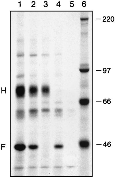
The expression of authentic F and H proteins of RPV by v2RVFH was demonstrated by radioimmunoprecipitation (Fig. 2). A diffuse band of 77 to 80 kDa, similar to that found in RPV-infected cells (11), was immunoprecipitated only from cells infected with rVVs expressing the H protein (Fig. 2, lanes 1, 2, and 3). As expected, the uncleaved F protein (F0) was not detected, but the larger subunit (F1) of this proteolytically cleaved polypeptide, which is nonglycosylated (11) and of a calculated molecular mass of 47 kDa (12), was immunoprecipitated only from cells infected with rVVs expressing the F protein (Fig. 2, lanes 1, 2, and 4). Both proteins were abundantly expressed by v2RVFH-infected cells under control of the strong synthetic promoters (6).
FIG. 2.
Radioimmunoprecipitation of F and H polypeptides of RPV expressed by rVVs. Lane 1, cells infected with v2RVFH expressing both F and H genes of RPV under the control of strong synthetic promoters (dsP); lane 2, cells infected with vRVFH expressing both F and H genes under control of the P7.5 promoters; lane 3, cells infected with vRVH expressing only the H gene under control of the P7.5 promoter; lane 4, cells infected with vRVF expressing only the F gene under control of the P7.5 promoter; lane 5, cells infected with the wild-type Copenhagen strain of VV; and lane 6, molecular size standards (shown in kilodaltons).
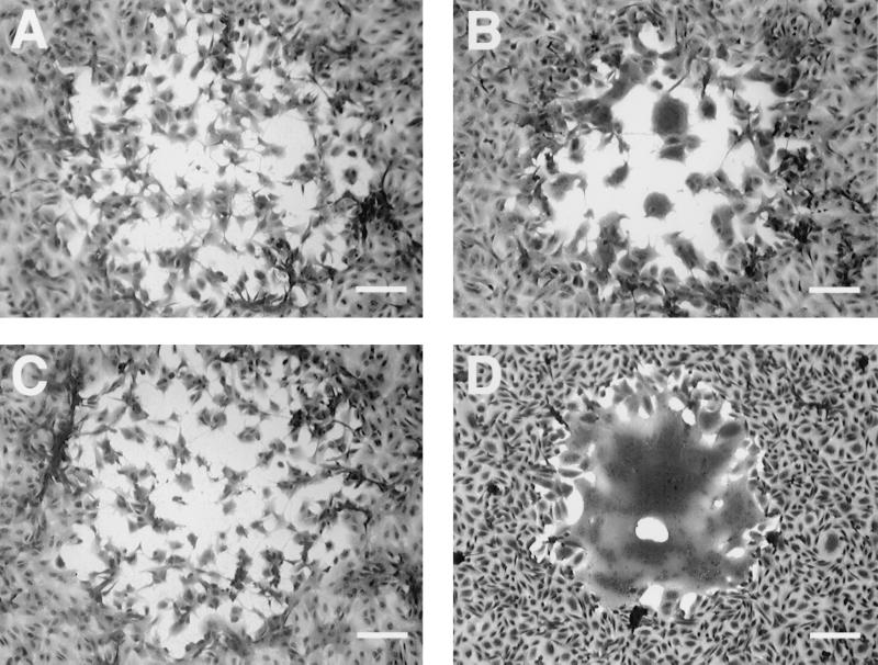
Quantification of the bands revealed that v2RVFH expresses at least three- and fourfold more H and F proteins, respectively, than the previously developed single and double rVV vaccines for rinderpest, which express the genes under the P7.5 early/late VV promoter. Higher levels of expression of the F and H genes by v2RVFH were further confirmed by the formation of plaques with large syncytia (Fig. 3). For morbilliviruses, fusion occurs preferentially when both the F and H glycoproteins are expressed (23, 31, 33, 40). Thus, plaques from the previous rVV expressing the F and H genes under the P7.5 promoter (vRVFH) showed some evidence of syncytium formation, containing only a few nuclei (Fig. 3B). However, v2RVFH caused extensive syncytium formation, usually comprising the whole plaque and containing several hundred nuclei (Fig. 3D).
FIG. 3.
Syncytium formation by rVVs expressing the F and H genes of RPV. BS-C-1 cell monolayers infected with VV were stained 2 days postinfection, and representative plaques were photographed. Cells infected with the wild-type Wyeth (A) and Copenhagen (C) strains of VV did not show any evidence of syncytium formation, while cells infected with vRVFH (B) showed some evidence of cell fusion. v2RVFH infection induced higher levels of syncytium formation, typically comprising the whole plaque and containing several hundred nuclei (D). Bars, 150 μm.
Protective immunity to cattle provided by v2RVFH.
A preliminary study to determine the 50% protective dose (PD50) of v2RVFH was performed at KARI. Groups of cattle, housed in separate stalls, were vaccinated intramuscularly with v2RVFH (102 to 106 PFU) or subcutaneously with PTCV (3.2 × 10² TCID50). In addition, to assess the transmissibility of v2RVFH from vaccinated to contact animals, sentinels were housed together with a group that received 108 PFU of v2RVFH. Daily examination of the animals over a period of 4 weeks revealed no adverse clinical reactions to vaccination and no pock lesions at the sites of inoculation (Table 1). The two sentinels did not show any evidence of lateral transmission of v2RVFH, as assessed by SN antibody titers to RPV (data not shown).
TABLE 1.
Determination of the PD50 of v2RVFH in African cattlea
| Vaccine | Dose | No. of cattle/no. tested
|
||
|---|---|---|---|---|
| Clinical response to vaccination | Clinical response to challengeb | Survival | ||
| v2RVFH | 102 PFU | 0/4 | 2/4 | 3/4 |
| v2RVFH | 103 PFU | 0/4 | 0/4 | 4/4 |
| v2RVFH | 104 PFU | 0/4 | 0/4 | 4/4 |
| v2RVFH | 105 PFU | 0/4 | 0/4 | 4/4 |
| v2RVFH | 106 PFU | 0/3 | 0/3 | 3/3 |
| v2RVFH | 108 PFU | 0/3 | 0/3 | 3/3 |
| PTCV | 3.2 × 10² TCID50 | 0/2 | 0/2 | 2/2 |
| Control | NAc | NA | 2/2 | 0/2 |
v2RVFH was given intramuscularly and PTCV was given subcutaneously. Controls (sentinels) are naive cattle housed together with animals that received 108 PFU of v2RVFH.
No. of cattle with fever, congestion, and mouth lesions. Animals were challenged 28 days postvaccination with RPV.
NA, not applicable.
All groups of vaccinated cattle that received PTCV or greater than 102 PFU of v2RVFH were protected from clinical rinderpest upon challenge (Table 1). In the group vaccinated with 102 PFU of v2RVFH, two of the four animals developed signs of rinderpest (fever, severe ocular and oral congestion, mucoid nasal discharges, and inappetence). One sick animal recovered before the onset of diarrhea, and the other died of acute rinderpest 10 days postchallenge, establishing 102 PFU as the PD50 for v2RVFH. The two sentinels died of rinderpest within 7 days postchallenge.
v2RVFH provides protective immunity equivalent to PTCV.
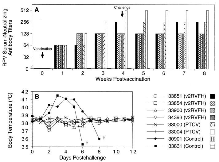
A study to determine the efficacy of v2RVFH was performed at NVI. Four cattle were vaccinated intramuscularly with 108 PFU of v2RVFH, two animals were vaccinated subcutaneously with 103 TCID50 of PTCV, and two animals were left unvaccinated. No pock lesions at the inoculation sites or adverse clinical reactions to vaccination were observed over a period of 4 weeks. All cattle vaccinated with v2RVFH developed SN antibody titers to RPV that were equivalent to the titers of animals vaccinated with PTCV (Fig. 4A). Four weeks postvaccination, all animals were challenged subcutaneously with RPV. All vaccinated animals resisted challenge, showing no clinical signs of rinderpest and exhibiting normal temperatures (Fig. 4B). Unvaccinated control animals developed high fevers and died 7 and 9 days postchallenge of rinderpest. No virus could be isolated from the nasal and ocular swabs taken from vaccinated animals at 2, 3, 4, and 7 days postchallenge. However, RPV was isolated from all samples taken from the unvaccinated control animals 3 days postchallenge and from all postmortem samples. SN titers to RPV after challenge did not increase significantly from titers on the day of challenge (1.0 ≤ P ≥ 0.8, repeated-measures ANOVA) (Fig. 4A). Thus, the absence of detectable virus replication and lack of anamnestic responses upon challenge of vaccinated animals indicate sterilizing immunity.
FIG. 4.
v2RVFH provides protective immunity equivalent to PTCV. Groups of cattle were vaccinated intramuscularly with 108 PFU of v2RVFH or subcutaneously with 103 TCID50 of PTCV or left untreated (controls). Animals were challenged 4 weeks later with RPV. (A) SN antibody titers to RPV were determined weekly. (B) Rectal temperatures were taken daily. The symbol † indicates death of an animal. Numbers in the lower right panel identify individual animals.
Long-term sterilizing immunity provided by v2RVFH.
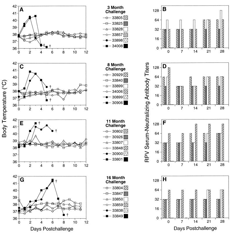
At NVI, groups of four cattle were vaccinated intramuscularly with 108 PFU of v2RVFH. At 3, 8, 11, and 16 months postvaccination, two naive control animals were added to each group, and all animals were challenged with RPV. SN titers to RPV in all vaccinated animals ranged from 32 to 128 on the day of challenge (Fig. 5B, D, F, and H). All vaccinated animals were completely protected from pathogenic RPV, while all control animals developed high fevers (Fig. 5A, C, E, and G) and died 4 to 9 days postchallenge from clinical rinderpest. Not a single vaccinated animal mounted an anamnestic antibody response, as measured by SN titers to RPV (Fig. 5B, D, F, and H), which did not increase significantly from levels recorded on the day of challenge (1.0 ≤ P > 0.1, repeated-measures ANOVA), indicating sterilizing immunity.
FIG. 5.
Long-term sterilizing immunity provided by v2RVFH. Groups of four cattle (open symbols in the left panels) were vaccinated intramuscularly with 108 PFU of v2RVFH and challenged 3 (A and B), 8 (C and D), 11 (E and F), or 16 (G and H) months later with RPV. Two naive cattle (solid symbols in the left panels) were added at the day of each challenge. Rectal temperatures were taken daily (left panels). The symbol † indicates death of an animal. SN antibody titers to RPV were determined weekly (right panels). Numbers in the center panels identify individual animals.
DISCUSSION
We have developed a second-generation rVV vaccine (v2RVFH) that induces both humoral immune responses and protective immunity comparable to those elicited by PTCV. We have enhanced efficacy by using strong synthetic promoters to increase the expression of the F and H genes of RPV. Moreover, we added the lacZ gene to serve as an easily identifiable marker to aid in rVV selection during the development of the vaccine and, more importantly, to distinguish the vaccine virus (v2RVFH) from other naturally occurring poxviruses during vaccination programs.
Compared to vRVFH (Fig. 3) or the single recombinants expressing F or H (data not shown), v2RVFH displays a distinct plaque morphology in cell culture that is characterized by the formation of massive syncytia due to higher levels of expression of both glycoproteins. Radioimmunoprecipitation studies showed that the band intensities for the F and H proteins expressed by v2RVFH were at least threefold higher than those generated by vRVFH or the single recombinants (Fig. 2). We conducted short- and long-term safety and efficacy studies in African cattle, and demonstrated that as little as 102 PFU of v2RVFH, given intramuscularly, protects 50% of the vaccinated cattle from virulent challenge with RPV. Unlike the intradermal route, the intramuscular route facilitates mass vaccination of cattle under field conditions. We also showed that a dose of 108 PFU of v2RVFH induced levels of SN antibodies to RPV comparable to those elicited by PTCV (Fig. 4) and that this dose provides sterilizing immunity to cattle for at least 16 months (Fig. 5). This level of protection suggests that v2RVFH confers long-term immunity equivalent to PTCV. Lastly, v2RVFH is not only highly efficacious but also safe, since it is attenuated by the insertional inactivation of the TK gene (4). As a result, no pock lesions were detected after vaccination, and there was no lateral transmission to contact animals, as observed with the highly attenuated modified VV Ankara (MVA) (32) and rVV vectors expressing cytokine genes (9, 41).
Live attenuated vaccines confer long-term protective immunity against infection with morbilliviruses. Although cell-mediated immune responses are required for clearance of the virus (10), antibody responses appear to be needed for sterilizing immunity (43). We have previously demonstrated that cattle vaccinated with rVVs expressing the F protein of RPV had low SN antibody titers and yet were protected from disease, although there were high levels of anamnestic antibody responses to RPV, indicating a lack of sterilizing immunity (42). We have also shown that cattle vaccinated with soluble F and H proteins of RPV expressed in baculovirus were not protected against disease, despite having higher SN antibody titers than those vaccinated with rVVs expressing the F protein (3). This establishes that antibodies alone are insufficient for protection. Since live virus vectors such as VV elicit both humoral and cell-mediated immune responses (21), the higher levels of expression of both F and H antigens by v2RVFH must have induced stronger immune responses, providing cattle with long-term sterilizing immunity to rinderpest.
Other investigators have developed single VV and capripoxvirus vectors expressing either the F or H protein of RPV; however, there were anamnestic responses that indicated replication of the challenge virus shortly after vaccination, and long-term studies showed only partial protection (13, 22, 24). In contrast, v2RVFH provided complete protection with sterilizing immunity during both short- and long-term studies.
Animals vaccinated with PTCV cannot be distinguished serologically from those infected with RPV. These animals are barred from export, significantly reducing their market value. We developed a rapid diagnostic kit for rinderpest based on the N protein of RPV, inexpensively produced in a baculovirus expression system. A single larva infected with a baculovirus expressing the N protein can be used for the diagnosis of more than 7,000 serum samples in duplicate (14). While animals exposed to the whole virus will test positive, those vaccinated with v2RVFH will test negative in this system. A recent outbreak of rinderpest in Kenya and Tanzania was caused by a strain of RPV (type 2 lineage) that is mild to cattle yet highly virulent to wildlife, killing up to 80% of wild ruminants (1, 16). Cycles of infection with strains that differ in virulence between livestock and wildlife may be the mechanism by which RPV is maintained in nature. Wild ruminants vaccinated orally with V-RG developed antibodies to rabies virus (34). Consequently, v2RVFH could be used as an oral vaccine in animal feed (pellets) for the immunization of both domestic and wild ruminants, and in conjunction with rapid diagnostic kits, v2RVFH could accomplish the eradication of rinderpest.
Acknowledgments
This work was supported by contracts awarded to T.D.Y. by the U.S. Agency for International Development (LAG-4178-A-00-3051-00 and PCE-G-00-99-00013) and the International Atomic Energy Agency of the United Nations (RAF5043-054-079k), as well as grants from the U.S. Department of Agriculture (94-39210-0532) and the National Institutes of Health (AI36197, AI37182, and AI47025). P.H.V. received support from Conselho Nacional de Desenvolvimento Científico e Tecnológico (CNPq), Brazil. F.H.A. was supported by the Floyd and Mary Schwall and UC Davis Biotechnology Program fellowships.
We are grateful to Robert Drillien for providing the Copenhagen strain of VV and to Bernard Moss for providing pJS5. We thank Fantahun Wondimagegne for technical assistance, Thomas Farver for advice on the statistical analysis of the data, Lael Brown for outstanding administrative support, the directors of NVI and KARI for their support and for the use of facilities, and the technical staff and animal caretakers at both institutes. We also thank Sally Owens, Kenneth Chan, and Yue Peng for critical reviews of the manuscript.
REFERENCES
- 1.Barrett, T., M. A. Forsyth, K. Inui, H. M. Wamwayi, R. Kock, J. Wambua, J. Mwanzia, and P. B. Rossiter. 1998. Rediscovery of the second African lineage of rinderpest virus: its epidemiological significance. Vet. Rec. 142:669–671. [DOI] [PubMed] [Google Scholar]
- 2.Barrett, T., and P. B. Rossiter. 1999. Rinderpest: the disease and its impact on humans and animals. Adv. Virus Res. 53:89–110. [DOI] [PubMed] [Google Scholar]
- 3.Bassiri, M., S. Ahmad, L. Giavedoni, L. Jones, J. T. Saliki, C. Mebus, and T. Yilma. 1993. Immunological responses of mice and cattle to baculovirus-expressed F and H proteins of rinderpest virus: lack of protection in the presence of neutralizing antibody. J. Virol. 67:1255–1261. [DOI] [PMC free article] [PubMed] [Google Scholar]
- 4.Buller, R. M., G. L. Smith, K. Cremer, A. L. Notkins, and B. Moss. 1985. Decreased virulence of recombinant vaccinia virus expression vectors is associated with a thymidine kinase-negative phenotype. Nature 317:813–815. [DOI] [PubMed] [Google Scholar]
- 5.Chakrabarti, S., K. Brechling, and B. Moss. 1985. Vaccinia virus expression vector: coexpression of β-galactosidase provides visual screening of recombinant virus plaques. Mol. Cell. Biol. 5:3403–3409. [DOI] [PMC free article] [PubMed] [Google Scholar]
- 6.Chakrabarti, S., J. R. Sisler, and B. Moss. 1997. Compact, synthetic, vaccinia virus early/late promoter for protein expression. BioTechniques 23:1094–1097. [DOI] [PubMed] [Google Scholar]
- 7.Earl, P. L., and B. Moss. 1994. Generation of recombinant vaccinia viruses, p.16.17.1–16.17.16. In F. M. Ausubel, R. Brent, R. E. Kingston, D. D. Moore, J. G. Seidman, J. A. Smith, and K. Struhl (ed.), Current protocols in molecular biology, vol. 2. John Wiley & Sons, New York, N.Y.
- 8.Giavedoni, L., L. Jones, C. Mebus, and T. Yilma. 1991. A vaccinia virus double recombinant expressing the F and H genes of rinderpest virus protects cattle against rinderpest and causes no pock lesions. Proc. Natl. Acad. Sci. USA 88:8011–8015. [DOI] [PMC free article] [PubMed] [Google Scholar]
- 9.Giavedoni, L. D., L. Jones, M. B. Gardner, H. L. Gibson, C. T. Ng, P. J. Barr, and T. Yilma. 1992. Vaccinia virus recombinants expressing chimeric proteins of human immunodeficiency virus and γ interferon are attenuated for nude mice. Proc. Natl. Acad. Sci. USA 89:3409–3413. [DOI] [PMC free article] [PubMed] [Google Scholar]
- 10.Good, R. A., and S. J. Zak. 1956. Disturbances in gamma globulin synthesis as “experiments of nature.” Pediatrics 18:109–149. [PubMed] [Google Scholar]
- 11.Grubman, M. J., C. Mebus, B. Dale, M. Yamanaka, and T. Yilma. 1988. Analysis of the polypeptides synthesized in rinderpest virus-infected cells. Virology 163:261–267. [DOI] [PubMed] [Google Scholar]
- 12.Hsu, D., M. Yamanaka, J. Miller, B. Dale, M. Grubman, and T. Yilma. 1988. Cloning of the fusion gene of rinderpest virus: comparative sequence analysis with other morbilliviruses. Virology 166:149–153. [DOI] [PubMed] [Google Scholar]
- 13.Inui, K., T. Barrett, R. P. Kitching, and K. Yamanouchi. 1995. Long-term immunity in cattle vaccinated with a recombinant rinderpest vaccine. Vet. Rec. 137:669–670. [PubMed] [Google Scholar]
- 14.Ismail, T., S. Ahmad, M. D’Souza-Ault, M. Bassiri, J. Saliki, C. Mebus, and T. Yilma. 1994. Cloning and expression of the nucleocapsid gene of virulent Kabete O strain of rinderpest virus in baculovirus: use in differential diagnosis between vaccinated and infected animals. Virology 198:138–147. [DOI] [PubMed] [Google Scholar]
- 15.Kieny, M. P., R. Lathe, R. Drillien, D. Spehner, S. Skory, D. Schmitt, T. Wiktor, H. Koprowski, and J. P. Lecocq. 1984. Expression of rabies virus glycoprotein from a recombinant vaccinia virus. Nature 312:163–166. [DOI] [PubMed] [Google Scholar]
- 16.Kock, R. A., J. M. Wambua, J. Mwanzia, H. Wamwayi, E. K. Ndungu, T. Barrett, N. D. Kock, and P. B. Rossiter. 1999. Rinderpest epidemic in wild ruminants in Kenya, 1993–97. Vet. Rec. 145:275–283. [DOI] [PubMed] [Google Scholar]
- 17.Lai, A. C., and Y. Chu. 1991. A rapid method for screening vaccinia virus recombinants. BioTechniques 10:564–565. [PubMed] [Google Scholar]
- 18.Mackowiak, M., J. Maki, L. Motes-Kreimeyer, T. Harbin, and K. van Kampen. 1999. Vaccination of wildlife against rabies: successful use of a vectored vaccine obtained by recombinant technology. Adv. Vet. Med. 41:571–583. [DOI] [PubMed] [Google Scholar]
- 19.Mariner, J. C., J. A. House, C. A. Mebus, A. Sollod, and C. Stem. 1991. Production of a thermostable Vero cell-adapted rinderpest vaccine. J. Tissue Culture Methods 13:253–258. [Google Scholar]
- 20.Mariner, J. C., J. A. House, A. E. Sollod, C. Stem, M. van den Ende, and C. A. Mebus. 1990. Comparison of the effect of various chemical stabilizers and lyophilization cycles on the thermostability of a Vero cell-adapted rinderpest vaccine. Vet. Microbiol. 21:195–209. [DOI] [PubMed] [Google Scholar]
- 21.Moss, B. 1996. Genetically engineered poxviruses for recombinant gene expression, vaccination, and safety. Proc. Natl. Acad. Sci. USA 93:11341–11348. [DOI] [PMC free article] [PubMed] [Google Scholar]
- 22.Ngichabe, C. K., H. M. Wamwayi, T. Barrett, E. K. Ndungu, D. N. Black, and C. J. Bostock. 1997. Trial of a capripoxvirus-rinderpest recombinant vaccine in African cattle. Epidemiol. Infect. 118:63–70. [DOI] [PMC free article] [PubMed] [Google Scholar]
- 23.Nussbaum, O., C. C. Broder, B. Moss, L. B. Stern, S. Rozenblatt, and E. A. Berger. 1995. Functional and structural interactions between measles virus hemagglutinin and CD46. J. Virol. 69:3341–3349. [DOI] [PMC free article] [PubMed] [Google Scholar]
- 24.Ohishi, K., K. Inui, T. Barrett, and K. Yamanouchi. 2000. Long-term protective immunity to rinderpest in cattle following a single vaccination with a recombinant vaccinia virus expressing the virus haemagglutinin protein. J. Gen. Virol. 81:1439–1446. [DOI] [PubMed] [Google Scholar]
- 25.Pastoret, P. P., and B. Brochier. 1996. The development and use of a vaccinia-rabies recombinant oral vaccine for the control of wildlife rabies: a link between Jenner and Pasteur. Epidemiol. Infect. 116:235–240. [DOI] [PMC free article] [PubMed] [Google Scholar]
- 26.Pastoret, P. P., and B. Brochier. 1999. Epidemiology and control of fox rabies in Europe. Vaccine 17:1750–1754. [DOI] [PubMed] [Google Scholar]
- 27.Plowright, W. 1984. The duration of immunity in cattle following inoculation of rinderpest cell culture vaccine. J. Hyg. 92:285–296. [DOI] [PMC free article] [PubMed] [Google Scholar]
- 28.Plowright, W. 1968. Rinderpest virus. Virol. Monogr. 3:25–110. [Google Scholar]
- 29.Plowright, W., and R. D. Ferris. 1962. Studies with rinderpest virus in tissue culture: the use of attenuated culture virus as a vaccine for cattle. Res. Vet. Sci. 3:172–182. [Google Scholar]
- 30.Rossiter, P. B., and D. M. Jessett. 1982. Microtitre techniques for the assay of rinderpest virus and neutralising antibody. Res. Vet. Sci. 32:253–256. [PubMed] [Google Scholar]
- 31.Schmid, A., P. Spielhofer, R. Cattaneo, K. Baczko, V. ter Meulen, and M. A. Billeter. 1992. Subacute sclerosing panencephalitis is typically characterized by alterations in the fusion protein cytoplasmic domain of the persisting measles virus. Virology 188:910–915. [DOI] [PubMed] [Google Scholar]
- 32.Sutter, G., L. S. Wyatt, P. L. Foley, J. R. Bennink, and B. Moss. 1994. A recombinant vector derived from the host range-restricted and highly attenuated MVA strain of vaccinia virus stimulates protective immunity in mice to influenza virus. Vaccine 12:1032–1040. [DOI] [PubMed] [Google Scholar]
- 33.Taylor, J., S. Pincus, J. Tartaglia, C. Richardson, G. Alkhatib, D. Briedis, M. Appel, E. Norton, and E. Paoletti. 1991. Vaccinia virus recombinants expressing either the measles virus fusion or hemagglutinin glycoprotein protect dogs against canine distemper virus challenge. J. Virol. 65:4263–4274. [DOI] [PMC free article] [PubMed] [Google Scholar]
- 34.U.S. Department of Agriculture Animal and Plant Health Inspection Service. 1989. Veterinary biologics authorized field trial of an experimental biologic: the Wistar Institute of Anatomy and Biology proposed field trial of a live experimental vaccinia rabies vaccine. Environmental assessment and preliminary finding of no significant impact. Animal and Plant Health Inspection Service, U.S. Department of Agriculture, Hyattsville, Md.
- 35.van Regenmortel, M. H. V., C. M. Fauquet, D. H. L. Bishop, E. B. Carstens, M. K. Estes, S. M. Lemon, J. Maniloff, M. A. Mayo, D. J. McGeoch, C. R. Pringle, and R. B. Wickner (ed.). 2000. Virus taxonomy: seventh report of the International Committee on Taxonomy of Viruses. Academic Press, San Diego, Calif.
- 36.Varsanyi, T. M., B. Morein, A. Löve, and E. Norrby. 1987. Protection against lethal measles virus infection in mice by immune-stimulating complexes containing the hemagglutinin or fusion protein. J. Virol. 61:3896–3901. [DOI] [PMC free article] [PubMed] [Google Scholar]
- 37.Verardi, P. H., L. A. Jones, F. H. Aziz, S. Ahmad, and T. D. Yilma. 2001. Vaccinia virus vectors with an inactivated gamma interferon receptor homolog gene (B8R) are attenuated in vivo without a concomitant reduction in immunogenicity. J. Virol. 75:11–18. [DOI] [PMC free article] [PubMed] [Google Scholar]
- 38.Wamwayi, H. M., and J. S. Wafula. 1987. Microtitre technique for detection and titration of rinderpest virus in infected bovine tissues and secretions. Bull. Anim. Health Prod. Afr. 35:318–320. [Google Scholar]
- 39.Wiktor, T. J., R. I. Macfarlan, K. J. Reagan, B. Dietzschold, P. J. Curtis, W. H. Wunner, M. P. Kieny, R. Lathe, J. P. Lecocq, M. Mackett, B. Moss, and H. Koprowski. 1984. Protection from rabies by a vaccinia virus recombinant containing the rabies virus glycoprotein gene. Proc. Natl. Acad. Sci. USA 81:7194–7198. [DOI] [PMC free article] [PubMed] [Google Scholar]
- 40.Wild, T. F., E. Malvoisin, and R. Buckland. 1991. Measles virus: both the haemagglutinin and fusion glycoproteins are required for fusion. J. Gen. Virol. 72:439–442. [DOI] [PubMed] [Google Scholar]
- 41.Yilma, T., K. Anderson, K. Brechling, and B. Moss. 1987. Expression of an adjuvant gene (interferon-γ) in infectious vaccinia virus recombinants, p.393–396. In R. M. Chanock (ed.), Vaccine 87: modern approaches to new vaccines: prevention of AIDS and other viral, bacterial, and parasitic diseases. Cold Spring Harbor Laboratory, Cold Spring Harbor, N.Y.
- 42.Yilma, T., D. Hsu, L. Jones, S. Owens, M. Grubman, C. Mebus, M. Yamanaka, and B. Dale. 1988. Protection of cattle against rinderpest with vaccinia virus recombinants expressing the HA or F gene. Science 242:1058–1061. [DOI] [PubMed] [Google Scholar]
- 43.Zhu, Y., P. Rota, L. Wyatt, A. Tamin, S. Rozenblatt, N. Lerche, B. Moss, W. Bellini, and M. McChesney. 2000. Evaluation of recombinant vaccinia virus-measles vaccines in infant rhesus macaques with preexisting measles antibody. Virology 276:202–213. [DOI] [PubMed] [Google Scholar]


