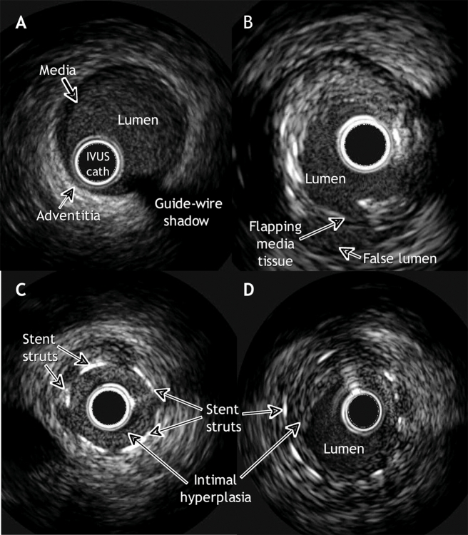
Fig. 1. Images of coronary arteries, revealed with intravascular ultrasonography (IVUS). A: A normal artery. The layers of the arterial wall, from innermost to outermost, are the intima, media and adventitia. The intima is an endothelial cell layer where atheroma accumulates; the external boundary of this layer is the internal elastic membrane. The media, composed of smooth muscle cells, elastin and collagen, is encircled by the external elastic membrane. The adventitia is mainly composed of fibrous tissue. In young people free of atherosclerotic disease, the 3 layers are difficult to see as separate structures because the media and intima are smaller than the resolution of IVUS (< 100 mm). B: Mixed plaque (fibrotic and calcific) with a dissection of the media at the “6 o'clock” position. C: In-stent restenosis occluding the lumen. D: In-stent restenosis without angiographically significant occlusion.

An official website of the United States government
Here's how you know
Official websites use .gov
A
.gov website belongs to an official
government organization in the United States.
Secure .gov websites use HTTPS
A lock (
) or https:// means you've safely
connected to the .gov website. Share sensitive
information only on official, secure websites.
