Abstract
Active visual perception relies on the ability to interpret correctly retinal motion signals induced either by moving objects viewed with static eyes or by stationary objects viewed with moving eyes. A motionless environment is not normally perceived as moving during saccadic eye movements. It is commonly believed that this phenomenon involves central oculomotor signals that inhibit intrasaccadic visual motion processing. The keystone of this extraretinal theory relies on experimental reports showing that physically stationary scenes displayed only during saccades, thus producing high retinal velocities, are never perceived as moving but appear as static blurred images. We, however, provide evidence that stimuli optimized for high-speed motion detection elicit clear motion perception against saccade direction, thus making the search for extraretinal suppression superfluous. The data indicate that visual motion is the main cue used by observers to perform the task independently of other perceptual factors covarying with intrasaccadic stimulation. By using stimuli of different durations, we show that the probability of perceiving the stimulus as static, rather than moving, increases when the intrasaccadic stimulation is preceded or followed by a significant extrasaccadic stimulation. We suggest that intrasaccadic motion perception is accomplished by motion-selective magnocellular neurons through temporal integration of rapidly increasing retinal velocities. The functional mechanism that usually prevents this intrasaccadic activity from being perceived seems to rely on temporal masking effects induced by the static retinal images present before and/or after the saccade.
The daily observation that we do not perceive our static environment as moving when we make saccadic eye movements does not imply that we are blind during saccades. Intrasaccadic vision can be revealed by displaying a static stimulus during a saccade made in the dark. Early studies showed that a brief stimulus (1 ms) minimizing any smearing across the retinal photoreceptors is perceived as a sharp image, whereas a slightly longer stimulus (e.g., 20 ms) appears as a smeared and blurred image (1–3). However, motion perception was not observed in these pioneering studies and has never been reported since. This absence of intrasaccadic motion perception was initially interpreted as evidence that motion detectors are unable to process the high speeds produced as the retina sweeps over the physically static stimulus, up to 500°/s for large saccades. This hypothesis was at odds, however, with the subsequent finding that observers with static eyes can perceive the motion of stimuli moving as fast as 800°/s (4). This apparent paradox therefore led to the now traditional view that we are motion-blind during saccades (5–9).
Experimental support for this theory is weak, however. Although high-speed motion perception, tested with static eyes, only occurs when the visual stimulus is a periodic grating of low spatial frequency (4, 10), the studies reporting intrasaccadic blurred perception (1–3) have never used this specific stimulus. We therefore reassessed the ability of the visual system to process intrasaccadic retinal motion by displaying static gratings of different spatial frequencies during saccadic flight.
Methods
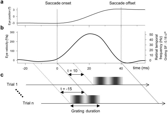
Human observers made voluntary horizontal saccades while a static vertical grating was displayed at random instants relative to the occurrence of saccades (Fig. 1). Only two percepts were reported across trials; the grating appeared either as static or as moving against the saccade direction. Observers indicated their percept by pressing one of two buttons. In all experiments, observers were encouraged to use a conservative criterion, that is, to respond “motion” only when the motion percept was conspicuous. To assess intrasaccadic perception of naïve observers (who were not aware that the stimulus was always stationary on the screen), we first run preliminary sessions in which the observers were not required to report any specific percept. At the end of each of these preliminary sessions, observers were simply asked to describe the appearance of the stimuli presented across trials. After a few hundred trials, the spontaneous reports were that on most trials the bars seemed to be static (or slightly jittering), whereas on a few trials they seemed to move against the direction of the saccade. Once this perceptual categorization was established, we started the experiments. Off-line analysis of ocular data allowed us to assess the dependence of intrasaccadic motion perception on the temporal relationship between saccade and grating onsets. The direction of saccades was constant within each experimental block (120 trials) and alternated across blocks.
Fig 1.
Methods. (a) Eye position signal for a typical 6° saccade. (b) Eye velocity profile (left ordinate) and corresponding retinal temporal frequency for a 0.18 c/° grating presented continuously (right ordinate). (c) Schematic of experimental method. A static grating is displayed for 19 ms on each trial around the time of saccade latency. The difference between grating and saccade onsets (t) is +10 ms in trial 1 and −15 ms in trial n. Gratings of optimal low spatial frequency appearing near saccade onset (t ≈ 0 ms) produce vivid motion perception against the saccade direction.
The grating, covering the whole screen, was displayed on each trial with an onset close to average saccadic latency initially determined for each observer. The grating was displayed for 3, 4, 6, or 8 frames (i.e., 18.75, 25, 37.5, or 50 ms) with a mean luminance of 18.4 cd/m2. Its spatial frequency was 0.04, 0.18, or 1.8 cycles per degree (c/°). Before and after the presentation of the grating, the screen was gray (18.4 cd/m2). Two continuously presented dots of different colors indicated the size (6°) and the direction of the horizontal saccade to be made.
Stimuli were displayed on a 21-inch Sony Trinitron color monitor (GDM-F500T9; Sony, Tokyo) driven by a display controller (Cambridge Research System VSG 2/3F; Cambridge Research Systems, Cambridge, U.K.) with a 160-Hz refresh rate (frame duration = 6.25 ms). At a viewing distance of 68 cm, the average separation between adjacent pixels subtended 0.04° of visual angle. The screen subtended 31.6° × 23.3°. A look-up table in the software was used to linearize the intensity response of the screen phosphors at an 8-bit luminance resolution.
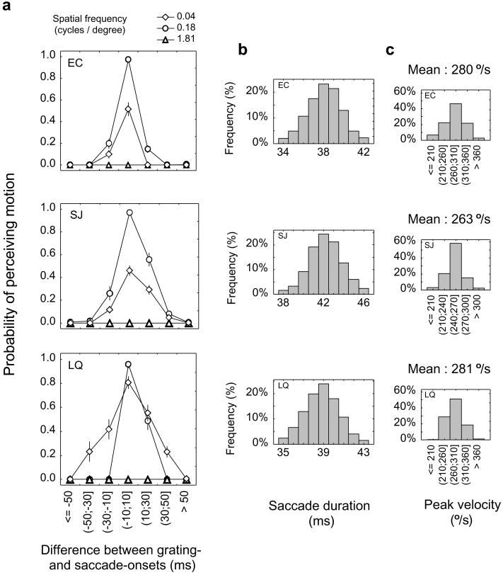
Horizontal movements of the left eye were measured with an infrared video eye tracker (Iscan RK-716) at a sampling rate of 240 Hz. To allow accurate alignment of eye movements and presentation of stimuli, acquisition of ocular data were triggered by a frame synchronization signal from the display controller. Head movements were minimized with chin and head rests. Calibrated eye position data were smoothed off-line with a spline function to reduce noise and allow interpolation with millisecond accuracy (11). Eye velocity and acceleration were computed with a two-point differentiation. Average peak-to-peak velocity noise was 15°/s. Saccades were detected by using 45°/s velocity and 1,000°/s2 acceleration criteria. Saccade onset and offset were defined, respectively, as 3 ms before and 3 ms after both criteria were crossed. For each observer, the distribution of saccade durations across sessions was assessed, and we only kept in the analysis the durations ranging from −4 ms to +4 ms around the modal class (Fig. 2b): on average, 92% of the saccades fell within this range. The saccade durations (Fig. 2b) and peak velocities (Fig. 2c) calculated with our technique met the main sequence criteria (12, 13).
Fig 2.
Results of experiment 1 for three observers (LQ was a naïve subject). (a) Probability of perceiving motion for static gratings presented at different times relative to saccade onset. Motion perception occurs with low spatial frequencies (circles and diamonds), whereas it is abolished with high spatial frequencies (triangles). Error bars show standard errors ( ). (b) Distributions of the durations of saccades kept in the analysis. (c) Distributions of peak velocities.
Results
Our major finding was observed with a low spatial frequency grating (0.18 c/°) of short duration (19 ms) and high contrast (100%). In some trials, motion was clearly perceived in the direction opposite to the saccade. In all other trials, the grating appeared as a static flash. These percepts were spontaneously reported by four naïve observers with no training in psychophysical observing. The results, plotted in Fig. 2a for three observers (including naïve observer LQ), show that motion perception occurs only when the grating is displayed during some significant portion of the saccade. Motion is consistently perceived when grating onset lies within the (−10 ms, +10 ms) bin, that is, when it is close to saccade onset. Because the duration of saccades (Fig. 2b and Methods) is on average twice the grating's duration, this finding implies that motion perception is strongly elicited by gratings stimulating the retina during the first half of the saccade. In contrast, data for gratings whose onset lies within the (+10 ms, +30 ms) bin show that the grating's presence during the second half of the saccade produces little motion perception. These findings are best described by looking at the temporal course of retinal stimulation. In a typical saccade, the speed of the eyes across time monotonically increases until it reaches a peak in the middle of the saccade and then decreases again (Fig. 1b). The retinal speed of a static stimulus therefore varies in the same way: for a grating stimulating the first half of a saccade, the retinal speed changes from a null velocity to the peak saccadic velocity (distributions of peak velocities are shown in Fig. 2c). In our experiment, such a grating has a retinal velocity increasing from 0°/s to a mean peak velocity of 280°/s, or equivalently a retinal temporal frequency (TF = speed × spatial frequency) changing from 0 to about 50 Hz (Fig. 1b, right scale). A symmetrically decreasing profile is produced for gratings displayed during the second half of the saccade. Our results therefore show that intrasaccadic motion perception optimally occurs when the retinal stimulation is an increasing profile of temporal frequencies lying within a 0- to 50-Hz range.
The observers consistently reported that their performance relied on visual motion cues. It could be argued, however, that they actually used cues not related to motion per se, such as the synchrony of saccade and grating onsets. If this were true, performance should remain unaffected by manipulating the motion information available during intrasaccadic stimulation. We found, however, that degrading motion information, either by increasing or decreasing the grating's spatial frequency, had a dramatic impact on motion perception. First, we used a spatial frequency of 1.8 c/° and found that motion perception was never observed (Fig. 2a), presumably because the resulting retinal temporal frequencies (TF at the saccadic peak ≅ 500 Hz) are not resolvable by motion detectors. We then used a grating whose spatial frequency was much less (0.04 c/°) and found a drop in performance. For observers SJ and EC, a large overall decrease occurred in the probability of perceiving motion. Observer LQ had both a lower performance at the peak of the curve and an increase of motion responses in trials not producing any intrasaccadic stimulation [false alarms in bins (−50 ms, −30 ms) and (+30 ms, +50 ms)]. This stimulus, compared with the 0.18 c/° grating, seems less optimal for motion processing either because the distance traveled by the retina (6°) is only a fraction of the grating's spatial period, or because the saccade-induced retinal temporal frequencies are lower (from 0 to ≈10 Hz).
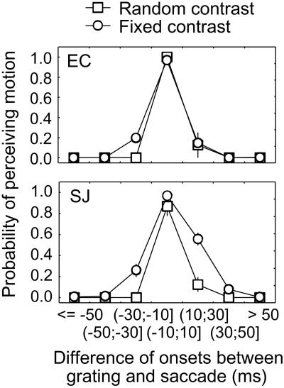
These results suggest that motion information is the relevant cue used by observers to perform the task. However, high-speed retinal motion is always accompanied by a reduction in apparent contrast, known as motion blur (14), especially for stimuli of very brief durations similar to ours (15). It is possible that motion perception was sometimes reported by observers when they actually only perceived blur, thus overestimating the ability to perceive intrasaccadic motion. In the second experiment, the contrast of the grating was randomized across trials (from 10% to 100%) to disrupt perceptual judgments based on apparent contrast and to maximize the use of a pure motion cue. We investigated whether this manipulation would reduce intrasaccadic motion detection of the optimal low spatial frequency grating tested previously (0.18 c/°). We only kept the trials in which the contrast was 100% in the analysis to allow comparison with the fixed 100% contrast condition. Fig. 3 shows that motion perception is still very high for gratings appearing near saccade onset, whereas it is almost abolished for gratings appearing elsewhere. The high probability of perceiving motion is unlikely to reflect a perceptual strategy relying on apparent contrast. If observers were reporting motion whenever apparent contrast is low, the probability of “motion” responses for trials that do not elicit any intrasaccadic stimulation (i.e., the false alarms rate) should monotonically increase as physical contrast of the grating is decreased. However, the false alarms rate was constant (≈0%) across the 10 different levels of contrast (except for a slight increase in false alarms, 8%, for SJ with the 10% contrast). Altogether, these results confirm that gratings stimulating the first half of the saccade elicit a vivid motion percept which is almost never confused with blur. Gratings displayed in the second half of the saccade seem to produce blur rather than motion, thus leading to some confusions in the fixed contrast condition except for the highly trained observer EC. Moreover, this asymmetry rules out the possibility that observers confuse motion with temporal modulation of apparent contrast, as equal performance would have been expected for gratings displayed either in the first or in the second half of the saccade.
Fig 3.
Results of experiment 2 for two observers. Contrast of the 0.18 c/° grating was randomized across trials (from 10% to 100% in steps of 10%). Only 100% contrast data are presented here (squares) to allow comparison with fixed 100% contrast data replotted from Fig. 2a (circles). Error bars show standard errors.
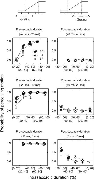
In the third experiment, gratings of longer durations (25, 37.5, or 50 ms) and optimal spatial frequency (0.18 c/°) were used to study the effect of extrasaccadic stimulation. We found again that the stimulus seemed to be moving (against saccade direction) or to be static depending on the timing between stimulus and saccade. Data, including those from the first experiment (duration = 19 ms), were grouped into two categories (Fig. 4). For gratings appearing before saccade onset and ending within the saccadic flight (left graphs), overall probability of perceiving motion is high. In contrast, for gratings appearing during the saccadic flight and extending further into the period after the saccade (right graphs), motion is almost never perceived. This asymmetry shows that postsaccadic stimulations are more effective than presaccadic stimulations in suppressing motion perception. In both cases, an interaction clearly occurs between the duration of the extrasaccadic stimulation and the duration of the intrasaccadic stimulation (expressed as a percentage of the saccade duration): motion perception is highest with long intrasaccadic and short extrasaccadic stimulations.
Fig 4.
Results of experiment 3 for three observers (EP was a naïve subject). Stimuli and methods were the same as in experiment 1 except for the duration of the grating (25, 37.5, or 50 ms). Data from experiment 1 (duration = 19 ms) have been included in this analysis. Grating onset could be either before saccade onset (Left) or within the saccade (Right). Abscissa indicates the proportion of the saccade duration that was stimulated by the grating. Note the reversed axis for right-hand graphs. Error bars show standard errors.
Discussion
Clear psychophysical evidence now exists for an intact ability of the visual system to process visual motion when the intrasaccadic retinal profile is either accelerating, as in the present work, or approximately constant (16, 17), provided that extrasaccadic stimulation is absent. In both cases, low spatial frequency stimuli are required to elicit optimal intrasaccadic motion perception, as would be expected from studies investigating high-speed motion processing with static eyes (4, 10). Low-level motion detectors in the magnocellular stream (18), which have spatiotemporal characteristics allowing them to detect very brief and fast moving stimuli (19, 20), are most likely to underlie intrasaccadic motion perception. Electrophysiological evidence shows that neurons in the middle temporal cortex, an area devoted to visual motion analysis, are able to encode the fine temporal structure of moving stimuli varying in direction on a timescale as short as 30 ms (21, 22). Moreover, middle temporal cortex neurons can be transiently activated by the visual flow created by fixational saccades (23). Our technique could be fruitfully used to find out whether these neurons underlie conscious intrasaccadic motion perception.
What are the factors that may cause the ordinary absence of intrasaccadic motion perception? Our previous study suggested two possibilities (16, 24). First, on the basis of evidence that activation of motion detectors requires a relatively constant speed over a sufficiently long period (25–28), we thought that the rapidly changing retinal velocity induced by static stimuli during saccades should not be able to produce reliable velocity signals. This idea is ruled out by the present results. Second, we proposed that pre- and postsaccadic static retinal images were able to mask intrasaccadic retinal motion. Experiment 3 supports this hypothesis and complements early studies showing that temporal masking is responsible for the elimination of the blurred perception (greyout) elicited by a contoured environment displayed only during a saccade (2, 29). Temporal masking seems sufficient, therefore, to explain why intrasaccadic high-speed retinal slip is usually not consciously perceived either as a moving or as a blurred image.
The theoretical possibility that extraretinal signals modulate visual processing during saccades with the purpose of preventing motion perception cannot be ruled out. However, our work indicates that this putative mechanism is clearly not able to achieve the desired suppression of motion perception, suggesting either that this extraretinal mechanism does not exist or that it is very inefficient. It should also be noted that current evidence for active extraretinal suppression is controversial. In one influential theory, it is proposed that the activity of the magnocellular stream, the motion-processing pathway, is suppressed in the lateral geniculate nucleus during saccades (5–7). This hypothesis relies on one major psychophysical finding: contrast sensitivity for horizontal gratings flashed during horizontal saccades is reduced, and this reduction seems specific to the magnocellular system (7, 8). As already underlined (16), the main flaw in these studies is that no retinal motion is induced by the horizontal grating, so that the data cannot be easily related to motion processing. Moreover, these results can be interpreted exclusively in terms of retinal processes (24). In a more recent study based on visual neuronal responses in the middle temporal cortex and medial superior temporal cortex of behaving monkeys (9), it is claimed that extraretinal signals might affect a restricted subpopulation of neurons by reversing their direction selectivity. The authors suggest that the activity of this subpopulation would be used to cancel out the activity of neurons that respond to retinal motion against the saccade direction. The putative suppression mechanism would thus rely on the absence of a net motion direction at the population level. This theory, however, is difficult to admit for two main reasons. First, the link between the reported patterns of neural response and intrasaccadic motion processing is rather obscure because monkeys' motion perception was not assessed. Consequently, the reversal of direction selectivity may be related to various sorts of sensory or sensory-motor functions associated with saccades. It could, for instance, reflect remapping processes (30), spatial shifts of attention (31), or different types of attentional modulation known to arise in extrastriate areas (32). Second, the reversal in direction selectivity is not observed in the initial part of the neuronal responses, but only after a relatively long delay (about 50 ms). In other words, the net population response during the early period is clearly signaling the retinal motion induced by the saccade. This activity, whose duration is close to saccadic duration, seems to correspond to a pure visual response caused by intrasaccadic retinal motion without any extraretinal influence. This early neural response would not be predicted by active suppression theories because their main requirement is that extraretinal influence takes place very early (usually before the saccade). Therefore, the delayed reversal in direction selectivity does not seem to reflect an anticipatory mechanism used to prevent intrasaccadic motion processing.
We show that temporal masking more parsimoniously accounts for the usual absence of intrasaccadic motion perception. Our results may help characterize the mechanisms underlying these masking effects. The finding in experiment 3 that probability of perceiving motion depends on the ratio between the durations of intra- and extrasaccadic stimulations suggests a form of energy-dependent integration masking (33). The greater efficiency of backward- vs. forward-masking further suggests that masking by interruption (34) may be concurrently functional, thus potentiating the masking effect of trailing visual signals. This would also account for our finding that gratings displayed in the first half of the saccade are perceived as moving, whereas those displayed in the second half appear motionless. It is currently difficult to draw firmer conclusions because very little is known about the response of the visual system when primates with static eyes are faced with retinal stimulations similar to the retinal profiles induced by saccades in our experiments. Investigating the mechanisms underlying temporal integration of such short and complex visual stimulations will be necessary to understand intrasaccadic perception fully.
Acknowledgments
We thank D. Keeble, M. J. Morgan, and R. H. Wurtz for helpful comments on the manuscript.
This paper was submitted directly (Track II) to the PNAS office.
References
- 1.Matin E., Clymer, A. B. & Matin, L. (1972) Science 178, 179-182. [DOI] [PubMed] [Google Scholar]
- 2.Campbell F. W. & Wurtz, R. H. (1978) Vision Res. 18, 1297-1303. [DOI] [PubMed] [Google Scholar]
- 3.Mitrani L., Mateeff, S. & Yakimoff, N. (1970) Vision Res. 10, 405-409. [DOI] [PubMed] [Google Scholar]
- 4.Burr D. C. & Ross, J. (1982) Vision Res. 22, 479-484. [DOI] [PubMed] [Google Scholar]
- 5.Ross J., Burr, D. & Morrone, C. (1996) Behav. Brain Res. 80, 1-8. [DOI] [PubMed] [Google Scholar]
- 6.Ross J., Morrone, M. C., Goldberg, M. E. & Burr, D. C. (2001) Trends Neurosci. 24, 113-121. [DOI] [PubMed] [Google Scholar]
- 7.Burr D. C., Morrone, M. C. & Ross, J. (1994) Nature 371, 511-513. [DOI] [PubMed] [Google Scholar]
- 8.Diamond M. R., Ross, J. & Morrone, M. C. (2000) J. Neurosci. 20, 3449-3455. [DOI] [PMC free article] [PubMed] [Google Scholar]
- 9.Thiele A., Henning, P., Kubischik, M. & Hoffmann, K. P. (2002) Science 295, 2460-2462. [DOI] [PubMed] [Google Scholar]
- 10.Morgan M. J. & Castet, E. (1995) Nature 378, 380-383. [DOI] [PubMed] [Google Scholar]
- 11.Busettini C., Miles, F. A. & Schwarz, U. (1991) J. Neurophysiol. 66, 865-878. [DOI] [PubMed] [Google Scholar]
- 12.Collewijn H., Erkelens, C. J. & Steinman, R. M. (1988) J. Physiol. 404, 157-182. [DOI] [PMC free article] [PubMed] [Google Scholar]
- 13.van der Geest J. N. & Frens, M. A. (2002) J. Neurosci. Methods 114, 185-195. [DOI] [PubMed] [Google Scholar]
- 14.Land M. F. (1999) J. Comp. Physiol. A 185, 341-352. [DOI] [PubMed] [Google Scholar]
- 15.Burr D. (1980) Nature 284, 164-165. [DOI] [PubMed] [Google Scholar]
- 16.Castet E. & Masson, G. S. (2000) Nat. Neurosci. 3, 177-183. [DOI] [PubMed] [Google Scholar]
- 17.Garcia-Perez M. A. & Peli, E. (2001) J. Neurosci. 21, 7313-7322. [DOI] [PMC free article] [PubMed] [Google Scholar]
- 18.Yabuta N. H., Sawatari, A. & Callaway, E. M. (2001) Science 292, 297-300. [DOI] [PubMed] [Google Scholar]
- 19.Movshon J. A. & Newsome, W. T. (1996) J. Neurosci. 16, 7733-7741. [DOI] [PMC free article] [PubMed] [Google Scholar]
- 20.Hawken M. J., Shapley, R. M. & Grosof, D. H. (1996) Visual Neurosci. 13, 477-492. [DOI] [PubMed] [Google Scholar]
- 21.Buracas G. T., Zador, A. M., DeWeese, M. R. & Albright, T. D. (1998) Neuron 20, 959-969. [DOI] [PubMed] [Google Scholar]
- 22.Buracas G. T. & Albright, T. D. (1999) Trends Neurosci. 22, 303-309. [DOI] [PubMed] [Google Scholar]
- 23.Bair W. & O'Keefe, L. P. (1998) Visual Neurosci. 15, 779-786. [DOI] [PubMed] [Google Scholar]
- 24.Castet E., Jeanjean, S. & Masson, G. S. (2001) Trends Neurosci. 24, 316-317. [DOI] [PubMed] [Google Scholar]
- 25.Burr D. C. (1981) Proc. R. Soc. London Ser. B 211, 321-339. [DOI] [PubMed] [Google Scholar]
- 26.van Santen J. P. & Sperling, G. (1985) J. Opt. Soc. Am. 2, 300-321. [DOI] [PubMed] [Google Scholar]
- 27.Watson A. B. & Turano, K. (1995) Vision Res. 35, 325-336. [DOI] [PubMed] [Google Scholar]
- 28.Morgan M. J. (1980) Philos. Trans. R. Soc. London B 290, 117-135. [DOI] [PubMed] [Google Scholar]
- 29.Judge S. J., Wurtz, R. H. & Richmond, B. J. (1980) J. Neurophysiol. 43, 1133-1155. [DOI] [PubMed] [Google Scholar]
- 30.Nakamura K. & Colby, C. L. (2002) Proc. Natl. Acad. Sci. USA 99, 4026-4031. [DOI] [PMC free article] [PubMed] [Google Scholar]
- 31.Deubel H. & Schneider, W. X. (1996) Vision Res. 36, 1827-1837. [DOI] [PubMed] [Google Scholar]
- 32.Treue S. (2001) Trends Neurosci. 24, 295-300. [DOI] [PubMed] [Google Scholar]
- 33.Breitmeyer B. G., (1984) Visual Masking: An Integrative Approach (Oxford Univ. Press, London).
- 34.Scheerer E. (1973) Psychol. Forsch. 36, 71-93. [DOI] [PubMed] [Google Scholar]


