

Cisplatin [cis-diamminedichloroplatinum (II)] is a highly active anticancer agent. This agent is curative against most testicular cancers and is highly active against a wide range of other tumor types, notably ovarian, bladder carcinoma, and non-small-cell lung cancer (1). Treatment failure is frequently caused by the development of resistance to cisplatin. Although the insensitive tumors generally exhibit only low-level resistance, the use of cisplatin at close to its maximally tolerated dose implies that the developed resistance eliminates cisplatin as an active compound. Resistance to cisplatin has been widely studied in a variety of models and in clinical samples, but it has been difficult to identify the molecular changes leading to drug resistance. Ishida et al. (2) report in this issue of PNAS the identification of an important pathway for uptake of cisplatin into yeast and mammalian cells, uptake mediated by a high-affinity copper transporter.
The action of cisplatin in cell killing is now well established (3). In serum, the high concentration of chloride ions enhances cisplatin stability. The lower chloride concentration in cells favors rapid hydrolysis of the chloride ligands of cisplatin, leading to an activated molecule that is capable of reacting bifunctionally. Although cisplatin can react with a variety of cellular macromolecules, there is strong evidence that the most important target is DNA (3, 4). An important line of evidence that DNA is an essential target of cisplatin is the observation that bacterial and yeast mutants that are defective in various DNA-repair pathways are also hypersensitive to cisplatin. Cisplatin can form both intrastrand and interstrand DNA crosslinks, with intrastrand purine:purine representing the majority of the adducts.
Figure a.
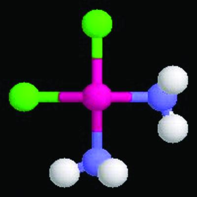
Cisplatin structure courtesy of Mitch Miller (NetGenics, Cleveland).
As is the case with other anticancer agents, reduced accumulation of cisplatin is frequently observed in cisplatin-resistant cell lines. Although drug efflux has been intensively studied as a mechanism of drug resistance, relatively few studies have demonstrated a role for reduced drug uptake in acquired drug resistance. However, cisplatin has been an exception to this generalization, and several authors have suggested that decreased uptake of cisplatin is an important factor that can result in drug resistance (5). Whereas some considerations have led to the suggestion that a major mechanism for cisplatin uptake is passive diffusion, other studies have suggested a role for active transport. For example, reactive aldehydes, such as benzaldehyde, are able to block cisplatin accumulation, suggesting modification of a membrane protein required for cisplatin uptake (reviewed in ref. 6).
To identify yeast genes that play a role in sensitivity to cisplatin, Ishida et al. started with a simple genetic approach. By using the transposon mutagenesis approach of Snyder and coworkers (7), they screened yeast loss-of-function mutants for cisplatin resistance. An advantage of the transposon mutagenesis approach is that in general only one gene is mutated per cell and the mutation is usually a complete loss of function. In addition, the mutated gene is marked by the transposon, greatly simplifying identification of the mutated gene. This approach is similar to that used in recent studies that have employed a set of yeast strains carrying deletions in all ORFs encoding nonessential genes. The deletion set has been used to characterize, on a genome scale, genes required for repair of DNA damage caused by a variety of different DNA-damaging agents (8–10). Both approaches allow efficient identification of all loss-of-function mutations that result in a specific phenotype.
The strain that generated the highest level of cisplatin resistance carried a mutation in the MAC1 gene, a transcription factor that regulates the catalase genes, as well as genes required for the uptake of iron and copper (11). Each of the known genes regulated by MAC1 were sequentially deleted, and it was observed that deletion of the gene encoding the high-affinity copper transporter CTR1 nearly recapitulated the cisplatin resistance that was observed in mac1-deficient cells. This result indicated that the resistance seen in mac1Δ mutants was principally caused by a failure to express CTR1. Interestingly, deletion of a second yeast high-affinity copper transporter CTR3 produced only a minor effect on cisplatin sensitivity.
Ishida et al. carefully exclude the possibility that cisplatin resistance caused by ctr1Δ arises from indirect effects, rather than the ability of CTRp to transport cisplatin into cells. For example, because mutation of ctr1Δ results in an impairment of copper uptake, activities of enzymes, such as superoxide dismutase, that rely on copper or iron can be impaired. A series of deletion strains carrying defects in genes such as SOD1 (superoxide dismutase), FET3 (a protein encoding a ferro-O2-oxidoreductase that is part of the high-affinity iron transport system), LYS7 (a copper chaperone for superoxide dismutase that is important for protection from oxidative stress and which also confers an auxotrophy for lysine), and others were examined, but none of the single mutants resulted in significant enhancement of cisplatin resistance. The final key point in the demonstration that CTR1p plays an important role in cisplatin influx was that ctr1Δ results in a reduction of cisplatin levels in yeast cells, but does not alter efflux of cisplatin. Taken together, a very strong case is made for CTR1 as the major protein for cisplatin uptake in yeast.
The next obvious question is whether mammalian homologs of yCTR1 also are able to import cisplatin into cells, and whether the mammalian homologs play an important role in cisplatin levels. Mammalian homologs of yCTR1 have been identified (12, 13), in part through their ability to complement yeast ctr1Δ mutants. Interestingly, murine CTR1 plays an essential role in embryonic development; mouse embryos lacking mCTR1 die at mid-gestation, presumably from the lack of functioning of copper-dependent enzymes (14, 15). Ishida et al. used mouse cells carrying two, one, or no functional copies of mCTR1 and assessed cisplatin sensitivity and drug accumulation. Their results showed cisplatin accumulation and sensitivity was proportional to the number of functional CTR1 alleles. Homozygous ctr1 mutant cells were 8-fold resistant to cisplatin and exhibited a 70% reduction in cisplatin accumulation (2). Cells heterozygous for ctr1 also showed a reduction in cisplatin accumulation and intermediate cisplatin resistance. These results clearly establish CTR1p as a major factor in the uptake of cisplatin into mammalian cells and a potentially critical protein in cellular sensitivity to cisplatin.
Other investigators also have observed connections between cisplatin levels and copper-transport proteins. A member of the class of cation transporters called P-type ATPases plays important roles in copper homeostasis. Mutations in copper-transporting ATPases result in the syndromes called Wilson's disease and Menkes syndrome (16, 17). Komatsu et al. (18) have recently demonstrated that ectopic expression of the gene mutated in Wilson's disease, ATP7B results in an approximately 9-fold resistance to cisplatin and a somewhat lower level of resistance to copper. This result may suggest some shared properties among copper transporters that allow relatively efficient transport of cisplatin.
Regulation of copper levels in cells is a balancing act. Insufficient levels of copper lead to the inactivity of copper-dependent enzymes, but excessive levels of copper are toxic. Precise regulation of copper levels in yeast is attained by several mechanisms. The transcription factor MAC1 requires copper for transcriptional activation (19) but also senses copper levels and does not activate transcription when copper levels are high (20, 21). Regulation of Mac1p DNA binding by phosphorylation has also been suggested (22). Posttranslational mechanisms are also important in regulation of Ctr1p. High levels of copper lead to degradation of Ctr1p (23). Interestingly, the MAC1 transcription factor appears to be required for efficient degradation of Ctr1p (24).
Like copper, cisplatin could lead to Ctr1p degradation.
The details of the known regulation of CTR1 lead to the question of how copper levels influence cellular uptake of cisplatin, and whether cisplatin levels can alter the levels of Ctr1p. Not surprisingly, Ishida et al. (2) found that copper is a competitive inhibitor of cisplatin uptake. Somewhat surprisingly, they also found that, like copper, cisplatin could lead to Ctr1p degradation. This result would suggest that the highest intracellular levels of cisplatin would occur under conditions where Ctr1p expression is unlinked to its stability, i.e., under conditions where cisplatin (or copper) uptake does not lead to destabilization of Ctr1p. At present, regulation of mammalian Ctr1 remains to be elucidated. It seems likely that stability of mammalian Ctr1p will be an important regulatory mechanism.
There are several important avenues that the work of Ishida et al. opens up. There are the obvious questions about whether mutations of mammalian Ctr1 occur in platinum-insensitive tumors. A full understanding of the roles of Ctr1in drug resistance will require an understanding of how the transporter is regulated in mammalian cells. It will be equally interesting to determine whether Ctr1 expression influences the specific tissues that are especially sensitive to toxicity from cisplatin.
An additional set of questions relates to whether Ctr1 can transport other platinum compounds, such as carboplatin and oxaliplatin. If the level of Ctr1 expression is an important determinant of cisplatin resistance, it will be of interest to try to develop active platinum derivatives that have distinct uptake mechanisms. Although platinum compounds that can be efficiently accumulated in a Ctr1-independent fashion may be useful for overcoming resistance, such compounds also may have novel toxicity spectra. Nonetheless, the work of Ishida et al. has clearly opened up new avenues for the design of potentially active platinum anticancer agents.
Yeast cells are increasingly becoming applied as a model for the study of the action of anticancer drugs. Many of the experimental approaches described by Ishida et al. are still only feasible in yeast systems. Although this point has been long appreciated by cell biologists and biochemists interested in fundamental biological questions, workers with an interest in the mechanisms of anticancer only recently have appreciated the questions that can be addressed by using yeast. For example, yeast cells are being applied to study the action of anticancer drugs targeting DNA topoisomerases (25, 26), and to understand the action of drugs such as rapamycin that target the phosphatidylinositol-3 kinase TOR1 (27, 28).
The revolution in genomics has understandably generated a great deal of excitement over the potential for identifying new targets for cancer chemotherapy. An underappreciated point is that there is also great potential for better understanding the action of agents that we already know are clinically active. As elegantly demonstrated by Ishida et al., important insights await a careful analysis of already identified active anticancer agents.
Acknowledgments
This work was supported by National Cancer Institute Grants CA21765, CA52814, and CA82313 and the American Lebanese Syrian Associated Charities.
See companion article on page 14298.
References
- 1.Ardizzoni A., Antonelli, G., Grossi, F., Tixi, L., Cafferata, M. & Rosso, R. (1991) Ann. Oncol. 10, Suppl. 5, S13-S17. [DOI] [PubMed] [Google Scholar]
- 2.Ishida S., Lee, J., Thiele, D. J. & Herskowitz, I. (2002) Proc. Natl. Acad. Sci. USA 99, 14298-14302. [DOI] [PMC free article] [PubMed] [Google Scholar]
- 3.Cohen S. M. & Lippard, S. J. (2001) Prog. Nucleic Acid Res. Mol. Biol. 67, 93-130. [DOI] [PubMed] [Google Scholar]
- 4.Zamble D. B. & Lippard, S. J. (1995) Trends Biochem. Sci. 20, 435-439. [DOI] [PubMed] [Google Scholar]
- 5.Mese H., Sasaki, A., Alcalde, R. E., Nakayama, S. & Matsumura, T. (1998) Chemotherapy 44, 414-420. [DOI] [PubMed] [Google Scholar]
- 6.Kartalou M. & Essigmann, J. M. (2001) Mutat. Res. 478, 1-21. [DOI] [PubMed] [Google Scholar]
- 7.Ross-Macdonald P., Sheehan, A., Roeder, G. S. & Snyder, M. (1997) Proc. Natl. Acad. Sci. USA 94, 190-195. [DOI] [PMC free article] [PubMed] [Google Scholar]
- 8.Giaever G., Chu, A. M., Ni, L., Connelly, C., Riles, L., Veronneau, S., Dow, S., Lucau-Danila, A., Anderson, K., Andre, B., et al. (2002) Nature 418, 387-391. [DOI] [PubMed] [Google Scholar]
- 9.Birrell G. W., Giaever, G., Chu, A. M., Davis, R. W. & Brown, J. M. (2001) Proc. Natl. Acad. Sci. USA 98, 12608-12613. [DOI] [PMC free article] [PubMed] [Google Scholar]
- 10.Winzeler E. A., Shoemaker, D. D., Astromoff, A., Liang, H., Anderson, K., Andre, B., Bangham, R., Benito, R., Boeke, J. D., Bussey, H., et al. (1999) Science 285, 901-906. [DOI] [PubMed] [Google Scholar]
- 11.Jungmann J., Reins, H. A., Lee, J., Romeo, A., Hassett, R., Kosman, D. & Jentsch, S. (1993) EMBO J. 12, 5051-5056. [DOI] [PMC free article] [PubMed] [Google Scholar]
- 12.Lee J., Prohaska, J. R., Dagenais, S. L., Glover, T. W. & Thiele, D. J. (2000) Gene 254, 87-96. [DOI] [PubMed] [Google Scholar]
- 13.Zhou B. & Gitschier, J. (1997) Proc. Natl. Acad. Sci. USA 94, 7481-7486. [DOI] [PMC free article] [PubMed] [Google Scholar]
- 14.Kuo Y. M., Zhou, B., Cosco, D. & Gitschier, J. (2001) Proc. Natl. Acad. Sci. USA 98, 6836-6841. [DOI] [PMC free article] [PubMed] [Google Scholar]
- 15.Lee J., Prohaska, J. R. & Thiele, D. J. (2001) Proc. Natl. Acad. Sci. USA 98, 6842-6847. [DOI] [PMC free article] [PubMed] [Google Scholar]
- 16.Bingham M. J., Ong, T. J., Summer, K. H., Middleton, R. B. & McArdle, H. J. (1998) Am. J. Clin. Nutr. 67, 982S-987S. [DOI] [PubMed] [Google Scholar]
- 17.Seidel J., Caca, K., Schwab, S. G., Berr, F., Wildenauer, D. B., Mentzel, H. J., Horn, N. & Kauf, E. (2001) Cell Mol. Biol. 47, 149-157. [PubMed] [Google Scholar]
- 18.Komatsu M., Sumizawa, T., Mutoh, M., Chen, Z. S., Terada, K., Furukawa, T., Yang, X. L., Gao, H., Miura, N., Sugiyama, T. & Akiyama, S. (2000) Cancer Res. 60, 1312-1316. [PubMed] [Google Scholar]
- 19.Jensen L. T. & Winge, D. R. (1998) EMBO J. 17, 5400-5408. [DOI] [PMC free article] [PubMed] [Google Scholar]
- 20.Dancis A., Haile, D., Yuan, D. S. & Klausner, R. D. (1994) J. Biol. Chem. 269, 25660-25667. [PubMed] [Google Scholar]
- 21.Yamaguchi-Iwai Y., Serpe, M., Haile, D., Yang, W., Kosman, D. J., Klausner, R. D. & Dancis, A. (1997) J. Biol. Chem. 272, 17711-17718. [DOI] [PubMed] [Google Scholar]
- 22.Heredia J., Crooks, M. & Zhu, Z. (2001) J. Biol. Chem. 276, 8793-8797. [DOI] [PubMed] [Google Scholar]
- 23.Ooi C. E., Rabinovich, E., Dancis, A., Bonifacino, J. S. & Klausner, R. D. (1996) EMBO J. 15, 3515-3523. [PMC free article] [PubMed] [Google Scholar]
- 24.Yonkovich J., McKenndry, R., Shi, X. & Zhu, Z. (2002) J. Biol. Chem. 277, 23981-23984. [DOI] [PubMed] [Google Scholar]
- 25.Nitiss J. L. (1996) Anticancer Drugs 7, Suppl. 3, 27-34.8742095 [Google Scholar]
- 26.Jensen L. H., Nitiss, K. C., Rose, A., Dong, J., Zhou, J., Hu, T., Osheroff, N., Jensen, P. B., Sehested, M. & Nitiss, J. L. (2000) J. Biol. Chem. 275, 2137-2146. [DOI] [PubMed] [Google Scholar]
- 27.Alarcon C. M., Cardenas, M. E. & Heitman, J. (1996) Genes Dev. 10, 279-288. [DOI] [PubMed] [Google Scholar]
- 28.Rohde J., Heitman, J. & Cardenas, M. E. (2001) J. Biol. Chem. 275, 9583-9586. [DOI] [PubMed] [Google Scholar]