Abstract
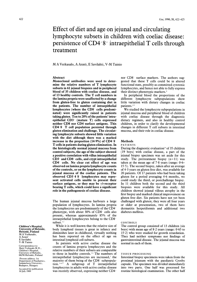
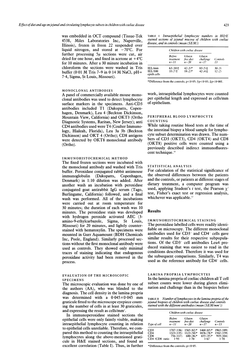
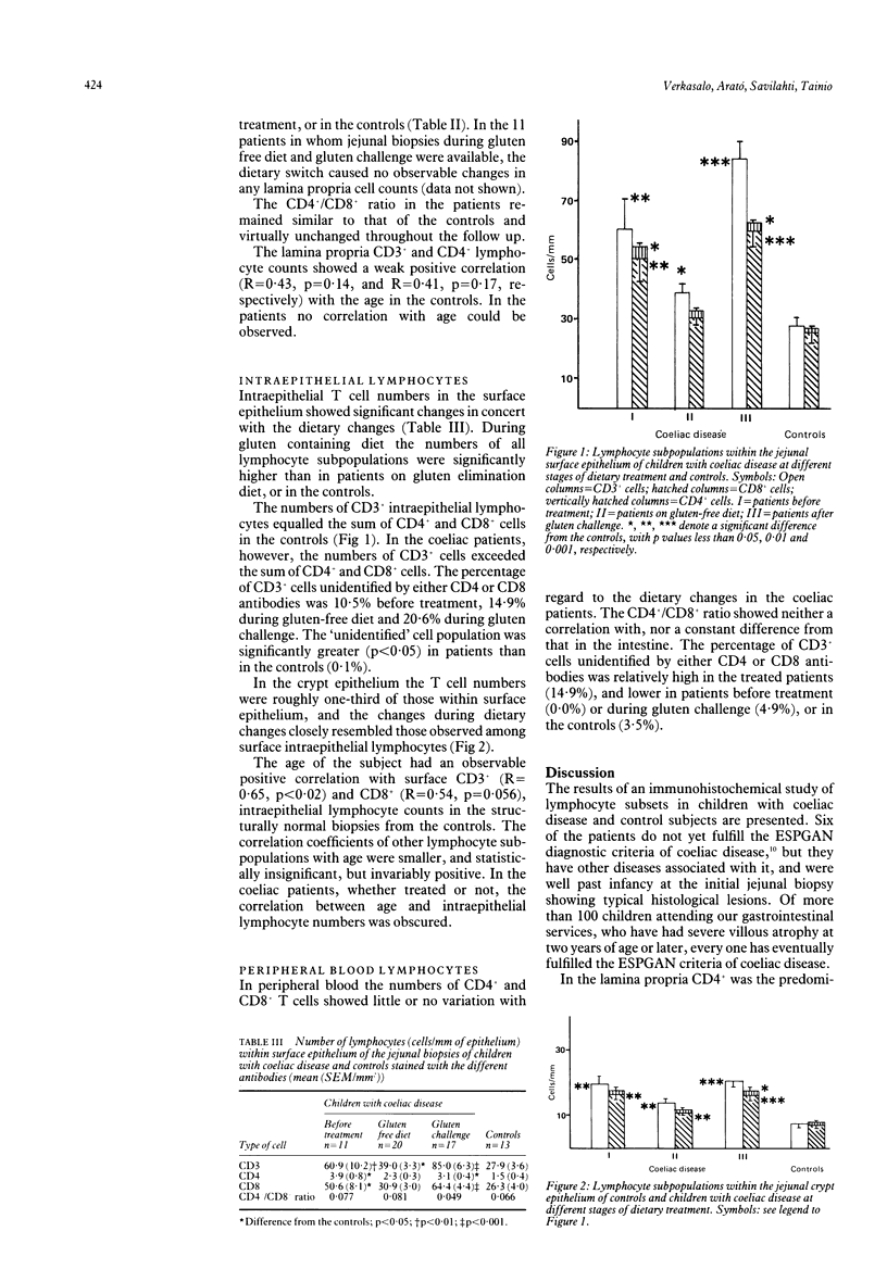
Monoclonal antibodies were used to determine the relative numbers of T lymphocyte subsets in 61 jejunal biopsies and in peripheral blood of 35 children with coeliac disease, and of 13 healthy controls. The T cell numbers in the lamina propria were unaffected by a change from gluten-free to gluten containing diet in the patients. The number of intraepithelial lymphocytes (where the CD8 cells predominated) were significantly raised in patients taking gluten. Ten to 20% of the patients' intraepithelial CD3 (mature T) cells expressed neither CD8 nor CD4 surface antigens. This CD4 8 T cell population persisted through gluten elimination and challenge. The circulating lymphocyte subsets showed little variation with the diet although there was a marked increase in the proportion (14.9%) of CD4 8 T cells in patients during gluten elimination. In the histologically normal jejunal mucosa from control subjects, the age of the subject showed a positive correlation with villus intraepithelial CD3+ and CD8+ cells, and crypt intraepithelial CD4+ cells. No clear cut effect of age was observed on lamina propria lymphocyte counts of the controls, or on the lymphocyte counts in jejunal mucosa of the coeliac patients. The observed CD3+4-8- lymphocytes may represent activated cells unable to present their surface antigens, or they may be gamma delta-receptor bearing T cells, which could have a significant role in the pathogenesis of coeliac disease.
Full text
PDF


Selected References
These references are in PubMed. This may not be the complete list of references from this article.
- Borst J., van Dongen J. J., Bolhuis R. L., Peters P. J., Hafler D. A., de Vries E., van de Griend R. J. Distinct molecular forms of human T cell receptor gamma/delta detected on viable T cells by a monoclonal antibody. J Exp Med. 1988 May 1;167(5):1625–1644. doi: 10.1084/jem.167.5.1625. [DOI] [PMC free article] [PubMed] [Google Scholar]
- Brenner M. B., McLean J., Dialynas D. P., Strominger J. L., Smith J. A., Owen F. L., Seidman J. G., Ip S., Rosen F., Krangel M. S. Identification of a putative second T-cell receptor. Nature. 1986 Jul 10;322(6075):145–149. doi: 10.1038/322145a0. [DOI] [PubMed] [Google Scholar]
- Cerf-Bensussan N., Schneeberger E. E., Bhan A. K. Immunohistologic and immunoelectron microscopic characterization of the mucosal lymphocytes of human small intestine by the use of monoclonal antibodies. J Immunol. 1983 Jun;130(6):2615–2622. [PubMed] [Google Scholar]
- James S. P., Fiocchi C., Graeff A. S., Strober W. Phenotypic analysis of lamina propria lymphocytes. Predominance of helper-inducer and cytolytic T-cell phenotypes and deficiency of suppressor-inducer phenotypes in Crohn's disease and control patients. Gastroenterology. 1986 Dec;91(6):1483–1489. [PubMed] [Google Scholar]
- Jenkins D., Goodall A., Scott B. B. T-lymphocyte populations in normal and coeliac small intestinal mucosa defined by monoclonal antibodies. Gut. 1986 Nov;27(11):1330–1337. doi: 10.1136/gut.27.11.1330. [DOI] [PMC free article] [PubMed] [Google Scholar]
- Kuitunen P., Kosnai I., Savilahti E. Morphometric study of the jejunal mucosa in various childhood enteropathies with special reference to intraepithelial lymphocytes. J Pediatr Gastroenterol Nutr. 1982;1(4):525–531. doi: 10.1097/00005176-198212000-00012. [DOI] [PubMed] [Google Scholar]
- Malizia G., Trejdosiewicz L. K., Wood G. M., Howdle P. D., Janossy G., Losowsky M. S. The microenvironment of coeliac disease: T cell phenotypes and expression of the T2 'T blast' antigen by small bowel lymphocytes. Clin Exp Immunol. 1985 May;60(2):437–446. [PMC free article] [PubMed] [Google Scholar]
- Rodgers V. D., Fassett R., Kagnoff M. F. Abnormalities in intestinal mucosal T cells in homosexual populations including those with the lymphadenopathy syndrome and acquired immunodeficiency syndrome. Gastroenterology. 1986 Mar;90(3):552–558. doi: 10.1016/0016-5085(86)91108-x. [DOI] [PubMed] [Google Scholar]
- Selby W. S., Janossy G., Bofill M., Jewell D. P. Lymphocyte subpopulations in the human small intestine. The findings in normal mucosa and in the mucosa of patients with adult coeliac disease. Clin Exp Immunol. 1983 Apr;52(1):219–228. [PMC free article] [PubMed] [Google Scholar]
- Selby W. S., Janossy G., Goldstein G., Jewell D. P. T lymphocyte subsets in human intestinal mucosa: the distribution and relationship to MHC-derived antigens. Clin Exp Immunol. 1981 Jun;44(3):453–458. [PMC free article] [PubMed] [Google Scholar]
- Tainio V. M. Lymphocyte subsets in infants: relationships to feeding, atopy, atopic heredity and infections. Int Arch Allergy Appl Immunol. 1985;78(3):305–310. doi: 10.1159/000233902. [DOI] [PubMed] [Google Scholar]
- Vetvicka V., Tlaskalová-Hogenová H., Fric P., Brochier J. Subpopulations of circulating lymphocytes in adults with coeliac disease. Immunol Lett. 1986 Aug;13(1-2):15–18. doi: 10.1016/0165-2478(86)90119-7. [DOI] [PubMed] [Google Scholar]


