Abstract
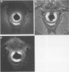
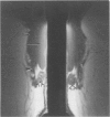
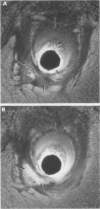
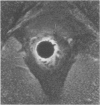
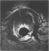
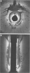
An internal receiver coil was used to obtain high resolution transverse and oblique coronal magnetic resonance images of the anal sphincter in five normal volunteers and five patients. The internal sphincter had a high signal intensity on T1 weighted, T2 weighted, and STIR sequences whereas the conjoined longitudinal muscle and external sphincter had a low signal intensity. The internal sphincter (but not the external sphincter) showed contrast enhancement after administration of intravenous gadopentetate dimeglumine. The oblique coronal plane was particularly useful for showing the thickness and the relations of the external sphincter. Sphincteric abscesses as well as muscle defects, hypertrophy, and atrophy were clearly shown. The coil was well tolerated by most subjects. It has considerable potential for improving the diagnosis of anorectal disease.
Full text
PDF
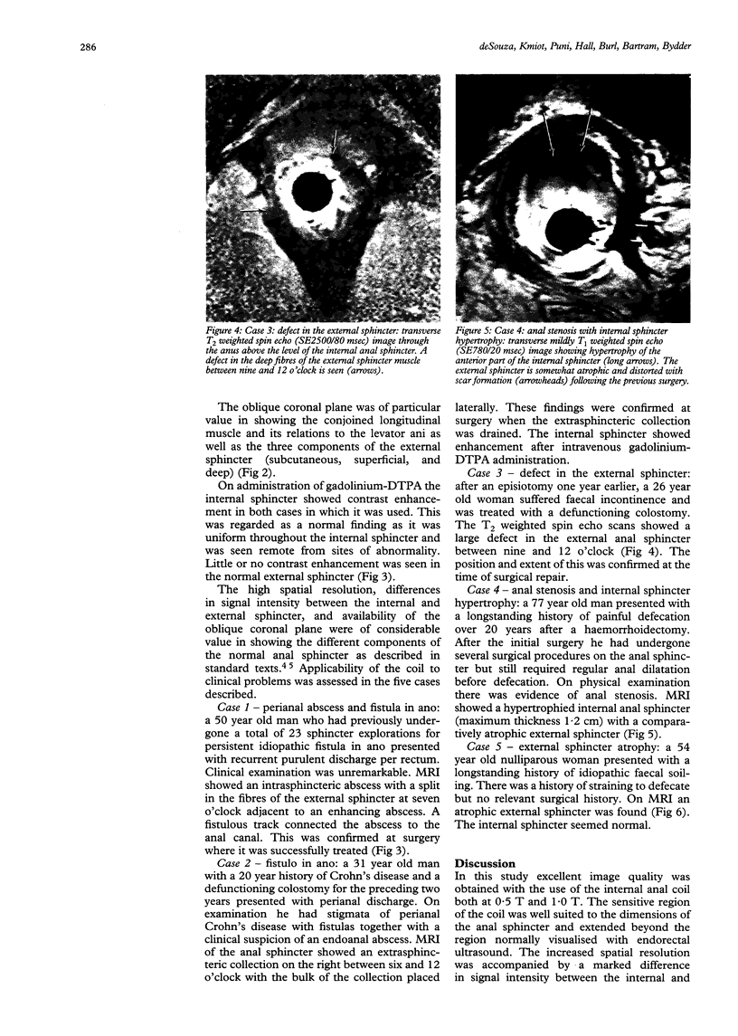
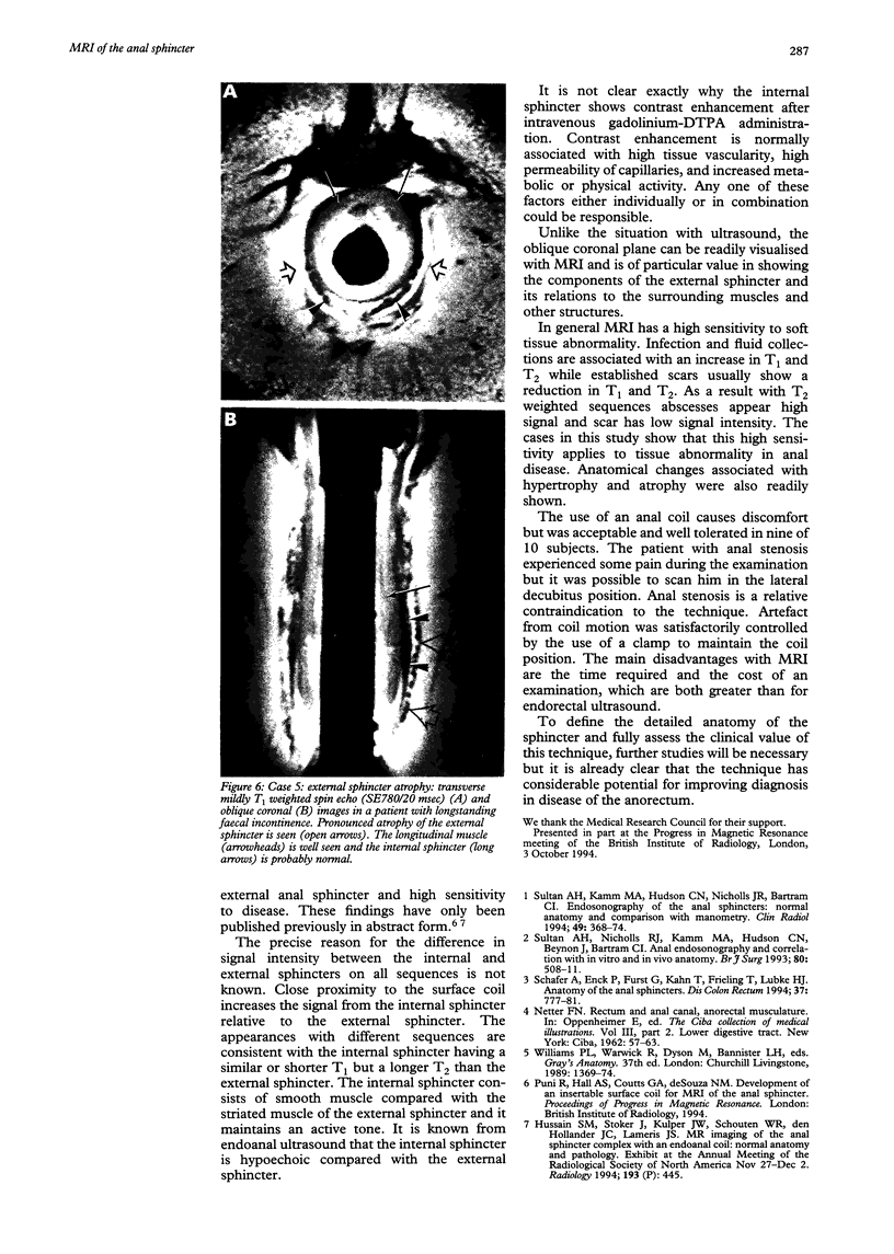
Images in this article
Selected References
These references are in PubMed. This may not be the complete list of references from this article.
- Schäfer A., Enck P., Fürst G., Kahn T., Frieling T., Lübke H. J. Anatomy of the anal sphincters. Comparison of anal endosonography to magnetic resonance imaging. Dis Colon Rectum. 1994 Aug;37(8):777–781. doi: 10.1007/BF02050142. [DOI] [PubMed] [Google Scholar]
- Sultan A. H., Kamm M. A., Hudson C. N., Nicholls J. R., Bartram C. I. Endosonography of the anal sphincters: normal anatomy and comparison with manometry. Clin Radiol. 1994 Jun;49(6):368–374. doi: 10.1016/s0009-9260(05)81819-7. [DOI] [PubMed] [Google Scholar]
- Sultan A. H., Nicholls R. J., Kamm M. A., Hudson C. N., Beynon J., Bartram C. I. Anal endosonography and correlation with in vitro and in vivo anatomy. Br J Surg. 1993 Apr;80(4):508–511. doi: 10.1002/bjs.1800800435. [DOI] [PubMed] [Google Scholar]


