Abstract
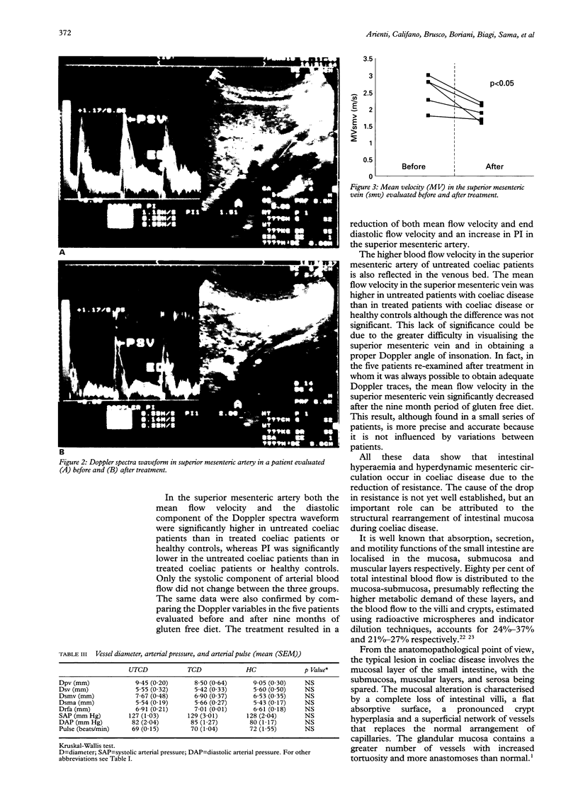
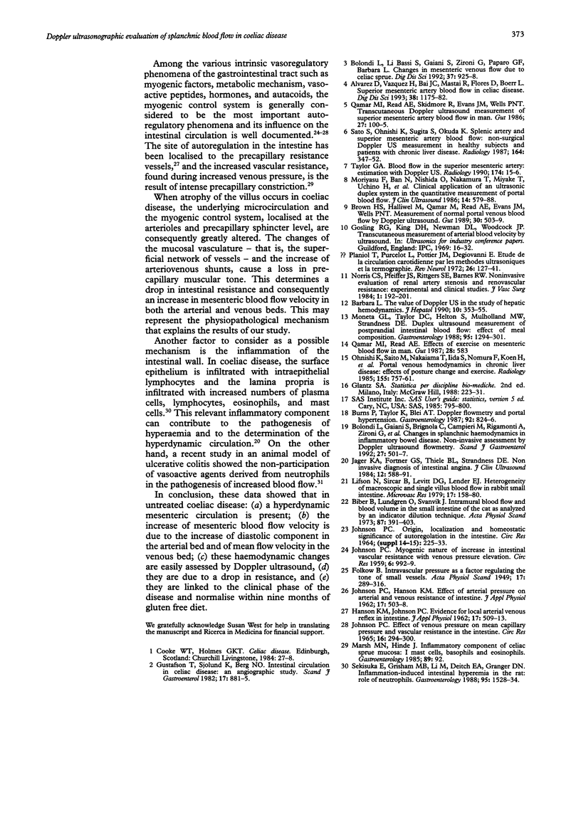
BACKGROUND: Current knowledge on splanchnic haemodynamics in coeliac disease is limited and incomplete. AIM: To evaluate splanchnic arterial and venous blood flow in coeliac disease. METHODS: In 22 coeliac (13 untreated, nine treated) patients and in nine healthy subjects the following variables were assessed: vessel diameter and mean flow velocity in portal vein, splenic vein, superior mesenteric vein, and superior mesenteric artery. Peak systolic velocity, end diastolic velocity and pulsatility index were also determined in the superior mesenteric artery. Five patients of the untreated group were re-evaluated after nine months on a gluten free diet. RESULTS: Significant differences in haemodynamic variables between the three groups were shown only in the superior mesenteric artery. An increase in both mean flow velocity and end diastolic velocity and a reduction in pulsatility index occurred in untreated patients compared with treated patients (p < 0.002; p < 0.04; p < 0.035) and with healthy controls (p < 0.001; p < 0.025; p < 0.0003). Similar results were obtained for the five patients evaluated before and after treatment (p < 0.03; p < 0.02; p < 0.03), in whom the mean flow velocity in the superior mesenteric vein also decreased after treatment (p < 0.05). No significant differences were noted between treated coeliac patients and healthy controls. CONCLUSIONS: In untreated coeliac disease there is a hyperdynamic mesenteric circulation that decreases after treatment.
Full text
PDF
Images in this article
Selected References
These references are in PubMed. This may not be the complete list of references from this article.
- Alvarez D., Vazquez H., Bai J. C., Mastai R., Flores D., Boerr L. Superior mesenteric artery blood flow in celiac disease. Dig Dis Sci. 1993 Jul;38(7):1175–1182. doi: 10.1007/BF01296064. [DOI] [PubMed] [Google Scholar]
- Biber B., Lundgren O., Svanvik J. Intramural blood flow and blood volume in the small intestine of the cat as analyzed by an indicator-dilution technique. Acta Physiol Scand. 1973 Mar;87(3):391–403. doi: 10.1111/j.1748-1716.1973.tb05403.x. [DOI] [PubMed] [Google Scholar]
- Bolondi L., Gaiani S., Brignola C., Campieri M., Rigamonti A., Zironi G., Gionchetti P., Belloli C., Miglioli M., Barbara L. Changes in splanchnic hemodynamics in inflammatory bowel disease. Non-invasive assessment by Doppler ultrasound flowmetry. Scand J Gastroenterol. 1992 Jun;27(6):501–507. doi: 10.3109/00365529209000112. [DOI] [PubMed] [Google Scholar]
- Bolondi L., Li Bassi S., Gaiani S., Zironi G., Paparo G. F., Barbara L. Changes in mesenteric venous flow due to celiac sprue. Dig Dis Sci. 1992 Jun;37(6):925–928. doi: 10.1007/BF01300392. [DOI] [PubMed] [Google Scholar]
- Brown H. S., Halliwell M., Qamar M., Read A. E., Evans J. M., Wells P. N. Measurement of normal portal venous blood flow by Doppler ultrasound. Gut. 1989 Apr;30(4):503–509. doi: 10.1136/gut.30.4.503. [DOI] [PMC free article] [PubMed] [Google Scholar]
- Burns P., Taylor K., Blei A. T. Doppler flowmetry and portal hypertension. Gastroenterology. 1987 Mar;92(3):824–826. doi: 10.1016/0016-5085(87)90040-0. [DOI] [PubMed] [Google Scholar]
- Gustafson T., Sjölund K., Berg N. O. Intestinal circulation in coeliac disease: an angiographic study. Scand J Gastroenterol. 1982 Oct;17(7):881–885. doi: 10.3109/00365528209181110. [DOI] [PubMed] [Google Scholar]
- HANSON K. M., JOHNSON P. C. Evidence for local arteriovenous reflex in intestine. J Appl Physiol. 1962 May;17:509–513. doi: 10.1152/jappl.1962.17.3.509. [DOI] [PubMed] [Google Scholar]
- JOHNSON P. C. EFFECT OF VENOUS PRESSURE ON MEAN CAPILLARY PRESSURE AND VASCULAR RESISTANCE IN THE INTESTINE. Circ Res. 1965 Mar;16:294–300. doi: 10.1161/01.res.16.3.294. [DOI] [PubMed] [Google Scholar]
- JOHNSON P. C., HANSON K. M. Effect of arterial pressure on arterial and venous resistance of intestine. J Appl Physiol. 1962 May;17:503–508. doi: 10.1152/jappl.1962.17.3.503. [DOI] [PubMed] [Google Scholar]
- JOHNSON P. C. Myogenic nature of increase in intestinal vascular resistance with venous pressure elevation. Circ Res. 1959 Nov;7:992–999. doi: 10.1161/01.res.7.6.992. [DOI] [PubMed] [Google Scholar]
- Jäger K. A., Fortner G. S., Thiele B. L., Strandness D. E. Noninvasive diagnosis of intestinal angina. J Clin Ultrasound. 1984 Nov-Dec;12(9):588–591. doi: 10.1002/jcu.1870120913. [DOI] [PubMed] [Google Scholar]
- Lifson N., Sircar B., Levitt D. G., Lender E. J. Heterogeneity of macroscopic and single villus blood flow in rabbit small intestine. Microvasc Res. 1979 Mar;17(2):158–180. doi: 10.1016/0026-2862(79)90404-7. [DOI] [PubMed] [Google Scholar]
- Marsh M. N., Hinde J. Inflammatory component of celiac sprue mucosa. I. Mast cells, basophils, and eosinophils. Gastroenterology. 1985 Jul;89(1):92–101. doi: 10.1016/0016-5085(85)90749-8. [DOI] [PubMed] [Google Scholar]
- Moneta G. L., Taylor D. C., Helton W. S., Mulholland M. W., Strandness D. E., Jr Duplex ultrasound measurement of postprandial intestinal blood flow: effect of meal composition. Gastroenterology. 1988 Nov;95(5):1294–1301. doi: 10.1016/0016-5085(88)90364-2. [DOI] [PubMed] [Google Scholar]
- Moriyasu F., Ban N., Nishida O., Nakamura T., Miyake T., Uchino H., Kanematsu Y., Koizumi S. Clinical application of an ultrasonic duplex system in the quantitative measurement of portal blood flow. J Clin Ultrasound. 1986 Oct;14(8):579–588. doi: 10.1002/jcu.1870140802. [DOI] [PubMed] [Google Scholar]
- Norris C. S., Pfeiffer J. S., Rittgers S. E., Barnes R. W. Noninvasive evaluation of renal artery stenosis and renovascular resistance. Experimental and clinical studies. J Vasc Surg. 1984 Jan;1(1):192–201. doi: 10.1067/mva.1984.avs0010192. [DOI] [PubMed] [Google Scholar]
- Ohnishi K., Saito M., Nakayama T., Iida S., Nomura F., Koen H., Okuda K. Portal venous hemodynamics in chronic liver disease: effects of posture change and exercise. Radiology. 1985 Jun;155(3):757–761. doi: 10.1148/radiology.155.3.3890004. [DOI] [PubMed] [Google Scholar]
- Planiol T., Pourcelot L., Pottier J. M., Degiovanni E. Etude de la circulation carotidienne par les méthodes ultrasoniques et la thermographie. Rev Neurol (Paris) 1972 Feb;126(2):127–141. [PubMed] [Google Scholar]
- Qamar M. I., Read A. E. Effects of exercise on mesenteric blood flow in man. Gut. 1987 May;28(5):583–587. doi: 10.1136/gut.28.5.583. [DOI] [PMC free article] [PubMed] [Google Scholar]
- Qamar M. I., Read A. E., Skidmore R., Evans J. M., Wells P. N. Transcutaneous Doppler ultrasound measurement of superior mesenteric artery blood flow in man. Gut. 1986 Jan;27(1):100–105. doi: 10.1136/gut.27.1.100. [DOI] [PMC free article] [PubMed] [Google Scholar]
- Sato S., Ohnishi K., Sugita S., Okuda K. Splenic artery and superior mesenteric artery blood flow: nonsurgical Doppler US measurement in healthy subjects and patients with chronic liver disease. Radiology. 1987 Aug;164(2):347–352. doi: 10.1148/radiology.164.2.2955448. [DOI] [PubMed] [Google Scholar]
- Sekizuka E., Grisham M. B., Li M. A., Deitch E. A., Granger D. N. Inflammation-induced intestinal hyperemia in the rat: role of neutrophils. Gastroenterology. 1988 Dec;95(6):1528–1534. doi: 10.1016/s0016-5085(88)80073-8. [DOI] [PubMed] [Google Scholar]
- Taylor G. A. Blood flow in the superior mesenteric artery: estimation with Doppler US. Radiology. 1990 Jan;174(1):15–16. doi: 10.1148/radiology.174.1.2403677. [DOI] [PubMed] [Google Scholar]
- The value of Doppler US in the study of hepatic hemodynamics. Consensus conference (Bologna, Italy, 12 September, 1989). J Hepatol. 1990 May;10(3):353–355. doi: 10.1016/0168-8278(90)90146-i. [DOI] [PubMed] [Google Scholar]


