Abstract
SH3PX1 [SNX9 (sorting nexin 9)] is a member of SNX super-family that is recognized by sharing a PX (phox homology) domain. We have previously shown that SH3PX1, phosphorylated by ACK2 (activated Cdc42-associated tyrosine kinase 2), regulates the degradation of EGF (epidermal growth factor) receptor. In mapping the tyrosine phosphorylation region, we found that the C-terminus of SH3PX1 is required for its tyrosine phosphorylation. Further analysis indicates that this region, known as the coiled-coil domain or the BAR (Bin–amphiphysin–Rvs homology) domain, is the dimerization domain of SH3PX1. Truncation of as little as 13 amino acid residues at the very C-terminus in the coiled-coil/BAR domain of SH3PX1 resulted in no dimerization, no ACK2-catalysed and EGF-stimulated tyrosine phosphorylation and no interaction with ACK2. The intracellular localization of SH3PX1 became dysfunctional upon truncation in the BAR domain. Taken together, our results indicate that the dimerization, which is mediated by the BAR domain, is essential for the intracellular function of SH3PX1.
Keywords: Bin–amphiphysin–Rvs homology domain (BAR domain), dimerization, epidermal growth factor (EGF), SH3PX1, sorting nexin 9 (SNX9), tyrosine phosphorylation
Abbreviations: ACK, activated Cdc42-associated tyrosine kinase; BAR domain, Bin–amphiphysin–Rvs homology domain; EGF, epidermal growth factor; EGFR, EGF receptor; GFP, green fluorescent protein; GST, glutathione S-transferase; HA, haemagglutinin; PX domain, phox homology domain; PY, phosphotyrosine; SH3 domain, Src homology 3 domain; SNX, sorting nexin
INTRODUCTION
Sorting nexins (SNXs) constitute more than 30 members that contain a PX (phox homology) domain [1]. The PX domain has been recently identified as an inositol phospholipid-binding module [2–4]. This feature connects the function of SNXs to interaction with membrane vesicles and regulation of intracellular vesicle trafficking. Previous studies have indicated that SNX1, SNX2, SNX9 and SNX16 participate in regulation of EGFR [EGF (epidermal growth factor) receptor] degradation [5–8]. SNX6 and SNX27 may regulate TGF-β receptor or α-HT receptor trafficking [9,10]. Many other SNXs have been shown to interact with endosomal vesicles and may regulate various steps of vesicle transport [11–14]. Although many studies have been pursued, the exact function of SNXs in vesicle trafficking is still not clear.
SH3PX1 (SNX9) was initially identified as an interactive protein of the metalloprotease disintegrins MDC9 (metalloprotease, disintegrin, cysteine-rich protein 9) and MDC15 [15]. In Drosophila, SH3PX1 may function in connection of the cytoskeletal structure to the receptor-like molecule Dscam trafficking [16]. Biochemically, SH3PX1 interacts with the clathrin-coated vesicle adaptor protein AP-2, clathrin and, recently, dynamin-2 [17,18], suggesting that SH3PX1 may be directly involved in clathrin-coated vesicle endocytosis. Nevertheless, the role of SH3PX1 in clathrin-coated vesicle endocytosis or receptor trafficking has not been defined.
Our previous studies have shown that SH3PX1 was tyrosine-phosphorylated in response to EGF signalling [7]. ACK2 (activated Cdc42-associated tyrosine kinase 2) is the tyrosine kinase that catalyses the tyrosine phosphorylation of SH3PX1 [7]. Furthermore, co-expression of SH3PX1 with ACK2 but not SH3PX1 alone in COS7 cells facilitated EGFR degradation, suggesting a role for the tyrosine phosphorylation of SH3PX1 in regulation of EGFR degradation. In the present study, we show that the tyrosine phosphorylation of SH3PX1 catalysed by ACK2 or stimulated by EGF signalling is controlled by dimerization of SH3PX1. Dimerization is also required for the interaction of SH3PX1 with ACK2 and the localization of SH3PX1 in cells.
MATERIALS AND METHODS
Cell culture, transfection and preparation of cell lysates
COS7 cells were cultured in Dulbecco's modified Eagle's medium plus 10% (v/v) fetal bovine serum. All cells were maintained at 37 °C in 5% CO2. For transfection, cells were cultured overnight to 90% confluence. DNA was transfected into cells using either Lipofectamine™ (Invitrogen) or Cytopure (Q-biogene) transfection reagents according to the manufacturer's instructions. After 36 h, cells requiring EGF stimulation were starved with serum-free media followed by continued incubation for 12 h. Cells were then stimulated with EGF at indicated time points before lysis. Cells not requiring EGF stimulation were incubated for 48 h after transfection. Cells were lysed using chilled mammalian cell lysis buffer (40 mM Hepes, pH 7.4, 100 mM NaCl, 1% Triton X-100, 25 mM glycerol phosphate, 1 mM sodium orthovanadate, 1 mM EDTA, 10 μg/ml aprotinin and 10 μg/ml leupeptin) on rocking plates at 4 °C for 30 min. Cell lysates were cleared by centrifugation at 14000 g in a microcentrifuge for 5 min at 4 °C before use.
Construction of the C-terminus truncation mutants of SH3PX1
The C-terminus truncation mutants ΔC1, ΔC1-3, ΔC2 and ΔC3 were made by PCR using pcDNA3-HA-SH3PX1 as a template. The truncation mutants were directly cloned into BamHI/EcoRI sites of pcDNA3 vector and tagged with an HA (haemagglutinin) tag at the N-terminus. The mutations were confirmed by DNA sequencing. For GST (glutathione S-transferase) fusion proteins, SH3PX1 was directly cloned from pCDNA3-SH3PX1 using BamHI/EcoRI into the GST-fusion protein vector pGEX-4T3. The C-terminus portion construct C400–595 (amino acid residues 400–595) was made by using PCR and digesting the fragments with BamHI/EcoRI and cloned into pGEX-4T3. Constructs were transformed into Escherichia coli (JM109).
GST protein affinity binding assays
GST-fusion proteins were expressed in E. coli (JM109) and affinity-purified with glutathione–agarose beads as described previously [19]. The GST–SH3PX1, the SH3 (Src homology 3) domain or the C-terminus portion (C400–595), bound to glutathione-conjugated agarose beads, was incubated with precleared lysates from COS7 cells that overexpressed Myc-tagged ACK2, at 4 °C for 3–4 h with rotation. The beads were washed three times with lysis buffer. The bound proteins were then resolved by SDS/PAGE. Co-precipitated ACK2 was then detected by immunoblotting with an anti-Myc antibody.
Immunoprecipitation and immunoblots
For immunoprecipitation, the precleared cell lysates were incubated with primary antibody for 30 min on ice. Protein A beads were added and the mixture was incubated at 4 °C for 3 h with rotation. After incubation, the lysates were removed and the beads were washed with lysis buffer three times. The immunoprecipitation products were directly dissolved in SDS/PAGE sample buffer (63 mM Tris/HCl, pH 6.8, 2% SDS, 0.0025% Bromophenol Blue, 5% 2-mercaptoethanol and 10% glycerol) and resolved with SDS/PAGE. The immunoblots were performed as outlined in the Western Lightning immunoblot kit (PerkinElmer LifeSciences).
Immunofluorescence
Myc-tagged SH3PX1-WT or Myc-tagged ΔC1-3 was co-transfected with GFP–ACK2 (where GFP is green fluorescent protein) into COS7 cells. After 24 h of incubation, cells were moved to glass bottom microwell dishes (MatTek Corp.) in preparation for immunostaining. Cells were incubated for another 24 h. For EGF stimulation, the cells were serum starved for 12 h before treating with EGF. The cells were washed three times with PBS, then fixed with 4% (w/v) paraformaldehyde in PBS for 10 min, followed by permeabilization with 0.02% Triton X-100 in PBS for 10 min. Immunostaining was performed by incubating the cells with primary antibody (anti-Myc) at 22 °C for 1 h followed by incubation with Texas Red-conjugated anti-mouse IgG at 22 °C for 1 h. The fluorescent staining of SH3PX1 and ΔC1-3 and green fluorescence of GFP–ACK2 were visualized under a Zeiss fluorescent microscope.
RESULTS
Deletion of the C-terminus diminished the tyrosine phosphorylation of SH3PX1 by ACK2
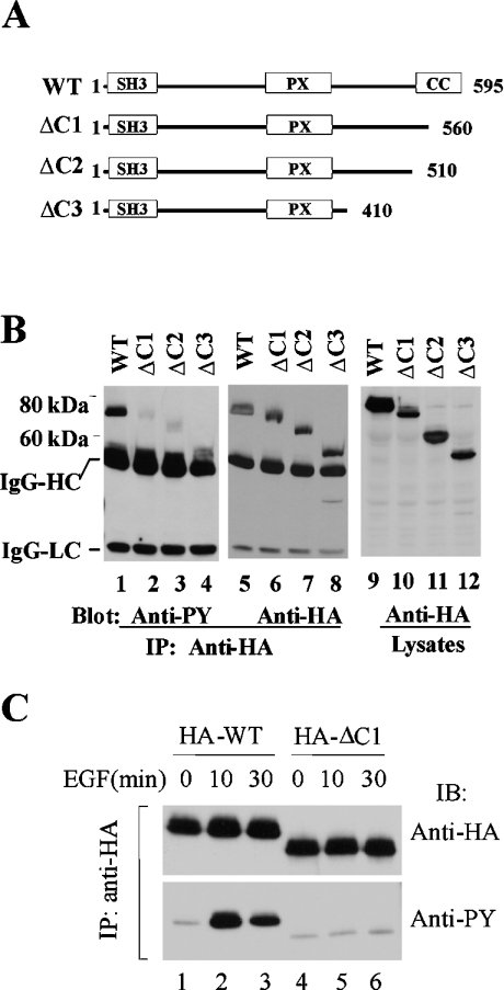
Our previous studies have shown that SH3PX1 is phosphorylated by ACK2 [7]. To map the tyrosine phosphorylation sites, we made a series of HA-tagged C-terminus truncation mutants of SH3PX1, as indicated in Figure 1(A). After co-transfection with ACK2 into COS7 cells, the mutants were immunoprecipitated by anti-HA antibody. The tyrosine phosphorylation of the mutants was detected by immunoblotting with anti-PY (phosphotyrosine) antibody. As shown in Figures 1(B) and 1(C), the truncation of the C-terminus eliminated both ACK2-catalysed (Figure 1B) and EGF-stimulated (Figure 1C) tyrosine phosphorylation in all truncation mutants, suggesting that the C-terminus is required for tyrosine phosphorylation of SH3PX1.
Figure 1. The C-terminus of SH3PX1 is required for its tyrosine phosphorylation.
(A) A schematic representation of the C-terminus truncation mutants of SH3PX1. CC, the coiled-coil domain that is the region for dimerization of SH3PX1. (B) Deletion of the coiled-coil domain of SH3PX1 eliminated the ACK2-catalysed phosphorylation. The HA-tagged wild-type or truncation mutants of SH3PX1 were co-transfected with Myc-tagged ACK2 (all lanes) into COS7 cells for 48 h. The SH3PX1 and its truncation mutants were immunoprecipitated with anti-HA and immunoblotted with anti-PY (4G10) (lanes 1–4) and anti-HA (lanes 5–8). The expression of the proteins was determined by immunoblotting the cell lysates with anti-HA (lanes 9–12). IgG-HC, IgG heavy chain; IgG-LC, IgG light chain. IP, immunoprecipitation. (C) The C-terminus is required for EGF-stimulated tyrosine phosphorylation of SH3PX1. The COS7 cells were transfected with HA-tagged SH3PX1 or ΔC1 for 36 h followed by 12 h serum starvation and subsequently subjected to EGF stimulation (100 ng/ml EGF) for the indicated times. SH3PX1 or ΔC1 was immunoprecipitated and immunoblotted with anti-HA (upper panel) and anti-PY (4G10) (lower panel). IB, immunoblotting.
One possible cause for elimination of tyrosine phosphorylation of SH3PX1 by truncation of the C-terminus is that the phosphorylation sites are located at the C-terminus. To examine this possibility, we made another C-terminus truncation mutant ΔC1-3, in which the last 13 amino acid residues at the C-terminus were deleted. No tyrosine residue is in the deleted portion. If deletion of the phosphorylation sites is the cause for elimination of the tyrosine phosphorylation in the C-terminus truncation mutants ΔC1, ΔC2 and ΔC3, the tyrosine phosphorylation of ΔC1-3 should be the same as full-length SH3PX1. To test this possibility, we co-transfected ΔC1-3 with ACK2 into COS7 cells and detected the tyrosine phosphorylation of ΔC1-3 by immunoblotting with anti-PY antibody. Surprisingly, ΔC1-3, just like other C-terminus truncation mutants, was not phosphorylated by ACK2 (Figure 2A). Furthermore, deletion of the last 13 C-terminus amino acid residues eliminated the tyrosine phosphorylation of SH3PX1 by EGF stimulation (Figure 2B). These results indicate that the elimination of the tyrosine phosphorylation of SH3PX1 by truncation of the C-terminus is not due to missing the phosphorylation sites, but rather, may be caused by structural changes.
Figure 2. Diminution of the tyrosine phosphorylation of SH3PX1 by deletion of the C-terminus did not result from losing the tyrosine phosphorylation sites.
(A) HA-tagged SH3PX1, ΔC1 or ΔC1-3 was co-transfected with Myc-tagged ACK2 into COS7 cells. SH3PX1 and the truncation mutants were immunoprecipitated with anti-HA and immunoblotted with anti-HA (the top panel) and anti-PY (the middle panel). The expression of ACK2 was detected by immunoblotting the cell lysates with anti-Myc (the bottom panel). IP, immunoprecipitation; IB, immunoblotting. (B) COS7 cells were transfected with HA-tagged SH3PX1 or its truncation mutants for 36 h followed by serum starvation for 12 h and subsequently stimulated with EGF (100 ng/ml) for the indicated times. SH3PX1 and the truncation mutants were immunoprecipitated with anti-HA, and immunoblotted with anti-HA (the bottom panel) and anti-PY (the top panel).
The C-terminus truncation mutants are dimerization-defective
Analysis of SH3PX1 molecular structure by the program Coils [20] indicates that amino acids 571–592 at the very C-terminus form a coiled-coil domain. This domain of Drosophila SH3PX1 has been shown to be critical for dimerization [16]. Structural analysis suggests that the coiled-coil domain of SH3PX1 is similar to the BAR (Bin–amphiphysin–Rvs homology) domain in amphiphysin, a known domain for dimerization in endocytic proteins [21]. To examine the function of the C-terminus coiled-coil domain in dimerization, we analysed the in vitro and in vivo dimerization capacity of both full-length and C-terminus-truncated SH3PX1. In in vitro assays, we attempted to determine the dimerization or oligomerization of purified bacterially expressed GST–SH3PX1 and GST–SH3PX1ΔC1-3 using the chemical cross-linking method. However, we failed to distinguish the difference in dimerization between GST–SH3PX1 and GST–SH3PX1ΔC1-3 and observed a high degree of oligomerization in all the samples, probably due to non-specific cross-linking. In in vivo assays, we co-transfected Myc-tagged SH3PX1 with HA-tagged full-length or C-terminus-truncated SH3PX1 into COS7 cells. The HA-tagged full-length or the truncation mutants of SH3PX1 were immunoprecipitated by anti-HA antibody; the co-immunoprecipitated Myc-tagged SH3PX1 was detected by immunoblotting with anti-Myc antibody. As shown in Figure 3, while wild-type SH3PX1 had dimerization capacity (Figure 3A, lane 3; Figure 3B, lane 1), the C-terminus truncation mutants ΔC1, ΔC2 and ΔC1-3 were not able to dimerize with full-length SH3PX1 (Figure 3A, lanes 1 and 2; Figure 3B, lane 4), nor ΔC1-3 with ΔC1-3 (Figure 3B, lane 3). These results indicate that the last 13 amino acid residues at the C-terminus are required for SH3PX1 dimerization. Taken together, the C-terminus truncation mutants are both tyrosine phosphorylation-defective and dimerization-defective. The next question is whether dimerization determines the tyrosine phosphorylation or the tyrosine phosphorylation determines the dimerization in SH3PX1.
Figure 3. The C-terminus of SH3PX1 is required for its dimerization.
(A) HA-tagged SH3PX1, ΔC1 or ΔC2 was co-transfected with Myc-tagged SH3PX1 into COS7 cells. The HA-tagged SH3PX1 and its truncation mutants were immunoprecipitated and immunoblotted by anti-HA (the top panel). The co-immunoprecipitated Myc-tagged SH3PX1 was detected by immunoblotting with anti-Myc (the middle panel). The expression level of Myc-tagged SH3PX1 was determined by immunoblotting the cell lysates with anti-Myc (the bottom panel). IP, immunoprecipitation. (B) HA-tagged SH3PX1 or ΔC1-3, a deletion mutant that lacks the last 13 amino acid residues at the C-terminus, was co-transfected with Myc-tagged SH3PX1 or ΔC1-3 into COS7 cells. The dimerization between SH3PX1, ΔC1-3 or SH3PX1 and ΔC1-3 was determined by co-immunoprecipitation. The immunoprecipitated Myc–SH3PX1 or Myc–ΔC1-3 and co-immunoprecipitated HA–SH3PX1 or HA–ΔC1-3 were detected by immunoblotting with anti-Myc (the second panel from top) and anti-HA (the top panel) respectively. The expression level of SH3PX1 or ΔC1-3 was determined by immunoblotting the cell lysates with anti-Myc (the bottom panel) or anti-HA (the second panel from bottom).
The dimerization of SH3PX1 is independent of EGF-induced tyrosine phosphorylation
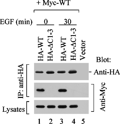
To further determine the relationship between tyrosine phosphorylation and dimerization of SH3PX1, we performed the dimerization assay under EGF stimulation. Our studies have shown that EGF stimulation induces tyrosine phosphorylation of SH3PX1 (Figures 1C and 2B). We co-transfected Myc-tagged full-length SH3PX1 with HA-tagged full-length SH3PX1 or the truncation mutant ΔC1-3 into COS7 cells, then stimulated the cells with EGF to generate the tyrosine phosphorylation of SH3PX1, and determined the dimerization of SH3PX1 by co-immunoprecipitation assays. As shown in Figure 4, the dimerization of the full-length SH3PX1 or the truncation mutant ΔC1-3 was not affected by EGF stimulation, suggesting that EGF-induced tyrosine phosphorylation of SH3PX1 does not regulate the dimerization.
Figure 4. Dimerization of SH3PX1 is independent of EGF signalling.
HA-tagged SH3PX1 or ΔC1-3 was co-transfected with Myc-tagged SH3PX1 into COS7 cells for 36 h. The cells were serum starved for 12 h and subsequently stimulated by EGF for 0 and 30 min. HA-tagged SH3PX1 and the truncation mutants were immunoprecipitated and immunoblotted with anti-HA (the top panel). The co-immunoprecipitated Myc–SH3PX1 was detected by immunoblotting with anti-Myc (the middle panel). The expression level of Myc-tagged SH3PX1 was determined by immunoblotting the cell lysates with anti-Myc (the bottom panel). IP, immunoprecipitation.
The dimerization of SH3PX1 is independent of ACK2
To address the relationship between the dimerization and the binding to ACK2, we utilized an SH3 domain deletion mutant ΔSH3, which is ACK2-binding-defective (Figure 5A), to perform the dimerization assay. Wild-type SH3PX1 was tyrosine-phosphorylated when co-transfected with ACK2 (Figure 5B, lane 1), while ΔSH3 was not tyrosine-phosphorylated by ACK2 (Figure 5B, lane 2), indicating that ΔSH3 is ACK2-phosphorylation-defective. However, both wild-type SH3PX1 and ΔSH3 have equal capacity of dimerization (Figure 5C), indicating that the dimerization of SH3PX1 is not regulated by interaction with ACK2. In summary, the dimerization of SH3PX1 is independent of EGF-induced tyrosine phosphorylation and the interaction with ACK2, suggesting that dimerization is an upstream process to the tyrosine phosphorylation and the interaction with ACK2. Considering that ΔC1-3, the dimerization-defective mutant that has the same tyrosine residues as full-length SH3PX1, is tyrosine phosphorylation-defective (Figures 2A and 2B), we conclude that the dimerization of SH3PX1 is required for its tyrosine phosphorylation, including ACK2-catalysed and EGF-stimulated tyrosine phosphorylation.
Figure 5. Dimerization of SH3PX1 is ACK2-independent.
(A) The SH3 domain deletion mutant of SH3PX1 (ΔSH3) is ACK2-binding-defective. HA-tagged SH3PX1 or ΔSH3 was co-transfected with Myc-tagged ACK2 into COS7 cells. ACK2 was immunoprecipitated and immunoblotted with anti-Myc (the top panel). The co-immunoprecipitated SH3PX1 or ΔSH3 was detected by immunoblotting with anti-HA (the middle panel). The expression level of SH3PX1 or ΔSH3 was determined by immunoblotting the cell lysates with anti-HA (the bottom panel). (B) The SH3 domain deletion mutant ΔSH3 is not phosphorylated by ACK2. HA-tagged SH3PX1 or ΔSH3 was co-transfected with Myc-tagged ACK2 into COS7 cells. SH3PX1 or ΔSH3 was immunoprecipitated and immunoblotted with anti-HA antibody. The tyrosine phosphorylation of SH3PX1 or ΔSH3 was detected by immunoblotting with anti-PY (the middle panel). The expression level of ACK2 was determined by immunoblotting the cell lysates with anti-Myc (the bottom panel). (C) The SH3 domain deletion mutant ΔSH3 is capable of dimerization. HA-tagged SH3PX1 or ΔSH3 was co-transfected with Myc-tagged SH3PX1. The HA-tagged SH3PX1 and the SH3 domain deletion mutant were immunoprecipitated and immunoblotted with anti-HA (the top panel). The co-immunoprecipitated Myc-tagged SH3PX1 was detected by immunoblotting with anti-Myc (the middle panel). The expression level of Myc-tagged SH3PX1 was determined by immunoblotting the cell lysates with anti-Myc (the bottom panel). IP, immunoprecipitation.
Dimerization is required for SH3PX1 to interact with ACK2
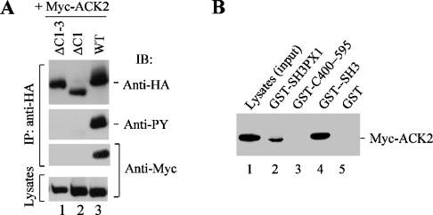
Next question is the mechanism underlying the effect of dimerization on the tyrosine phosphorylation of SH3PX1. We notice that the interaction of SH3PX1 with ACK2 is necessary for ACK2-catalysed tyrosine phosphorylation (Figure 5). Therefore we suspect that the dimerization of SH3PX1 may be involved in the interaction with ACK2. We co-transfected the HA-tagged full-length SH3PX1 or C-terminus truncation mutants ΔC1 and ΔC1-3 with Myc-tagged ACK2 and determined the interaction of SH3PX1 or the mutants with ACK2 by co-immunoprecipitation assays. As shown in Figure 6(A), while wild-type SH3PX1 was co-immunoprecipitated with ACK2 (lane 3), the C-terminus truncation mutant ΔC1 or ΔC1-3 did not interact with ACK2 (lanes 1 and 2). This result indicates that the C-terminus is required for the interaction of SH3PX1 with ACK2. The incapability of interaction with ACK2 may be the mechanism underlying the diminishment of tyrosine phosphorylation of SH3PX1 by the C-terminus truncation.
Figure 6. Dimerization is required for the interaction of SH3PX1 with ACK2.
(A) The dimerization-defective mutants do not bind to ACK2. HA-tagged SH3PX1, ΔC1 or ΔC1-3 was co-transfected with Myc-tagged ACK2 into COS7 cells. SH3PX1 and the truncation mutants were immunoprecipitated and immunoblotted with anti-HA (the top panel). The tyrosine phosphorylation of SH3PX1 and the truncation mutants were detected by immunoblotting with anti-PY (the second panel from top). The co-immunoprecipitated Myc-tagged ACK2 was detected by immunoblotting with anti-Myc (the second panel from bottom). The expression of ACK2 was detected by immunoblotting the cell lysates with anti-Myc (the bottom panel). (B) The C-terminus does not contain ACK-binding sites. The GST-fusion proteins that contain full-length SH3PX1, the SH3 domain of SH3PX1 or the C-terminus portion of SH3PX1 (C400–595) were expressed in E. coli and purified by affinity precipitation with glutathione-conjugated agarose beads. The bead-immobilized GST-fusion proteins were incubated with Myc–ACK2 expressed COS7 cell lysates. The co-precipitated ACK2 with the GST-fusion proteins was detected by immunoblotting with anti-Myc. IP, immunoprecipitation; IB, immunoblotting.
To determine whether defect in the dimerization or loss of the binding site to ACK2 in the C-terminus truncation mutant ΔC1 or ΔC1-3 eliminates the binding to ACK2, we made a series of GST-fusion proteins that contain the SH3 domain (GST–SH3), the C-terminus portion (amino acid residues 400–595, GST–C400–595) or the full-length SH3PX1, and performed ACK2-pulldown assay with these GST-fusion proteins. As shown in Figure 6(B), the full-length SH3PX1 or the SH3 domain was able to pull down ACK2 from ACK2-transfected COS7 cell lysates (lanes 2 and 4), while the C-terminus portion (amino acid residues 400–595) was not (lane 3). These results suggest that the SH3 domain is the ACK2-interactive domain, as we have shown previously [7], and the C-terminus of SH3PX1 does not interact with ACK2. Therefore the defect in interaction with ACK2 in the C-terminus truncation mutants ΔC1 and ΔC1-3 is caused by losing the capacity of dimerization, not the ACK2-binding sites.
Dimerization is required for intracellular localization of SH3PX1
To determine effects of the dimerization on intracellular localization of SH3PX1 and the interaction of SH3PX1 with ACK2 in cells, we co-transfected Myc-tagged wild-type SH3PX1 and the dimerization-defective mutant ΔC1-3 with GFP–ACK2 into COS7 cells, and immunostained SH3PX1 and ΔC1-3 with anti-Myc antibody. As shown in Figure 7(A), while wild-type SH3PX1 was co-localized with ACK2 on punctate structures (Figure 7A, panels a and b), the dimerization-defective mutant ΔC1-3 was not localized with ACK2 (Figure 7A, panels c and d). These results suggest that dimerization is required for its intracellular localization or its co-localization with ACK.
Figure 7. The intracellular localization of SH3PX1 is regulated by dimerization.
(A) The dimerization domain is required for co-localization of SH3PX1 with ACK2. Myc-tagged SH3PX1 or ΔC1-3 was co-transfected with GFP–ACK2 into COS7 cells. The localization of SH3PX1 (panel a) and ΔC1-3 (panel c) was determined by immunostaining with anti-Myc. ACK2 was visualized by GFP (panels b and d). (B) The dimerization domain is required for EGF-stimulated translocation of SH3PX1 and co-localization with ACK2 upon EGF stimulation. Myc-tagged SH3PX1 or ΔC1-3 was co-transfected with GFP–ACK2 into COS7 cells. After 36 h of transfection, the cells were serum starved for 12 h and then stimulated with EGF (100 ng/ml) for 30 min. The intracellular localization of SH3PX1 and ΔC1-3 was determined by immunostaining with anti-Myc (panels a, c, e and g). The localization of ACK2 was visualized by GFP fluorescence (panels b, d, f and h). No EGF stimulation (panels a–d); EGF stimulation for 30 min (panels e–h).
To determine effects of the dimerization on intracellular localization of SH3PX1 in response to EGF signalling, we cotransfected Myc-tagged SH3PX1 and the dimerization-defective mutant ΔC1-3 with GFP–ACK2 into COS7 cells. After 36 h transfection, the cells were serum starved for 12 h and then stimulated with EGF (100 ng/ml) for 30 min. The intracellular localization of SH3PX1 and ΔC1-3 was determined by immunostaining with anti-Myc antibody. The localization of ACK2 was visualized by GFP fluorescence. As shown in Figure 7(B), wild-type SH3PX1 was co-localized with ACK2 on vesicular and punctate structures upon EGF stimulation (Figure 7B, panels e and f). In addition, co-localization of wild-type SH3PX1 with ACK2 also occurred in the presence of fetal bovine serum (Figure 7A, panels a and b) and this co-localization was eliminated by serum starvation (Figure 7B, panels a and b). However, the intracellular localization of ΔC1-3 was not responsive to EGF signalling (Figure 7B, panels c and g). No co-localization of ΔC1-3 with ACK2 was detected (Figure 7B, panel h). These results further confirm that dimerization is essential for co-localization of SH3PX1 with ACK2 and suggest that dimerization is required for EGF-stimulated translocation of SH3PX1.
DISCUSSION
SH3PX1 is a phosphorylation substrate of the ACK tyrosine kinase family [7]. The tyrosine phosphorylation of SH3PX1 is in response to EGF signal. We have shown that tyrosine phosphorylation of SH3PX1 by ACK2 is critical for the function of SH3PX1 in regulation of EGFR degradation [7]. However, very little is known about the regulation of the tyrosine phosphorylation of SH3PX1.
In the present study, we report that the dimerization of SH3PX1 is required for the tyrosine phosphorylation and interaction with ACK2. Our studies indicate that deletion of the C-terminus coiled-coil/BAR domain of SH3PX1, which is the domain for dimerization, eliminates the interaction of SH3PX1 with ACK2. However, the dimerization domain does not contain ACK-binding sites (Figure 6B). Therefore dimerization is the essential structure for SH3PX1 to interact with ACK2. Given that the SH3 domain is the ACK-interactive domain [7] (Figure 6B), how does dimerization affect the binding of SH3PX1 to ACK2? Our hypothesis is that the SH3 domain of monomeric SH3PX1 is inhibited by either an intramolecular or an intermolecular component. Dimerization releases the SH3 domain by disrupting the interaction of the inhibitory component with the SH3 domain. We notice that a putative SH3 domain-binding motif (amino acid residues 306–311) resides in the PX domain of SH3PX1. The interaction of this putative SH3 domain-binding motif with the SH3 domain might affect binding of the SH3 domain to ACK and the PX domain to inositol phospholipids. It would be intriguing to test the interaction of this putative SH3 domain-binding motif with the SH3 domain in future experiments.
Dimerization is a molecular characteristic of SNXs. Many SNXs possess a coiled-coil/BAR domain at the C-terminus [21]. The interaction between the coiled-coil domains produces not only homodimers but also heterodimers of SNXs. Dimerization in SNXs was observed 7 years ago [22]. However, the function of dimerization in SNXs is still not clear. Recently, the dimerization domain of SNX1 has been found to regulate the binding affinity of the PX domain for inositol phospholipids [23], suggesting that dimerization affects the structure of other domains in SNX1. Our studies in the present paper suggest that the dimerization through the C-terminus of SH3PX1 regulates the function of its SH3 domain at the N-terminus. From these results, we propose that the dimerization determines the structural co-ordinations in SNXs that yield a favourable conformation to interact with inositol phospholipids or binding partners. To further understand the function of dimerization of SNXs, we need to look into the regulatory mechanisms of the structural co-ordinations.
Our previous studies suggest a role of SH3PX1 in regulation of EGFR degradation [7]. In the present study, we show that the translocation of SH3PX1 to vesicular and punctate structures was in response to EGF signalling (Figure 7B), further supporting the role of SH3PX1 in EGFR degradation. Dimerization seems essential for this function of SH3PX1 because the intracellular localization of the dimerization-defective mutant ΔC1-3 did not respond to EGF signalling. However, we still do not know how dimerization of SH3PX1 regulates EGFR degradation. Many other SNXs, such as SNX1, SNX2 and SNX16, also regulate EGFR endocytosis and degradation [5,6,8]. The C-terminus coiled-coil/BAR domain of SNX1 and SNX2 is highly conserved with that of SH3PX1. It is possible that SH3PX1, SNX1 and SNX2 form heterodimers through their coiled-coil/BAR domain and co-regulate EGFR degradation. In addition, SNX18 and a novel SNX in the Mammalian Gene Collection of the National Institutes of Health (MGC:32065) are highly homologous with SH3PX1 and contain the same domain structure as SH3PX1. These uncharacterized SNXs may also form heterodimers with SH3PX1 in cells and exert a similar function as SH3PX1. Taken together, dimerization through the coiled-coil/BAR domain may play an essential role in the function of SNXs. Further characterization of the dimerization will help us to understand the cellular function of SNXs.
Acknowledgments
We thank Lisa M. Stutzman of Lycoming College (Williamsport, PA, U.S.A.) for her excellent technical assistance.
References
- 1.Teasdale R. D., Loci D., Houghton F., Karlsson L., Gleeson P. A. A large family of endosome-localized proteins related to sorting nexin 1. Biochem. J. 2001;358:7–16. doi: 10.1042/0264-6021:3580007. [DOI] [PMC free article] [PubMed] [Google Scholar]
- 2.Bravo J., Karathanassis D., Pacold C. M., Pacold M. E., Ellson C. D., Anderson K. E., Butler P. J., Lavenir I., Perisic O., Hawkins P. T., et al. The crystal structure of the PX domain from p40phox bound to phosphatidylinositol 3-phosphate. Mol. Cell. 2001;8:829–839. doi: 10.1016/s1097-2765(01)00372-0. [DOI] [PubMed] [Google Scholar]
- 3.Kanai F., Liu H., Field S. J., Akbary H., Matsuo T., Brown G. E., Cantley L. C., Yaffe M. B. The PX domains of p47phox and p40phox bind to lipid products of PI(3)K. Nat. Cell Biol. 2001;3:675–678. doi: 10.1038/35083070. [DOI] [PubMed] [Google Scholar]
- 4.Xu Y., Hortsman H., Seet L., Wong S. H., Hong W. SNX3 regulates endosomal function through its PX-domain-mediated interaction with PtdIns(3)P. Nat. Cell Biol. 2001;3:658–666. doi: 10.1038/35083051. [DOI] [PubMed] [Google Scholar]
- 5.Kurten R. C., Cadena D. L., Gill G. N. Enhanced degradation of EGFRs by a sorting nexin, SNX1. Science. 1996;272:1008–1010. doi: 10.1126/science.272.5264.1008. [DOI] [PubMed] [Google Scholar]
- 6.Gullapalli A., Garrett T. A., Paing M. M., Griffin C. T., Yang Y., Trejo J. A role for sorting nexin 2 in epidermal growth factor receptor down-regulation: evidence for distinct functions of sorting nexin 1 and 2 in protein trafficking. Mol. Biol. Cell. 2004;15:2143–2155. doi: 10.1091/mbc.E03-09-0711. [DOI] [PMC free article] [PubMed] [Google Scholar]
- 7.Lin Q., Lo C. G., Cerione R. A., Yang W. The Cdc42-target ACK2 interacts with SH3PX1 (sorting nexin 9) to regulate EGFR degradation. J. Biol. Chem. 2002;277:10134–10138. doi: 10.1074/jbc.M110329200. [DOI] [PubMed] [Google Scholar]
- 8.Choi J. H., Hong W. P., Kim M. J., Kim J. H., Ryu S. H., Suh P. G. Sorting nexin 16 regulates EGF receptor trafficking by phosphatidylinositol-3-phosphate interaction with the Phox domain. J. Cell Sci. 2004;117:4209–4218. doi: 10.1242/jcs.01233. [DOI] [PubMed] [Google Scholar]
- 9.Parks W. T., Frank D. B., Huff C., Haft C. R., Martin J., Meng X., de Caestecker M. P., McNally J. G., Reddi A., Taylor S. I., et al. Sorting nexin 6, a novel SNX, interacts with the transforming growth factor-β family of receptor serine-threonine kinases. J. Biol. Chem. 2001;276:19332–19339. doi: 10.1074/jbc.M100606200. [DOI] [PubMed] [Google Scholar]
- 10.Joubert L., Hanson B., Barthet G., Sebben M., Claeysen S., Hong W., Marin P., Dumuis A., Bockaert J. New sorting nexin (SNX27) and NHERF specifically interact with the 5-HT4a receptor splice variant: roles in receptor targeting. J. Cell Sci. 2004;117:5367–5379. doi: 10.1242/jcs.01379. [DOI] [PubMed] [Google Scholar]
- 11.Merino-Trigo A., Kerr M. C., Houghton F., Lindberg A., Mitchell C., Teasdale R. D., Gleeson P. A. Sorting nexin 5 is localized to a subdomain of the early endosomes and is recruited to the plasma membrane following EGF stimulation. J. Cell Sci. 2004;117:6413–6424. doi: 10.1242/jcs.01561. [DOI] [PubMed] [Google Scholar]
- 12.Williams R., Schluter T., Roberts M. S., Knauth P., Bohnensack R., Cutler D. F. Sorting nexin 17 accelerates internalization yet retards degradation of P-selectin. Mol. Biol. Cell. 2004;15:3095–3105. doi: 10.1091/mbc.E04-02-0143. [DOI] [PMC free article] [PubMed] [Google Scholar]
- 13.Leprince C., Le Scolan E., Meunier B., Fraisier V., Brandon N., De Gunzburg J., Camonis J. Sorting nexin 4 and amphiphysin 2, a new partnership between endocytosis and intracellular trafficking. J. Cell Sci. 2003;116:1937–1948. doi: 10.1242/jcs.00403. [DOI] [PubMed] [Google Scholar]
- 14.Zheng B., Ma Y. C., Ostrom R. S., Lavoie C., Gill G. N., Insel P. A., Huang X. Y., Farquhar M. G. RGS-PX1, a GAP for Gαs and sorting nexin in vesicular trafficking. Science. 2001;294:1939–1942. doi: 10.1126/science.1064757. [DOI] [PubMed] [Google Scholar]
- 15.Howard L., Nelson K. K., Maciewicz R. A., Blobel C. P. Interaction of the metalloprotease disintegrins MDC9 and MDC15 with two SH3 domain-containing proteins, endophilin I and SH3PX1. J. Biol. Chem. 1999;274:31693–31699. doi: 10.1074/jbc.274.44.31693. [DOI] [PubMed] [Google Scholar]
- 16.Worby C. A., Simonson-Leff N., Clemens J. C., Kruger R. P., Muda M., Dixon J. E. The sorting nexin, DSH3PX1, connects the axonal guidance receptor, Dscam, to the actin cytoskeleton. J. Biol. Chem. 2001;276:41782–41789. doi: 10.1074/jbc.M107080200. [DOI] [PubMed] [Google Scholar]
- 17.Lundmark R., Carlsson S. R. Sorting nexin 9 participates in clathrin-mediated endocytosis through interactions with the core components. J. Biol. Chem. 2003;278:46772–46781. doi: 10.1074/jbc.M307334200. [DOI] [PubMed] [Google Scholar]
- 18.Lundmark R., Carlsson S. R. Regulated membrane recruitment of dynamin-2 mediated by sorting nexin 9. J. Biol. Chem. 2004;279:42694–42702. doi: 10.1074/jbc.M407430200. [DOI] [PubMed] [Google Scholar]
- 19.Yang W., Lo C. G., Despenza T., Cerione R. A. ACK2 directly interacts with clathrin and inhibits AP-2 mediated receptor endocytosis. J. Biol. Chem. 2001;276:17468–17473. doi: 10.1074/jbc.M010893200. [DOI] [PubMed] [Google Scholar]
- 20.Lupas A., Van Dyke M., Stock J. Predicting coiled coils from protein sequences. Science. 1991;252:1162–1164. doi: 10.1126/science.252.5009.1162. [DOI] [PubMed] [Google Scholar]
- 21.Habermann B. The BAR-domain family of proteins: a case of bending and binding? EMBO Rep. 2004;5:250–255. doi: 10.1038/sj.embor.7400105. [DOI] [PMC free article] [PubMed] [Google Scholar]
- 22.Haft C. R., de la Luz Sierra M., Barr V. A., Haft D. H., Taylor S. I. Identification of a family of sorting nexin molecules and characterization of their association with receptors. Mol. Cell. Biol. 1998;18:7278–7287. doi: 10.1128/mcb.18.12.7278. [DOI] [PMC free article] [PubMed] [Google Scholar]
- 23.Zhong Q., Watson M. J., Lazar C. S., Hounslow A. M., Waltho J. P., Gill G. N. Determinants of the endosomal localization of sorting nexin 1. Mol. Biol. Cell. 2005;16:2049–2057. doi: 10.1091/mbc.E04-06-0504. [DOI] [PMC free article] [PubMed] [Google Scholar]


