

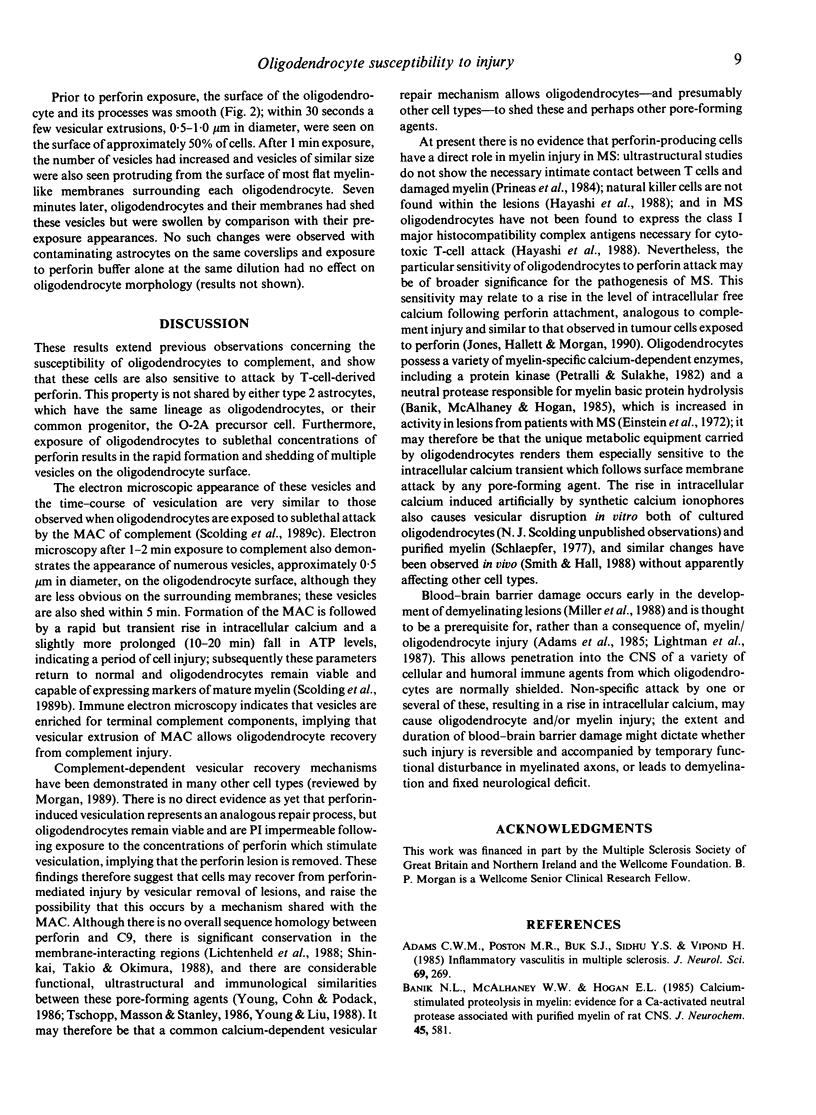
Abstract
Myelin injury in multiple sclerosis (MS) appears to be immune mediated, but the mechanisms remain unidentified. It has been shown previously that oligodendrocytes, which synthesize and maintain myelin in the central nervous system (CNS), are susceptible to attack by homologous complement and that injury may be reversible when lysis is resisted by vesicular removal of membrane attack complexes. Here it is reported that oligodendrocytes are also highly susceptible to attack by T-cell perforin, and respond similarly to sublethal attack by shedding membrane vesicles. These findings imply that oligodendrocytes are vulnerable to lysis and/or reversible injury by a variety of closely related pore-forming immune effectors; following blood-brain barrier disruption, attack by one or several of these molecules may cause transient or irreversible myelin injury. Furthermore, these results provide the first indication that vesicular repair mechanisms may allow nucleated cells to recover from perforin-mediated cell injury.
Full text
PDF


Images in this article

Selected References
These references are in PubMed. This may not be the complete list of references from this article.
- Adams C. W., Poston R. N., Buk S. J., Sidhu Y. S., Vipond H. Inflammatory vasculitis in multiple sclerosis. J Neurol Sci. 1985 Jul;69(3):269–283. doi: 10.1016/0022-510x(85)90139-x. [DOI] [PubMed] [Google Scholar]
- Banik N. L., McAlhaney W. W., Hogan E. L. Calcium-stimulated proteolysis in myelin: evidence for a Ca2+-activated neutral proteinase associated with purified myelin of rat CNS. J Neurochem. 1985 Aug;45(2):581–588. doi: 10.1111/j.1471-4159.1985.tb04026.x. [DOI] [PubMed] [Google Scholar]
- Bignami A., Eng L. F., Dahl D., Uyeda C. T. Localization of the glial fibrillary acidic protein in astrocytes by immunofluorescence. Brain Res. 1972 Aug 25;43(2):429–435. doi: 10.1016/0006-8993(72)90398-8. [DOI] [PubMed] [Google Scholar]
- Bottenstein J. E., Sato G. H. Growth of a rat neuroblastoma cell line in serum-free supplemented medium. Proc Natl Acad Sci U S A. 1979 Jan;76(1):514–517. doi: 10.1073/pnas.76.1.514. [DOI] [PMC free article] [PubMed] [Google Scholar]
- Einstein E. R., Csejtey J., Dalal K. B., Adams C. W., Bayliss O. B., Hallpike J. F. Proteolytic activity and basic protein loss in and around multiple sclerosis plaques: combined biochemical and histochemical observations. J Neurochem. 1972 Mar;19(3):653–662. doi: 10.1111/j.1471-4159.1972.tb01382.x. [DOI] [PubMed] [Google Scholar]
- Eisenbarth G. S., Walsh F. S., Nirenberg M. Monoclonal antibody to a plasma membrane antigen of neurons. Proc Natl Acad Sci U S A. 1979 Oct;76(10):4913–4917. doi: 10.1073/pnas.76.10.4913. [DOI] [PMC free article] [PubMed] [Google Scholar]
- Hayashi T., Morimoto C., Burks J. S., Kerr C., Hauser S. L. Dual-label immunocytochemistry of the active multiple sclerosis lesion: major histocompatibility complex and activation antigens. Ann Neurol. 1988 Oct;24(4):523–531. doi: 10.1002/ana.410240408. [DOI] [PubMed] [Google Scholar]
- Lightman S., McDonald W. I., Bird A. C., Francis D. A., Hoskins A., Batchelor J. R., Halliday A. M. Retinal venous sheathing in optic neuritis. Its significance for the pathogenesis of multiple sclerosis. Brain. 1987 Apr;110(Pt 2):405–414. doi: 10.1093/brain/110.2.405. [DOI] [PubMed] [Google Scholar]
- Miller D. H., Rudge P., Johnson G., Kendall B. E., Macmanus D. G., Moseley I. F., Barnes D., McDonald W. I. Serial gadolinium enhanced magnetic resonance imaging in multiple sclerosis. Brain. 1988 Aug;111(Pt 4):927–939. doi: 10.1093/brain/111.4.927. [DOI] [PubMed] [Google Scholar]
- Morgan B. P. Complement membrane attack on nucleated cells: resistance, recovery and non-lethal effects. Biochem J. 1989 Nov 15;264(1):1–14. doi: 10.1042/bj2640001. [DOI] [PMC free article] [PubMed] [Google Scholar]
- Petrali E. H., Sulakhe P. V. Calcium ion stimulated protein kinases in myelin. Prog Brain Res. 1982;56:125–144. doi: 10.1016/S0079-6123(08)63772-3. [DOI] [PubMed] [Google Scholar]
- Podack E. R., Young J. D., Cohn Z. A. Isolation and biochemical and functional characterization of perforin 1 from cytolytic T-cell granules. Proc Natl Acad Sci U S A. 1985 Dec;82(24):8629–8633. doi: 10.1073/pnas.82.24.8629. [DOI] [PMC free article] [PubMed] [Google Scholar]
- Prineas J. W., Kwon E. E., Cho E. S., Sharer L. R. Continual breakdown and regeneration of myelin in progressive multiple sclerosis plaques. Ann N Y Acad Sci. 1984;436:11–32. doi: 10.1111/j.1749-6632.1984.tb14773.x. [DOI] [PubMed] [Google Scholar]
- Raff M. C., Miller R. H., Noble M. A glial progenitor cell that develops in vitro into an astrocyte or an oligodendrocyte depending on culture medium. Nature. 1983 Jun 2;303(5916):390–396. doi: 10.1038/303390a0. [DOI] [PubMed] [Google Scholar]
- Schlaepfer W. W. Vesicular disruption of myelin simulated by exposure of nerve to calcium ionophore. Nature. 1977 Feb 24;265(5596):734–736. doi: 10.1038/265734a0. [DOI] [PubMed] [Google Scholar]
- Scolding N. J., Frith S., Linington C., Morgan B. P., Campbell A. K., Compston D. A. Myelin-oligodendrocyte glycoprotein (MOG) is a surface marker of oligodendrocyte maturation. J Neuroimmunol. 1989 May;22(3):169–176. doi: 10.1016/0165-5728(89)90014-3. [DOI] [PubMed] [Google Scholar]
- Scolding N. J., Houston W. A., Morgan B. P., Campbell A. K., Compston D. A. Reversible injury of cultured rat oligodendrocytes by complement. Immunology. 1989 Aug;67(4):441–446. [PMC free article] [PubMed] [Google Scholar]
- Scolding N. J., Morgan B. P., Houston A., Campbell A. K., Linington C., Compston D. A. Normal rat serum cytotoxicity against syngeneic oligodendrocytes. Complement activation and attack in the absence of anti-myelin antibodies. J Neurol Sci. 1989 Feb;89(2-3):289–300. doi: 10.1016/0022-510x(89)90030-0. [DOI] [PubMed] [Google Scholar]
- Scolding N. J., Morgan B. P., Houston W. A., Linington C., Campbell A. K., Compston D. A. Vesicular removal by oligodendrocytes of membrane attack complexes formed by activated complement. Nature. 1989 Jun 22;339(6226):620–622. doi: 10.1038/339620a0. [DOI] [PubMed] [Google Scholar]
- Shinkai Y., Takio K., Okumura K. Homology of perforin to the ninth component of complement (C9). Nature. 1988 Aug 11;334(6182):525–527. doi: 10.1038/334525a0. [DOI] [PubMed] [Google Scholar]
- Smith K. J., Hall S. M. Peripheral demyelination and remyelination initiated by the calcium-selective ionophore ionomycin: in vivo observations. J Neurol Sci. 1988 Jan;83(1):37–53. doi: 10.1016/0022-510x(88)90018-4. [DOI] [PubMed] [Google Scholar]
- Tschopp J., Jongeneel C. V. Cytotoxic T lymphocyte mediated cytolysis. Biochemistry. 1988 Apr 19;27(8):2641–2646. doi: 10.1021/bi00408a001. [DOI] [PubMed] [Google Scholar]
- Tschopp J., Masson D., Stanley K. K. Structural/functional similarity between proteins involved in complement- and cytotoxic T-lymphocyte-mediated cytolysis. 1986 Aug 28-Sep 3Nature. 322(6082):831–834. doi: 10.1038/322831a0. [DOI] [PubMed] [Google Scholar]
- Wren D. R., Noble M. Oligodendrocytes and oligodendrocyte/type-2 astrocyte progenitor cells of adult rats are specifically susceptible to the lytic effects of complement in absence of antibody. Proc Natl Acad Sci U S A. 1989 Nov;86(22):9025–9029. doi: 10.1073/pnas.86.22.9025. [DOI] [PMC free article] [PubMed] [Google Scholar]
- Young J. D., Cohn Z. A., Podack E. R. The ninth component of complement and the pore-forming protein (perforin 1) from cytotoxic T cells: structural, immunological, and functional similarities. Science. 1986 Jul 11;233(4760):184–190. doi: 10.1126/science.2425429. [DOI] [PubMed] [Google Scholar]
- Young J. D., Liu C. C. Multiple mechanisms of lymphocyte-mediated killing. Immunol Today. 1988 May;9(5):140–144. doi: 10.1016/0167-5699(88)91201-7. [DOI] [PubMed] [Google Scholar]