Abstract
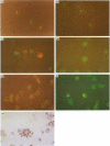
Following contact sensitization of mice there is a rapid accumulation of dendritic cells (DC) within lymph nodes draining the site of exposure. Previous studies have revealed that cells bearing high levels of contact allergen can also be identified within the low buoyant density fraction of draining lymph node cells, and it has been assumed that the majority of these are DC. The purpose of the present study was to establish the phenotypic characteristics of the antigen-bearing cells which appear in lymph nodes within hours of skin painting with contact allergens, including the contact sensitizing fluorochromes fluorescein isothiocyanate (FITC) and rhodamine B isothiocyanate (RITC). Indirect immunofluorescence and two-colour immunofluorescence analyses revealed that initially all antigen-bearing cells which arrive in the draining lymph nodes express class II MHC antigens and exhibit a dendritic morphology. Phagocytic cells, cells reactive with F4/80 and anti-Mac-1 antibodies and lymphoblasts are not associated with detectable levels of antigen. In addition, although Thy-1+ cells co-fractionate with lymph node DC they are not dendritic in nature and are not associated with antigen. These data are compatible with the hypothesis that following skin sensitization epidermal Langerhans' cells bind antigen and transport it to the regional lymph nodes. In addition, it is clear that the recently described population of Thy-1+, Ia- dendritic cells within the murine epidermis do not perform a similar function.
Full text
PDF


Images in this article
Selected References
These references are in PubMed. This may not be the complete list of references from this article.
- Asherson G. L., Mayhew B. Induction of cell-mediated immunity in the mouse: circumstantial evidence for highly immunogenic antigen in the regional lymph nodes following skin painting with contact sensitizing agents. Isr J Med Sci. 1976 Apr-May;12(4-5):454–467. [PubMed] [Google Scholar]
- Austyn J. M., Gordon S. F4/80, a monoclonal antibody directed specifically against the mouse macrophage. Eur J Immunol. 1981 Oct;11(10):805–815. doi: 10.1002/eji.1830111013. [DOI] [PubMed] [Google Scholar]
- Austyn J. M. Lymphoid dendritic cells. Immunology. 1987 Oct;62(2):161–170. [PMC free article] [PubMed] [Google Scholar]
- Bergstresser P. R., Fletcher C. R., Streilein J. W. Surface densities of Langerhans cells in relation to rodent epidermal sites with special immunologic properties. J Invest Dermatol. 1980 Feb;74(2):77–80. doi: 10.1111/1523-1747.ep12519909. [DOI] [PubMed] [Google Scholar]
- Bergstresser P. R., Tigelaar R. E., Dees J. H., Streilein J. W. Thy-1 antigen-bearing dendritic cells populate murine epidermis. J Invest Dermatol. 1983 Sep;81(3):286–288. doi: 10.1111/1523-1747.ep12518332. [DOI] [PubMed] [Google Scholar]
- Bigby M., Kwan T., Sy M. S. Ratio of Langerhans cells to Thy-1+ dendritic epidermal cells in murine epidermis influences the intensity of contact hypersensitivity. J Invest Dermatol. 1987 Nov;89(5):495–499. doi: 10.1111/1523-1747.ep12460983. [DOI] [PubMed] [Google Scholar]
- Chambers D. A. The Thy-1 epidermal cell: perspective and prospective. Br J Dermatol. 1985 Jul;113 (Suppl 28):24–33. doi: 10.1111/j.1365-2133.1985.tb15623.x. [DOI] [PubMed] [Google Scholar]
- Flotte T. J., Springer T. A., Thorbecke G. J. Dendritic cell and macrophage staining by monoclonal antibodies in tissue sections and epidermal sheets. Am J Pathol. 1983 Apr;111(1):112–124. [PMC free article] [PubMed] [Google Scholar]
- Haines K. A., Flotte T. J., Springer T. A., Gigli I., Thorbecke G. J. Staining of Langerhans cells with monoclonal antibodies to macrophages and lymphoid cells. Proc Natl Acad Sci U S A. 1983 Jun;80(11):3448–3451. doi: 10.1073/pnas.80.11.3448. [DOI] [PMC free article] [PubMed] [Google Scholar]
- Heufler C., Koch F., Schuler G. Granulocyte/macrophage colony-stimulating factor and interleukin 1 mediate the maturation of murine epidermal Langerhans cells into potent immunostimulatory dendritic cells. J Exp Med. 1988 Feb 1;167(2):700–705. doi: 10.1084/jem.167.2.700. [DOI] [PMC free article] [PubMed] [Google Scholar]
- Hume D. A., Robinson A. P., MacPherson G. G., Gordon S. The mononuclear phagocyte system of the mouse defined by immunohistochemical localization of antigen F4/80. Relationship between macrophages, Langerhans cells, reticular cells, and dendritic cells in lymphoid and hematopoietic organs. J Exp Med. 1983 Nov 1;158(5):1522–1536. doi: 10.1084/jem.158.5.1522. [DOI] [PMC free article] [PubMed] [Google Scholar]
- Inaba K., Steinman R. M. Resting and sensitized T lymphocytes exhibit distinct stimulatory (antigen-presenting cell) requirements for growth and lymphokine release. J Exp Med. 1984 Dec 1;160(6):1717–1735. doi: 10.1084/jem.160.6.1717. [DOI] [PMC free article] [PubMed] [Google Scholar]
- Jones D. A., Morris A. G., Kimber I. Assessment of the functional activity of antigen-bearing dendritic cells isolated from the lymph nodes of contact-sensitized mice. Int Arch Allergy Appl Immunol. 1989;90(3):230–236. doi: 10.1159/000235030. [DOI] [PubMed] [Google Scholar]
- Katz D. R., Mukherjee S., Maisey J., Miller K. Vitamin A acetate as a regulator of accessory cell function in delayed-type hypersensitivity responses. Int Arch Allergy Appl Immunol. 1987;82(1):53–56. doi: 10.1159/000234289. [DOI] [PubMed] [Google Scholar]
- Kimber I., Pierce B. B., Mitchell J. A., Kinnaird A. Depression of lymph node cell proliferation induced by oxazolone. Int Arch Allergy Appl Immunol. 1987;84(3):256–262. doi: 10.1159/000234432. [DOI] [PubMed] [Google Scholar]
- Kinnaird A., Peters S. W., Foster J. R., Kimber I. Dendritic cell accumulation in draining lymph nodes during the induction phase of contact allergy in mice. Int Arch Allergy Appl Immunol. 1989;89(2-3):202–210. doi: 10.1159/000234947. [DOI] [PubMed] [Google Scholar]
- Knight S. C., Bedford P., Hunt R. The role of dendritic cells in the initiation of immune responses to contact sensitizers. II. Studies in nude mice. Cell Immunol. 1985 Sep;94(2):435–439. doi: 10.1016/0008-8749(85)90267-9. [DOI] [PubMed] [Google Scholar]
- Knight S. C., Krejci J., Malkovsky M., Colizzi V., Gautam A., Asherson G. L. The role of dendritic cells in the initiation of immune responses to contact sensitizers. I. In vivo exposure to antigen. Cell Immunol. 1985 Sep;94(2):427–434. doi: 10.1016/0008-8749(85)90266-7. [DOI] [PubMed] [Google Scholar]
- Knight S. C. Veiled cells--"dendritic cells" of the peripheral lymph. Immunobiology. 1984 Dec;168(3-5):349–361. doi: 10.1016/S0171-2985(84)80122-9. [DOI] [PubMed] [Google Scholar]
- Koch F., Heufler C., Kämpgen E., Schneeweiss D., Böck G., Schuler G. Tumor necrosis factor alpha maintains the viability of murine epidermal Langerhans cells in culture, but in contrast to granulocyte/macrophage colony-stimulating factor, without inducing their functional maturation. J Exp Med. 1990 Jan 1;171(1):159–171. doi: 10.1084/jem.171.1.159. [DOI] [PMC free article] [PubMed] [Google Scholar]
- Kraal G., Breel M., Janse M., Bruin G. Langerhans' cells, veiled cells, and interdigitating cells in the mouse recognized by a monoclonal antibody. J Exp Med. 1986 Apr 1;163(4):981–997. doi: 10.1084/jem.163.4.981. [DOI] [PMC free article] [PubMed] [Google Scholar]
- Macatonia S. E., Edwards A. J., Knight S. C. Dendritic cells and the initiation of contact sensitivity to fluorescein isothiocyanate. Immunology. 1986 Dec;59(4):509–514. [PMC free article] [PubMed] [Google Scholar]
- Macatonia S. E., Knight S. C., Edwards A. J., Griffiths S., Fryer P. Localization of antigen on lymph node dendritic cells after exposure to the contact sensitizer fluorescein isothiocyanate. Functional and morphological studies. J Exp Med. 1987 Dec 1;166(6):1654–1667. doi: 10.1084/jem.166.6.1654. [DOI] [PMC free article] [PubMed] [Google Scholar]
- Mackenzie I. C., Squier C. A. Cytochemical identification of ATPase-positive langerhans cells in EDTA-separated sheets of mouse epidermis. Br J Dermatol. 1975 May;92(5):523–533. doi: 10.1111/j.1365-2133.1975.tb03120.x. [DOI] [PubMed] [Google Scholar]
- Oda T., Maeda H. A new simple fluorometric assay for phagocytosis. J Immunol Methods. 1986 Apr 17;88(2):175–183. doi: 10.1016/0022-1759(86)90004-9. [DOI] [PubMed] [Google Scholar]
- Okamoto H., Kripke M. L. Effector and suppressor circuits of the immune response are activated in vivo by different mechanisms. Proc Natl Acad Sci U S A. 1987 Jun;84(11):3841–3845. doi: 10.1073/pnas.84.11.3841. [DOI] [PMC free article] [PubMed] [Google Scholar]
- Picut C. A., Lee C. S., Dougherty E. P., Anderson K. L., Lewis R. M. Immunostimulatory capabilities of highly enriched Langerhans cells in vitro. J Invest Dermatol. 1988 Feb;90(2):201–206. doi: 10.1111/1523-1747.ep12462221. [DOI] [PubMed] [Google Scholar]
- Schuler G., Steinman R. M. Murine epidermal Langerhans cells mature into potent immunostimulatory dendritic cells in vitro. J Exp Med. 1985 Mar 1;161(3):526–546. doi: 10.1084/jem.161.3.526. [DOI] [PMC free article] [PubMed] [Google Scholar]
- Shelley W. B., Juhlin L. Selective uptake of contact allergens by the Langerhans cell. Arch Dermatol. 1977 Feb;113(2):187–192. [PubMed] [Google Scholar]
- Silberberg-Sinakin I., Thorbecke G. J., Baer R. L., Rosenthal S. A., Berezowsky V. Antigen-bearing langerhans cells in skin, dermal lymphatics and in lymph nodes. Cell Immunol. 1976 Aug;25(2):137–151. doi: 10.1016/0008-8749(76)90105-2. [DOI] [PubMed] [Google Scholar]
- Springer T., Galfré G., Secher D. S., Milstein C. Mac-1: a macrophage differentiation antigen identified by monoclonal antibody. Eur J Immunol. 1979 Apr;9(4):301–306. doi: 10.1002/eji.1830090410. [DOI] [PubMed] [Google Scholar]
- Steinman R. M., Cohn Z. A. Identification of a novel cell type in peripheral lymphoid organs of mice. I. Morphology, quantitation, tissue distribution. J Exp Med. 1973 May 1;137(5):1142–1162. doi: 10.1084/jem.137.5.1142. [DOI] [PMC free article] [PubMed] [Google Scholar]
- Steinman R. M., Nussenzweig M. C. Dendritic cells: features and functions. Immunol Rev. 1980;53:127–147. doi: 10.1111/j.1600-065x.1980.tb01042.x. [DOI] [PubMed] [Google Scholar]
- Stingl G., Wolff-Schreiner E. C., Pichler W. J., Gschnait F., Knapp W., Wolff K. Epidermal Langerhans cells bear Fc and C3 receptors. Nature. 1977 Jul 21;268(5617):245–246. doi: 10.1038/268245a0. [DOI] [PubMed] [Google Scholar]
- Streilein J. W., Bergstresser P. R. Langerhans cell function dictates induction of contact hypersensitivity or unresponsiveness to DNFB in Syrian hamsters. J Invest Dermatol. 1981 Sep;77(3):272–277. doi: 10.1111/1523-1747.ep12482453. [DOI] [PubMed] [Google Scholar]
- Sullivan S., Bergstresser P. R., Tigelaar R. E., Streilein J. W. Induction and regulation of contact hypersensitivity by resident, bone marrow-derived, dendritic epidermal cells: Langerhans cells and Thy-1+ epidermal cells. J Immunol. 1986 Oct 15;137(8):2460–2467. [PubMed] [Google Scholar]
- Toews G. B., Bergstresser P. R., Streilein J. W. Epidermal Langerhans cell density determines whether contact hypersensitivity or unresponsiveness follows skin painting with DNFB. J Immunol. 1980 Jan;124(1):445–453. [PubMed] [Google Scholar]
- Tschachler E., Schuler G., Hutterer J., Leibl H., Wolff K., Stingl G. Expression of Thy-1 antigen by murine epidermal cells. J Invest Dermatol. 1983 Sep;81(3):282–285. doi: 10.1111/1523-1747.ep12518326. [DOI] [PubMed] [Google Scholar]
- Witmer-Pack M. D., Olivier W., Valinsky J., Schuler G., Steinman R. M. Granulocyte/macrophage colony-stimulating factor is essential for the viability and function of cultured murine epidermal Langerhans cells. J Exp Med. 1987 Nov 1;166(5):1484–1498. doi: 10.1084/jem.166.5.1484. [DOI] [PMC free article] [PubMed] [Google Scholar]
- Witmer-Pack M. D., Valinsky J., Olivier W., Steinman R. M. Quantitation of surface antigens on cultured murine epidermal Langerhans cells: rapid and selective increase in the level of surface MHC products. J Invest Dermatol. 1988 Mar;90(3):387–394. doi: 10.1111/1523-1747.ep12456460. [DOI] [PubMed] [Google Scholar]


