

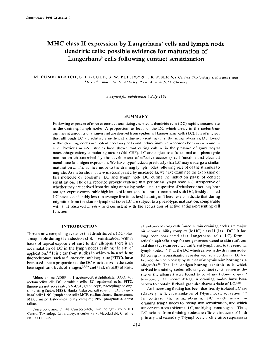
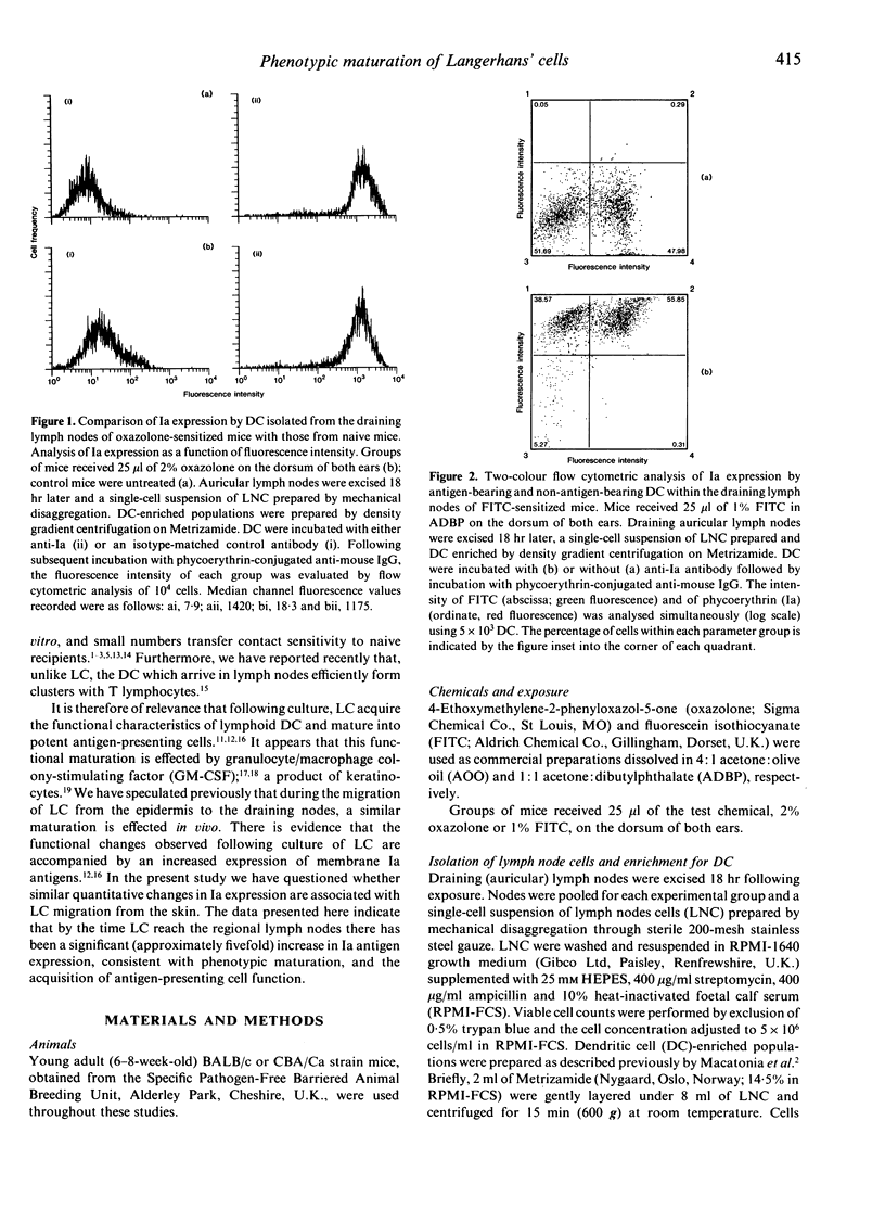
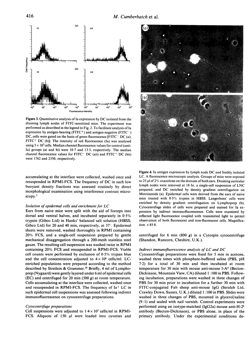
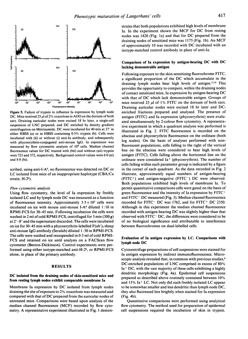
Abstract
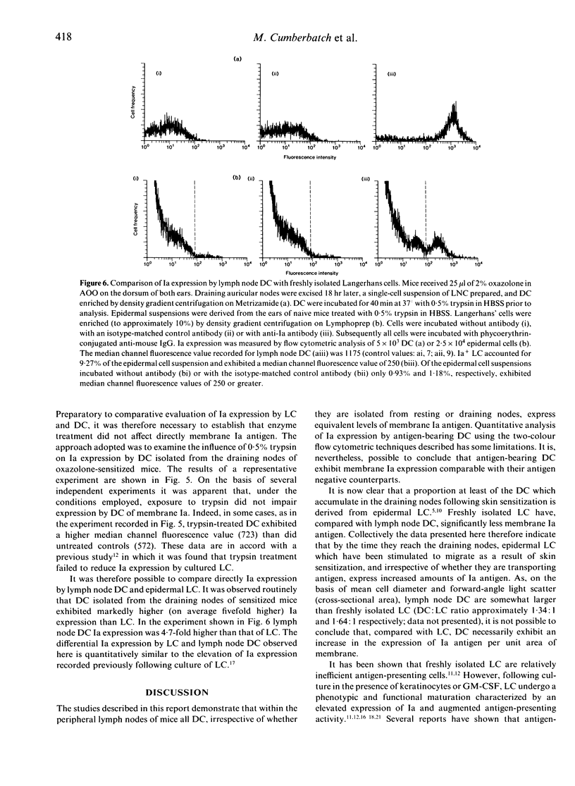
Following exposure of mice to contact sensitizing chemicals, dendritic cells (DC) rapidly accumulate in the draining lymph nodes. A proportion, at least, of the DC which arrive in the nodes bear significant amounts of antigen and are derived from epidermal Langerhans' cells (LC). It is of interest that although LC are relatively inefficient antigen-presenting cells, the antigen-bearing DC found within draining nodes are potent accessory cells and induce immune responses both in vitro and in vivo. Previous in vitro studies have shown that during culture in the presence of granulocyte/macrophage colony-stimulating factor (GM-CSF), LC are subject to a functional and phenotypic maturation characterized by the development of effective accessory cell function and elevated membrane Ia antigen expression. We have hypothesized previously that LC may undergo a similar maturation in vivo as they move to the draining lymph nodes following receipt of the stimulus to migrate. As maturation in vitro is accompanied by increased Ia, we have examined the expression of this molecule on epidermal LC and lymph node DC during the induction phase of contact sensitization. The data reported provide evidence that peripheral lymph node DC, irrespective of whether they are derived from draining or resting nodes, and irrespective of whether or not they bear antigen, express comparable high levels of Ia antigen. In contrast, compared with DC, freshly isolated LC have considerably less (on average five times less) Ia antigen. These results indicate that during migration from the skin to lymphoid tissue LC are subject to a phenotypic maturation, comparable with that observed in vitro, and consistent with the acquisition of active antigen-presenting cell function.
Full text
PDF
Images in this article
Selected References
These references are in PubMed. This may not be the complete list of references from this article.
- Cumberbatch M., Kimber I. Phenotypic characteristics of antigen-bearing cells in the draining lymph nodes of contact sensitized mice. Immunology. 1990 Nov;71(3):404–410. [PMC free article] [PubMed] [Google Scholar]
- Gerberick G. F., Ryan C. A., Fletcher E. R., Howard A. D., Robinson M. K. Increased number of dendritic cells in draining lymph nodes accompanies the generation of contact photosensitivity. J Invest Dermatol. 1991 Mar;96(3):355–361. [PubMed] [Google Scholar]
- Heufler C., Koch F., Schuler G. Granulocyte/macrophage colony-stimulating factor and interleukin 1 mediate the maturation of murine epidermal Langerhans cells into potent immunostimulatory dendritic cells. J Exp Med. 1988 Feb 1;167(2):700–705. doi: 10.1084/jem.167.2.700. [DOI] [PMC free article] [PubMed] [Google Scholar]
- Inaba K., Steinman R. M. Resting and sensitized T lymphocytes exhibit distinct stimulatory (antigen-presenting cell) requirements for growth and lymphokine release. J Exp Med. 1984 Dec 1;160(6):1717–1735. doi: 10.1084/jem.160.6.1717. [DOI] [PMC free article] [PubMed] [Google Scholar]
- Jones D. A., Morris A. G., Kimber I. Assessment of the functional activity of antigen-bearing dendritic cells isolated from the lymph nodes of contact-sensitized mice. Int Arch Allergy Appl Immunol. 1989;90(3):230–236. doi: 10.1159/000235030. [DOI] [PubMed] [Google Scholar]
- Kinnaird A., Peters S. W., Foster J. R., Kimber I. Dendritic cell accumulation in draining lymph nodes during the induction phase of contact allergy in mice. Int Arch Allergy Appl Immunol. 1989;89(2-3):202–210. doi: 10.1159/000234947. [DOI] [PubMed] [Google Scholar]
- Knight S. C., Krejci J., Malkovsky M., Colizzi V., Gautam A., Asherson G. L. The role of dendritic cells in the initiation of immune responses to contact sensitizers. I. In vivo exposure to antigen. Cell Immunol. 1985 Sep;94(2):427–434. doi: 10.1016/0008-8749(85)90266-7. [DOI] [PubMed] [Google Scholar]
- Koide S. L., Inaba K., Steinman R. M. Interleukin 1 enhances T-dependent immune responses by amplifying the function of dendritic cells. J Exp Med. 1987 Feb 1;165(2):515–530. doi: 10.1084/jem.165.2.515. [DOI] [PMC free article] [PubMed] [Google Scholar]
- Kripke M. L., Munn C. G., Jeevan A., Tang J. M., Bucana C. Evidence that cutaneous antigen-presenting cells migrate to regional lymph nodes during contact sensitization. J Immunol. 1990 Nov 1;145(9):2833–2838. [PubMed] [Google Scholar]
- Kupper T. S., Lee F., Coleman D., Chodakewitz J., Flood P., Horowitz M. Keratinocyte derived T-cell growth factor (KTGF) is identical to granulocyte macrophage colony stimulating factor (GM-CSF). J Invest Dermatol. 1988 Aug;91(2):185–188. doi: 10.1111/1523-1747.ep12464470. [DOI] [PubMed] [Google Scholar]
- Macatonia S. E., Edwards A. J., Knight S. C. Dendritic cells and the initiation of contact sensitivity to fluorescein isothiocyanate. Immunology. 1986 Dec;59(4):509–514. [PMC free article] [PubMed] [Google Scholar]
- Macatonia S. E., Knight S. C. Dendritic cells and T cells transfer sensitization for delayed-type hypersensitivity after skin painting with contact sensitizer. Immunology. 1989 Jan;66(1):96–99. [PMC free article] [PubMed] [Google Scholar]
- Macatonia S. E., Knight S. C., Edwards A. J., Griffiths S., Fryer P. Localization of antigen on lymph node dendritic cells after exposure to the contact sensitizer fluorescein isothiocyanate. Functional and morphological studies. J Exp Med. 1987 Dec 1;166(6):1654–1667. doi: 10.1084/jem.166.6.1654. [DOI] [PMC free article] [PubMed] [Google Scholar]
- Picut C. A., Lee C. S., Dougherty E. P., Anderson K. L., Lewis R. M. Immunostimulatory capabilities of highly enriched Langerhans cells in vitro. J Invest Dermatol. 1988 Feb;90(2):201–206. doi: 10.1111/1523-1747.ep12462221. [DOI] [PubMed] [Google Scholar]
- Schuler G., Steinman R. M. Murine epidermal Langerhans cells mature into potent immunostimulatory dendritic cells in vitro. J Exp Med. 1985 Mar 1;161(3):526–546. doi: 10.1084/jem.161.3.526. [DOI] [PMC free article] [PubMed] [Google Scholar]
- Shelley W. B., Juhlin L. Langerhans cells form a reticuloepithelial trap for external contact antigens. Nature. 1976 May 6;261(5555):46–47. doi: 10.1038/261046a0. [DOI] [PubMed] [Google Scholar]
- Shelley W. B., Juhlin L. Selective uptake of contact allergens by the Langerhans cell. Arch Dermatol. 1977 Feb;113(2):187–192. [PubMed] [Google Scholar]
- Shimada S., Caughman S. W., Sharrow S. O., Stephany D., Katz S. I. Enhanced antigen-presenting capacity of cultured Langerhans' cells is associated with markedly increased expression of Ia antigen. J Immunol. 1987 Oct 15;139(8):2551–2555. [PubMed] [Google Scholar]
- Silberberg-Sinakin I., Thorbecke G. J., Baer R. L., Rosenthal S. A., Berezowsky V. Antigen-bearing langerhans cells in skin, dermal lymphatics and in lymph nodes. Cell Immunol. 1976 Aug;25(2):137–151. doi: 10.1016/0008-8749(76)90105-2. [DOI] [PubMed] [Google Scholar]
- Steinman R. M. Cytokines amplify the function of accessory cells. Immunol Lett. 1988 Mar;17(3):197–202. doi: 10.1016/0165-2478(88)90028-4. [DOI] [PubMed] [Google Scholar]
- Streilein J. W., Grammer S. F. In vitro evidence that Langerhans cells can adopt two functionally distinct forms capable of antigen presentation to T lymphocytes. J Immunol. 1989 Dec 15;143(12):3925–3933. [PubMed] [Google Scholar]
- Streilein J. W., Grammer S. F., Yoshikawa T., Demidem A., Vermeer M. Functional dichotomy between Langerhans cells that present antigen to naive and to memory/effector T lymphocytes. Immunol Rev. 1990 Oct;117:159–183. doi: 10.1111/j.1600-065x.1990.tb00572.x. [DOI] [PubMed] [Google Scholar]
- Witmer-Pack M. D., Olivier W., Valinsky J., Schuler G., Steinman R. M. Granulocyte/macrophage colony-stimulating factor is essential for the viability and function of cultured murine epidermal Langerhans cells. J Exp Med. 1987 Nov 1;166(5):1484–1498. doi: 10.1084/jem.166.5.1484. [DOI] [PMC free article] [PubMed] [Google Scholar]