

Abstract
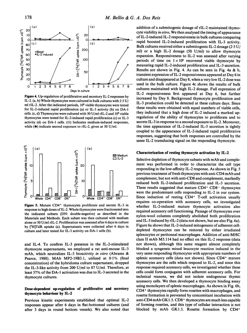
Addition of recombinant interleukin-2 (rIL-2) to normal adult murine thymocytes in vitro as the only exogenous stimulus leads to a dose-dependent mitogenic response characterized by two distinct dosage kinetic components. The high-affinity IL-2 thymocyte response is mounted by in vivo-activated (IL-2 receptor light chain positive) thymocytes, while the low-affinity IL-2 response, of larger amplitude, is carried out by resting thymocytes. Addition of IL-2 to thymocytes also triggers intense IL-3 secretory responses with both high and low IL-2 affinity components. Addition of high IL-2 dosages to thymocyte bulk cultures results in a dramatic increase in IL-2 responsiveness for both proliferation and IL-3 secretion on a per viable cell basis and with tightly coupled temporal kinetics. The low-affinity component of IL-2-proliferative and IL-3-secreting responses is carried out by resting mature CD4+ thymocytes, as assessed by negative selection with monoclonal antibodies (mAb) plus complement. The mechanism of resting thymocyte activation by high doses of IL-2 is partially characterized. Depletion of endogenous thymus-adherent cells abolished both proliferation and IL-3 secretion, and addition of splenic accessory cells or peritoneal macrophages to depleted thymocytes restored IL-2 responsiveness. Mature CD4+ thymocytes spontaneously form rosettes with adherent accessory cells, while CD8+ thymocytes do so with much less efficiency. Rosette formation of CD4+, but not of CD8+ thymocytes, can be blocked by anti-CD4 mAb GK1.5. At the same dosage as it prevents rosette formation, mAb GK1.5 also blocks the low-affinity thymocyte response to IL-2. The high-affinity IL-2 response is completely resistant to the action of cyclosporin A (CsA), but the low-affinity IL-2 response, although of much larger amplitude, can be almost completely suppressed by CsA. Together, these results demonstrate that resting CD4+ thymocytes can be induced to proliferation and lymphokine secretion by IL-2 alone in a process that is dependent on interaction with accessory cells, involves CD4 adhesion molecules and triggers activation through a CsA-sensitive pathway. In addition, the results demonstrate that IL-2 alone is able to enhance thymocyte IL-2 responsiveness and IL-3 secretory responses in vitro. The ability of IL-2 to induce and maintain thymocyte function is discussed in the light of these results.
Full text
PDF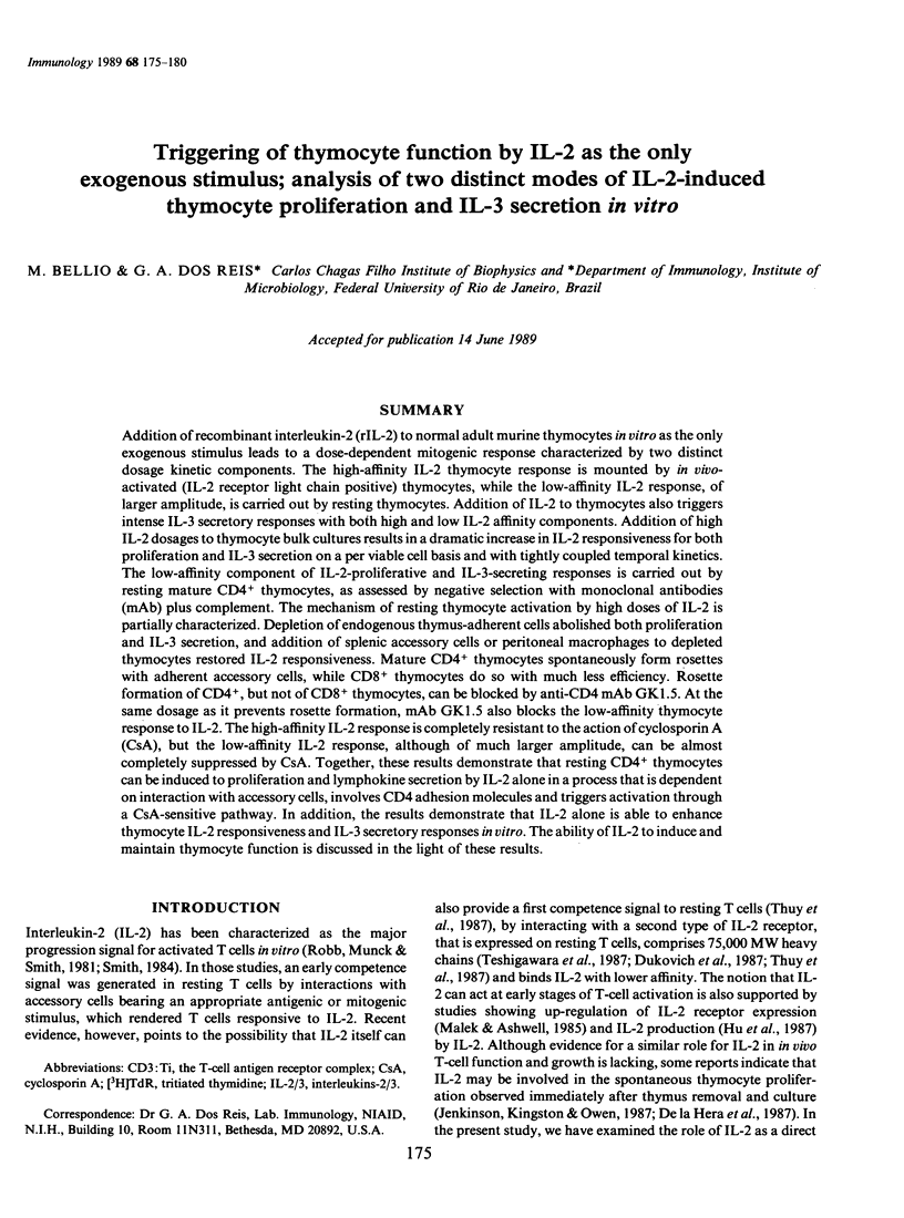


Selected References
These references are in PubMed. This may not be the complete list of references from this article.
- Abrams J. S., Pearce M. K. Development of rat anti-mouse interleukin 3 monoclonal antibodies which neutralize bioactivity in vitro. J Immunol. 1988 Jan 1;140(1):131–137. [PubMed] [Google Scholar]
- Bich-Thuy L. T., Dukovich M., Peffer N. J., Fauci A. S., Kehrl J. H., Greene W. C. Direct activation of human resting T cells by IL 2: the role of an IL 2 receptor distinct from the Tac protein. J Immunol. 1987 Sep 1;139(5):1550–1556. [PubMed] [Google Scholar]
- De la Hera A., Toribio M. L., Marcos M. A., Marquez C., Martinez C. Interleukin 2 pathway is autonomously activated in human T11+3-4-6-8- thymocytes. Eur J Immunol. 1987 May;17(5):683–687. doi: 10.1002/eji.1830170516. [DOI] [PubMed] [Google Scholar]
- Dos Reis G. A., Shevach E. M. Effect of cyclosporin A on T cell function in vitro: the mechanism of suppression of T cell proliferation depends on the nature of the T cell stimulus as well as the differentiation state of the responding T cell. J Immunol. 1982 Dec;129(6):2360–2367. [PubMed] [Google Scholar]
- Dukovich M., Wano Y., Le thi Bich Thuy, Katz P., Cullen B. R., Kehrl J. H., Greene W. C. A second human interleukin-2 binding protein that may be a component of high-affinity interleukin-2 receptors. Nature. 1987 Jun 11;327(6122):518–522. doi: 10.1038/327518a0. [DOI] [PubMed] [Google Scholar]
- Hu J., Vaquero C., Huet S., Bernard A., Sterkers G. Interleukin 2 up-regulates its own production. J Immunol. 1987 Dec 15;139(12):4109–4115. [PubMed] [Google Scholar]
- Jenkinson E. J., Kingston R., Owen J. J. Importance of IL-2 receptors in intra-thymic generation of cells expressing T-cell receptors. Nature. 1987 Sep 10;329(6135):160–162. doi: 10.1038/329160a0. [DOI] [PubMed] [Google Scholar]
- Johnson H. M., Vassallo T., Torres B. A. Interleukin 2-mediated events in gamma-interferon production are calcium dependent at more than one site. J Immunol. 1985 Feb;134(2):967–970. [PubMed] [Google Scholar]
- Julius M. H., Simpson E., Herzenberg L. A. A rapid method for the isolation of functional thymus-derived murine lymphocytes. Eur J Immunol. 1973 Oct;3(10):645–649. doi: 10.1002/eji.1830031011. [DOI] [PubMed] [Google Scholar]
- Karasuyama H., Melchers F. Establishment of mouse cell lines which constitutively secrete large quantities of interleukin 2, 3, 4 or 5, using modified cDNA expression vectors. Eur J Immunol. 1988 Jan;18(1):97–104. doi: 10.1002/eji.1830180115. [DOI] [PubMed] [Google Scholar]
- Malek T. R., Ashwell J. D. Interleukin 2 upregulates expression of its receptor on a T cell clone. J Exp Med. 1985 Jun 1;161(6):1575–1580. doi: 10.1084/jem.161.6.1575. [DOI] [PMC free article] [PubMed] [Google Scholar]
- Malek T. R., Ortega G., Jakway J. P., Chan C., Shevach E. M. The murine IL 2 receptor. II. Monoclonal anti-IL 2 receptor antibodies as specific inhibitors of T cell function in vitro. J Immunol. 1984 Oct;133(4):1976–1982. [PubMed] [Google Scholar]
- Mills G. B., Cheung R. K., Grinstein S., Gelfand E. W. Interleukin 2-induced lymphocyte proliferation is independent of increases in cytosolic-free calcium concentrations. J Immunol. 1985 Apr;134(4):2431–2435. [PubMed] [Google Scholar]
- Papiernik M., Penit C., el Rouby S. Control of prothymocyte proliferation by thymic accessory cells. Eur J Immunol. 1987 Sep;17(9):1303–1310. doi: 10.1002/eji.1830170913. [DOI] [PubMed] [Google Scholar]
- Robb R. J., Munck A., Smith K. A. T cell growth factor receptors. Quantitation, specificity, and biological relevance. J Exp Med. 1981 Nov 1;154(5):1455–1474. doi: 10.1084/jem.154.5.1455. [DOI] [PMC free article] [PubMed] [Google Scholar]
- Smith K. A. Interleukin 2. Annu Rev Immunol. 1984;2:319–333. doi: 10.1146/annurev.iy.02.040184.001535. [DOI] [PubMed] [Google Scholar]
- Teshigawara K., Wang H. M., Kato K., Smith K. A. Interleukin 2 high-affinity receptor expression requires two distinct binding proteins. J Exp Med. 1987 Jan 1;165(1):223–238. doi: 10.1084/jem.165.1.223. [DOI] [PMC free article] [PubMed] [Google Scholar]
- Toribio M. L., Alonso J. M., Bárcena A., Gutiérrez J. C., de la Hera A., Marcos M. A., Márquez C., Martínez C. Human T-cell precursors: involvement of the IL-2 pathway in the generation of mature T cells. Immunol Rev. 1988 Aug;104:55–79. doi: 10.1111/j.1600-065x.1988.tb00759.x. [DOI] [PubMed] [Google Scholar]
- Ythier A. A., Abbud-Filho M., Williams J. M., Loertscher R., Schuster M. W., Nowill A., Hansen J. A., Maltezos D., Strom T. B. Interleukin 2-dependent release of interleukin 3 activity by T4+ human T-cell clones. Proc Natl Acad Sci U S A. 1985 Oct;82(20):7020–7024. doi: 10.1073/pnas.82.20.7020. [DOI] [PMC free article] [PubMed] [Google Scholar]