Abstract
The immunophenotypes of the HLA-DR-positive leucocyte populations in normal human skin were studied using an extensive panel of monoclonal antibodies, which included antibodies from the Third International Leucocyte Differentiation Antigen Workshop (3rd LDAW). Langerhans' cells (LC) in the epidermis stained with antibodies from CD15c, Groups 10, 12a, 12b and 15, of the myeloid panel and from CD39 of the B-cell panel. However, LC did not react with CD14 antibodies or 63D3, which are frequently used to stain tissue macrophages. In addition to epidermal LC (26 cells/linear mm) a significant population of CD1a-positive cells was identified in the papillary dermis (7 cells/linear mm of overlying epidermis). The dermal HLA-DR-positive leucocytes consisted of three cell populations. The most numerous cell type stained with antibodies to monocytes/macrophages. There were fewer, though substantial, numbers of T lymphocytes (mainly CD7-negative) and the least numerous was the population of CD1a-positive cells. The CD1a-positive cells and the population of dermal cells that stain with monocyte/macrophage markers are both potential antigen-presenting cells for the skin-associated immune system.
Full text
PDF


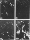
Images in this article
Selected References
These references are in PubMed. This may not be the complete list of references from this article.
- Ashmun R. A., Peiper S. C., Rebentisch M. B., Look A. T. Expression of the human monocyte membrane antigen gp55 by murine fibroblasts after DNA-mediated gene transfer. Blood. 1987 Mar;69(3):886–892. [PubMed] [Google Scholar]
- Bos J. D., Zonneveld I., Das P. K., Krieg S. R., van der Loos C. M., Kapsenberg M. L. The skin immune system (SIS): distribution and immunophenotype of lymphocyte subpopulations in normal human skin. J Invest Dermatol. 1987 May;88(5):569–573. doi: 10.1111/1523-1747.ep12470172. [DOI] [PubMed] [Google Scholar]
- Boyd A. W. Human leukocyte antigens: an update on structure, function and nomenclature. Pathology. 1987 Oct;19(4):329–337. doi: 10.3109/00313028709103879. [DOI] [PubMed] [Google Scholar]
- Drexhage H. A., Mullink H., de Groot J., Clarke J., Balfour B. M. A study of cells present in peripheral lymph of pigs with special reference to a type of cell resembling the Langerhans cell. Cell Tissue Res. 1979 Nov;202(3):407–430. doi: 10.1007/BF00220434. [DOI] [PubMed] [Google Scholar]
- Groh V., Tani M., Harrer A., Wolff K., Stingl G. Leu-3/T4 expression on epidermal Langerhans cells in normal and diseased skin. J Invest Dermatol. 1986 Feb;86(2):115–120. doi: 10.1111/1523-1747.ep12284090. [DOI] [PubMed] [Google Scholar]
- Harrist T. J., Muhlbauer J. E., Murphy G. F., Mihm M. C., Jr, Bhan A. K. T6 is superior to Ia (HLA-DR) as a marker for Langerhans cells and indeterminate cells in normal epidermis: a monoclonal antibody study. J Invest Dermatol. 1983 Feb;80(2):100–103. doi: 10.1111/1523-1747.ep12531695. [DOI] [PubMed] [Google Scholar]
- Hart D. N., McKenzie J. L. Isolation and characterization of human tonsil dendritic cells. J Exp Med. 1988 Jul 1;168(1):157–170. doi: 10.1084/jem.168.1.157. [DOI] [PMC free article] [PubMed] [Google Scholar]
- Hirschberg H., Braathen L. R., Thorsby E. Antigen presentation by vascular endothelial cells and epidermal Langerhans cells: the role of HLA-DR. Immunol Rev. 1982;66:57–77. doi: 10.1111/j.1600-065x.1982.tb00434.x. [DOI] [PubMed] [Google Scholar]
- Hogg N., MacDonald S., Slusarenko M., Beverley P. C. Monoclonal antibodies specific for human monocytes, granulocytes and endothelium. Immunology. 1984 Dec;53(4):753–767. [PMC free article] [PubMed] [Google Scholar]
- Hogg N., Takacs L., Palmer D. G., Selvendran Y., Allen C. The p150,95 molecule is a marker of human mononuclear phagocytes: comparison with expression of class II molecules. Eur J Immunol. 1986 Mar;16(3):240–248. doi: 10.1002/eji.1830160306. [DOI] [PubMed] [Google Scholar]
- Kaplan G., Nusrat A., Witmer M. D., Nath I., Cohn Z. A. Distribution and turnover of Langerhans cells during delayed immune responses in human skin. J Exp Med. 1987 Mar 1;165(3):763–776. doi: 10.1084/jem.165.3.763. [DOI] [PMC free article] [PubMed] [Google Scholar]
- Katz S. I., Tamaki K., Sachs D. H. Epidermal Langerhans cells are derived from cells originating in bone marrow. Nature. 1979 Nov 15;282(5736):324–326. doi: 10.1038/282324a0. [DOI] [PubMed] [Google Scholar]
- Lanier L. L., Engleman E. G., Gatenby P., Babcock G. F., Warner N. L., Herzenberg L. A. Correlation of functional properties of human lymphoid cell subsets and surface marker phenotypes using multiparameter analysis and flow cytometry. Immunol Rev. 1983;74:143–160. doi: 10.1111/j.1600-065x.1983.tb01088.x. [DOI] [PubMed] [Google Scholar]
- Link M., Warnke R., Finlay J., Amylon M., Miller R., Dilley J., Levy R. A single monoclonal antibody identifies T-cell lineage of childhood lymphoid malignancies. Blood. 1983 Oct;62(4):722–728. [PubMed] [Google Scholar]
- Liu H. H., Schroeter A. L., Muller S. A. OKT6 is not superior to HLA-DR or ATPase as a marker for Langerhans' cells in normal human epidermis. Clin Exp Dermatol. 1986 May;11(3):229–237. doi: 10.1111/j.1365-2230.1986.tb00453.x. [DOI] [PubMed] [Google Scholar]
- McMichael A. J., Pilch J. R., Galfré G., Mason D. Y., Fabre J. W., Milstein C. A human thymocyte antigen defined by a hybrid myeloma monoclonal antibody. Eur J Immunol. 1979 Mar;9(3):205–210. doi: 10.1002/eji.1830090307. [DOI] [PubMed] [Google Scholar]
- Murphy G. F., Bhan A. K., Harrist T. J., Mihm M. C., Jr In situ identification of T6-positive cells in normal human dermis by immunoelectron microscopy. Br J Dermatol. 1983 Apr;108(4):423–431. doi: 10.1111/j.1365-2133.1983.tb04594.x. [DOI] [PubMed] [Google Scholar]
- Murphy G. F., Bhan A. K., Sato S., Harrist T. J., Mihm M. C., Jr Characterization of Langerhans cells by the use of monoclonal antibodies. Lab Invest. 1981 Nov;45(5):465–468. [PubMed] [Google Scholar]
- Poulter L. W., Campbell D. A., Munro C., Janossy G. Discrimination of human macrophages and dendritic cells by means of monoclonal antibodies. Scand J Immunol. 1986 Sep;24(3):351–357. doi: 10.1111/j.1365-3083.1986.tb02104.x. [DOI] [PubMed] [Google Scholar]
- Rowden G., Lewis M. G., Sullivan A. K. Ia antigen expression on human epidermal Langerhans cells. Nature. 1977 Jul 21;268(5617):247–248. doi: 10.1038/268247a0. [DOI] [PubMed] [Google Scholar]
- Schmitt D., Faure M., Dambuyant-Dezutter C., Thivolet J. The semi-quantitative distribution of T4 and T6 surface antigens on human Langerhans cells. Br J Dermatol. 1984 Dec;111(6):655–661. doi: 10.1111/j.1365-2133.1984.tb14148.x. [DOI] [PubMed] [Google Scholar]
- Silberberg-Sinakin I., Baer R. L., Thorbecke G. J. Langerhans cells: a review of their nature with emphasis on their immunologic functions. Prog Allergy. 1978;24:268–294. [PubMed] [Google Scholar]
- Silberberg-Sinakin I., Thorbecke G. J., Baer R. L., Rosenthal S. A., Berezowsky V. Antigen-bearing langerhans cells in skin, dermal lymphatics and in lymph nodes. Cell Immunol. 1976 Aug;25(2):137–151. doi: 10.1016/0008-8749(76)90105-2. [DOI] [PubMed] [Google Scholar]
- Sontheimer R. D., Stastny P., Nũnez G. HLA-D region antigen expression by human epidermal Langerhans cells. J Invest Dermatol. 1986 Dec;87(6):707–710. doi: 10.1111/1523-1747.ep12456670. [DOI] [PubMed] [Google Scholar]
- Starling G. C., Davidson S. E., McKenzie J. L., Hart D. N. Inhibition of natural killer-cell mediated cytolysis with monoclonal antibodies to restricted and non-restricted epitopes of the leucocyte common antigen. Immunology. 1987 Jul;61(3):351–356. [PMC free article] [PubMed] [Google Scholar]
- Stingl G., Wolff-Schreiner E. C., Pichler W. J., Gschnait F., Knapp W., Wolff K. Epidermal Langerhans cells bear Fc and C3 receptors. Nature. 1977 Jul 21;268(5617):245–246. doi: 10.1038/268245a0. [DOI] [PubMed] [Google Scholar]
- Turbitt M. L., Mackie R. M. An assessment of the diagnostic value of the monoclonal antibodies Leu 8, OKT9, OKT10 and Ki67 in cutaneous lymphocytic infiltrates. Br J Dermatol. 1986 Aug;115(2):151–158. doi: 10.1111/j.1365-2133.1986.tb05711.x. [DOI] [PubMed] [Google Scholar]
- Ugolini V., Nunez G., Smith R. G., Stastny P., Capra J. D. Initial characterization of monoclonal antibodies against human monocytes. Proc Natl Acad Sci U S A. 1980 Nov;77(11):6764–6768. doi: 10.1073/pnas.77.11.6764. [DOI] [PMC free article] [PubMed] [Google Scholar]
- Werdelin O. Determinant protection. A hypothesis for the activity of immune response genes in the processing and presentation of antigens by macrophages. Scand J Immunol. 1986 Dec;24(6):625–636. doi: 10.1111/j.1365-3083.1986.tb02181.x. [DOI] [PubMed] [Google Scholar]
- Wood G. S., Abel E. A., Hoppe R. T., Warnke R. A. Leu-8 and Leu-9 antigen phenotypes: immunologic criteria for the distinction of mycosis fungoides from cutaneous inflammation. J Am Acad Dermatol. 1986 Jun;14(6):1006–1013. doi: 10.1016/s0190-9622(86)70124-2. [DOI] [PubMed] [Google Scholar]
- Wood G. S., Warner N. L., Warnke R. A. Anti-Leu-3/T4 antibodies react with cells of monocyte/macrophage and Langerhans lineage. J Immunol. 1983 Jul;131(1):212–216. [PubMed] [Google Scholar]
- Zelickson A. S. Langerhans cells in normal human skin capillaries. J Am Acad Dermatol. 1979 Sep;1(3):265–266. doi: 10.1016/s0190-9622(79)80006-7. [DOI] [PubMed] [Google Scholar]


