Abstract
Radial patterning in the urinary tract and gut depends on reciprocal signaling between epithelial cells, which form mucosa, and mesenchyme, which forms smooth muscle and connective tissue. These interactions depend on sonic hedgehog (Shh), which is secreted by epithelial cells and induces expression of bone morphogenetic protein 4 (Bmp4), a signaling molecule required for differentiation of smooth muscle progenitors. Patterning of the specialized mucosa lining the anterior-posterior (A-P) axis may be controlled independently by regionally expressed mesenchymal transcription factors. A study by Airik et al. in this issue of the JCI reveals that T-box 18 (Tbx18), a transcription factor selectively expressed in ureteral mesenchyme, regulates smooth muscle differentiation by maintaining Shh1 responsiveness in mesenchymal progenitors. Deletion of Tbx18 resulted in defective urothelial differentiation at the level of the ureter, suggesting that Tbx18 acts via mesenchyme as an important regulator of A-P patterning in the urinary tract.
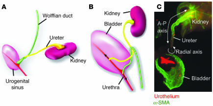
Efficient urine transport is crucial for renal function, preventing damage induced by buildup of urine in the kidney. Urinary tract defects that impede urine transport occur in 1–2% of the human population and can cause obstruction, renal scarring, and end-stage renal disease (1–3). Urine is normally transported from the kidney to the bladder via the renal pelvis and ureters, which are lined with a water-resistant transitional epithelium, the urothelium, and are surrounded by a muscular coat that mediates myogenic peristalsis (Figure 1).
Figure 1.
The embryonic urinary tract. (A and B) Schematic representations of the mouse urinary tract at E11.5 (A) and E15 (B). (C) Whole-mount urinary tract from an E15 mouse stained with uroplakin (red), a marker of the urothelium, and α-SMA (green), a marker of smooth muscle. The A-P and radial axes are shown with white arrows. Note that urothelial differentiation occurs in renal pelvis, ureters, and bladder, but not in the kidney, which is lined with a distinct mucosa. Magnification, ×10.
The kidneys and ureters are derived from the ureteric bud, a posterior branch of the Wolffian ducts, paired epithelial tubes that at late developmental stages differentiate into the vas deferens, seminal vesicles, and epididymis in males and regress in females. The ureteric bud tip invades kidney mesenchyme and undergoes successive rounds of branching morphogenesis that give rise to the renal collecting duct system. The posterior ureteric bud stalk differentiates into the ureter, a muscular tube that conducts urine from the kidney to the bladder (Figure 1). The bladder and urethra form from the cloaca, an endodermal pouch that is partitioned into the hindgut dorsally and the urogenital sinus ventrally. Initially the ureters drain into the Wolffian ducts and hence are not connected directly with the bladder. Formation of mature connections occurs prior to the onset of renal function, when the ureter orifice is transposed from the Wolffian duct to the urogenital sinus. Subsequent growth and expansion of the bladder then shifts the position of the ureter orifice anterior to its final insertion site in the bladder neck.
Mouse models of physical obstruction
Recently, mouse models have been generated that display various types of obstruction, enabling us to begin to understand how urinary tract defects occur in humans. For example, deletion of the genes Sprouty1, Slit2, or Foxc1/Foxc2 or maternal exposure to retinoic acid results in obstruction due to formation of supernumerary ureters that join the lower urinary tract abnormally (4–7). The configuration of these duplicated systems is typical to that in humans: Ureters that emerge at the proper site on the Wolffian duct close to the urogenital sinus generally undergo normal transposition, joining the bladder neck, while branches that form at abnormally high positions on the Wolffian duct fail to undergo transposition, joining the lower urinary tract outside the normal insertion site, for example in the genital tract or urethra (ref. 8; reviewed in ref. 9). Once renal function begins, “plumbing” problems linked to malpositioned ureters can disrupt urine transport and cause severe injury to the kidney. Proper differentiation of the specialized cell types that line the ureter is also crucial for efficient urine transport. Abnormalities in urothelial differentiation can cause physical or functional obstruction. Ureteral valves, an example of physical obstruction, are associated with overgrowth of the transitional epithelium that occludes the ureteral lumen, blocking urine transport (10). A similar phenotype was observed in mouse mutants lacking uroplakins, a family of proteins that form the hexagonal plaques lining the apical surface of the urothelium (11). Deletion of uroplakin 2 (Upk2) or Upk3 results in hyperplasia of the ureteral epithelium that effectively blocks urine flow, resulting in obstruction and, in some cases, renal failure (12, 13).
Mouse models of functional obstruction
Functional obstruction — defective urine transport in the absence of physical blockage — may be linked to defects in the ureter smooth muscle coat that interfere with peristalsis or result in inadequate structural integrity (14, 15). Recent studies suggest that ureteral smooth muscle differentiation depends on sonic hedgehog (Shh) and bone morphogenetic protein 4 (Bmp4) (14, 16, 17). Conditional inactivation of Shh in urinary tract epithelia results in renal hypoplasia, hydroureter, and hydronephrosis. Hydronephrosis and hydroureter were likely to be due to defective differentiation of periureteral mesenchyme (14). These studies further suggest that Shh acts in part by regulating proliferation of mesenchymal smooth muscle progenitors and in part by inducing expression of Bmp4 in ureteral mesenchyme, which is necessary for recruitment of periureteral mesenchyme and for its differentiation into smooth muscle (16, 17). A similar Shh- and Bmp4-dependent pathway operates in the gut, establishing basic epithelial and mesenchymal patterning along the radial axis (18, 19).
Tbx18, a mesenchymal transcription factor that controls radial and anterior-posterior patterning in the urinary tract
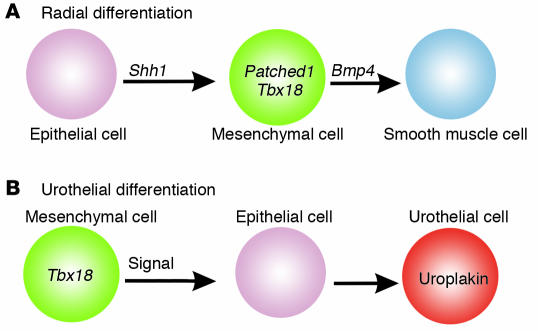
A study in this issue of the JCI by Airik et al. (20) suggests that T-box 18 (Tbx18), a T-box transcription factor selectively expressed in ureteral mesenchyme, is important for both radial and anterior-posterior (A-P) patterning, inducing terminal differentiation of the ureteral epithelium. Mouse mutants lacking Tbx18 displayed hydronephrosis and hydroureter without physical blockage and are hence to my knowledge a new model of functional obstruction. Histological analysis of mutant ureters reveals defects in both muscle and urothelial differentiation. In wild-type embryos, ureteral mesenchyme aggregated around the ureter and differentiated into the circular and longitudinal smooth muscle coat. Deletion of Tbx18 resulted in deficiency of smooth muscle; ureteral mesenchyme failed to condense around the ureter and was instead mislocalized to the surface of the kidney, forming a fibrous outer layer. Analysis of gene expression revealed that 2 Shh targets, Bmp4 and Patched1, were downregulated in the mesenchyme of Tbx18 mutants, suggesting that Tbx18 is likely to be important for controlling the response to hedgehog signaling in mesenchyme and for expression of Bmp4, an important mediator of smooth muscle differentiation (Figure 2A). Establishment of radial patterning in the gut is also controlled by an Shh- and Bmp4-dependent signaling pathway; however, further differentiation of specialized epithelia that line the A-P axis probably depends on regionally expressed mesenchymal transcription factors. For example, differentiation of the mucosal lining of the stomach requires BarH-like homeobox 1 (Barx1), a transcription factor that is selectively expressed in stomach mesenchyme (21).
Figure 2.
A model of Tbx18 action in radial patterning and urothelial differentiation. (A) Tbx18 is selectively expressed in ureteral mesenchyme, where it is required for smooth muscle differentiation and for differentiation of the urothelium, the specialized mucosa that provides barrier function. Tbx18 controls smooth muscle differentiation by regulating Shh responsiveness in subepithelial mesenchyme (3). Shh secreted from ureteral epithelia induces expression of Patched1 and Bmp4, a signaling molecule critical for recruitment of ureteral mesenchyme and for its differentiation into smooth muscle. (B) Tbx18 is selectively expressed in ureteral mesenchyme and is important for differentiation of the urothelium lining the ureters, but not the urothelium lining the bladder and urethra.
Differentiation of epithelia lining the renal collecting ducts, bladder, and urethra was apparently normal in Tbx18 mutants; however, differentiation of the mucosa lining the ureter was blocked, the ureteral epithelium remained undifferentiated (20), and uroplakins, markers of urothelial differentiation (22), were not expressed (20). The observation that expression of the Wnt inhibitor Sfrp2 was downregulated in mutant mesenchyme raises the intriguing possibility that Tbx18 may act in a manner similar to Barx1 in the stomach, inducing expression of signals that control urothelial patterning by suppressing Wnt signaling (Figure 2B). Whether this is or is not the case awaits further investigation to define the role of Wnt signaling during ureter differentiation and the requirement for Sfrp2 in urothelial morphogenesis. That epithelial differentiation in the urinary tract is controlled by signals from mesenchyme is well established; however, the identity of these mesenchymal signals has been unknown (23). The studies reported here by Airik et al. (20) suggest that Tbx18 may be such a mesenchymal signal, controlling ureter differentiation. Whether other transcription factors (or even Tbx family members) control patterning of specialized epithelia at other sites in the urinary tract is an interesting possibility.
Footnotes
See the related article beginning on page 663.
Nonstandard abbreviations used: A-P, anterior-posterior; Barx1, BarH-like homeobox 1; Bmp4, bone morphogenetic protein 4; Shh, sonic hedgehog; Tbx18, T-box 18; Upk, uroplakin.
Conflict of interest: The author has declared that no conflict of interest exists.
References
- 1.Chevalier RL. Obstructive nephropathy and the developing kidney: too little or too much angiotensin? Kidney Int. 2004;65:1517–1518. doi: 10.1111/j.1523-1755.2004.00533.x. [DOI] [PubMed] [Google Scholar]
- 2.Chevalier RL. Pathophysiology of obstructive nephropathy in the newborn. Semin. Nephrol. 1998;18:585–593. [PubMed] [Google Scholar]
- 3.Scott JE, Renwick M. Antenatal diagnosis of congenital abnormalities in the urinary tract. Results from the Northern Region Fetal Abnormality Survey. Br. J. Urol. 1988;62:295–300. doi: 10.1111/j.1464-410x.1988.tb04351.x. [DOI] [PubMed] [Google Scholar]
- 4.Kume T, Deng K, Hogan BL. Murine forkhead/winged helix genes Foxc1 (Mf1) and Foxc2 (Mfh1) are required for the early organogenesis of the kidney and urinary tract. Development. 2000;127:1387–1395. doi: 10.1242/dev.127.7.1387. [DOI] [PubMed] [Google Scholar]
- 5.Grieshammer U, et al. SLIT2-mediated ROBO2 signaling restricts kidney induction to a single site. Dev. Cell. 2004;6:709–717. doi: 10.1016/s1534-5807(04)00108-x. [DOI] [PubMed] [Google Scholar]
- 6.Basson MA, et al. Sprouty1 is a critical regulator of GDNF/RET-mediated kidney induction. Dev. Cell. 2005;8:229–239. doi: 10.1016/j.devcel.2004.12.004. [DOI] [PubMed] [Google Scholar]
- 7.Batourina E, et al. Apoptosis induced by vitamin A signaling is crucial for connecting the ureters to the bladder. Nat. Genet. 2005;37:1082–1089. doi: 10.1038/ng1645. [DOI] [PubMed] [Google Scholar]
- 8.Mackie GG, Stephens FD. Duplex kidneys: a correlation of renal dysplasia with position of the ureteral orifice. J. Urol. 1975;114:274–280. doi: 10.1016/s0022-5347(17)67007-1. [DOI] [PubMed] [Google Scholar]
- 9.Ichikawa I, Kuwayama F, Pope JC, Stephens FD, Miyazaki Y. Paradigm shift from classic anatomic theories to contemporary cell biological views of CAKUT. Kidney Int. 2002;61:889–898. doi: 10.1046/j.1523-1755.2002.00188.x. [DOI] [PubMed] [Google Scholar]
- 10.Wall B, Wachter E. Congenital ureteral valve: its role as a primary obstructive lesion: classification of the literature and report of an authentic case. J. Urol. 1952;68:684–690. doi: 10.1016/S0022-5347(17)68266-1. [DOI] [PubMed] [Google Scholar]
- 11.Wu XR, et al. Mammalian uroplakins. A group of highly conserved urothelial differentiation-related membrane proteins. J. Biol. Chem. 1994;269:13716–13724. [PubMed] [Google Scholar]
- 12.Kong XT, et al. Roles of uroplakins in plaque formation, umbrella cell enlargement, and urinary tract diseases. J. Cell Biol. 2004;167:1195–1204. doi: 10.1083/jcb.200406025. [DOI] [PMC free article] [PubMed] [Google Scholar]
- 13.Hu P, et al. Ablation of uroplakin III gene results in small urothelial plaques, urothelial leakage, and vesicoureteral reflux. Urology. 2001;57:117. doi: 10.1016/s0090-4295(01)01062-7. [DOI] [PubMed] [Google Scholar]
- 14.Yu J, Carroll TJ, McMahon AP. Sonic hedgehog regulates proliferation and differentiation of mesenchymal cells in the mouse metanephric kidney. Development. 2002;129:5301–5312. doi: 10.1242/dev.129.22.5301. [DOI] [PubMed] [Google Scholar]
- 15.Chang CP, et al. Calcineurin is required in urinary tract mesenchyme for the development of the pyeloureteral peristaltic machinery. J. Clin. Invest. 2004;113:1051–1058. doi:10.1172/JCI200420049. doi: 10.1172/JCI20049. [DOI] [PMC free article] [PubMed] [Google Scholar]
- 16.Miyazaki Y, Oshima K, Fogo A, Hogan BL, Ichikawa I. Bone morphogenetic protein 4 regulates the budding site and elongation of the mouse ureter. J. Clin. Invest. 2000;105:863–873. doi: 10.1172/JCI8256. [DOI] [PMC free article] [PubMed] [Google Scholar]
- 17.Raatikainen-Ahokas A, Hytonen M, Tenhunen A, Sainio K, Sariola H. BMP-4 affects the differentiation of metanephric mesenchyme and reveals an early anterior-posterior axis of the embryonic kidney. Dev. Dyn. 2000;217:146–158. doi: 10.1002/(SICI)1097-0177(200002)217:2<146::AID-DVDY2>3.0.CO;2-I. [DOI] [PubMed] [Google Scholar]
- 18.Sukegawa A, et al. The concentric structure of the developing gut is regulated by Sonic hedgehog derived from endodermal epithelium. Development. 2000;127:1971–1980. doi: 10.1242/dev.127.9.1971. [DOI] [PubMed] [Google Scholar]
- 19.Roberts DJ, Smith DM, Goff DJ, Tabin CJ. Epithelial-mesenchymal signaling during the regionalization of the chick gut. Development. 1998;125:2791–2801. doi: 10.1242/dev.125.15.2791. [DOI] [PubMed] [Google Scholar]
- 20.Airik R, Bussen M, Singh MK, Petry M, Kispert A. Tbx18 regulates the development of the ureteral mesenchyme. J. Clin. Invest. 2006;116:663–674. doi:10.1172/JCI26027. doi: 10.1172/JCI26027. [DOI] [PMC free article] [PubMed] [Google Scholar]
- 21.Kim BM, Buchner G, Miletich I, Sharpe PT, Shivdasani RA. The stomach mesenchymal transcription factor Barx1 specifies gastric epithelial identity through inhibition of transient Wnt signaling. Dev. Cell. 2005;8:611–622. doi: 10.1016/j.devcel.2005.01.015. [DOI] [PubMed] [Google Scholar]
- 22.Liang FX, et al. Cellular basis of urothelial squamous metaplasia: roles of lineage heterogeneity and cell replacement. J. Cell Biol. 2005;171:835–844. doi: 10.1083/jcb.200505035. [DOI] [PMC free article] [PubMed] [Google Scholar]
- 23.Lipschutz JH, Young P, Taguchi O, Cunha GR. Urothelial transformation into functional glandular tissue in situ by instructive mesenchymal induction. Kidney Int. 1996;49:59–66. doi: 10.1038/ki.1996.8. [DOI] [PubMed] [Google Scholar]


