Abstract
A dual-antigen enzyme-linked immunosorbent assay specific for human immunodeficiency virus type 2 (HIV-2) envelope proteins, ELISA-HIV2, was developed with two new recombinant polypeptides, rpC2-C3 and rgp36, derived from the HIV-2 envelope. The diagnostic performance was determined with HIV-2, HIV-1, and HIV-1/2 samples. Both polypeptides showed 100% specificity. Clinical sensitivity was 100% for rgp36 and 93.4% for rpC2-C3. ELISA-HIV2 may be used for the specific diagnosis and confirmation of HIV-2 infection.
Human immunodeficiency virus type 2 (HIV-2), the second AIDS virus isolated from west African patients in 1985 (8), is now present in all continents (20, 21, 23). The highest prevalence of HIV-2 in west Africa is found in Guinea-Bissau, where prevalence rates of between 5 and 10% of the adult urban population have been reported (39). The highest prevalence of HIV-2 outside west Africa is found in Portugal, where a prevalence rate of 3.4% has been reported among AIDS cases (9).
Six immunogenic regions were identified in the HIV-2 envelope glycoproteins: three in gp125 (amino acids 234 to 248 in C2, 296 to 337 in V3, and 472 to 507 in C5) and three in the gp36 ectodomain (amino acids 573 to 595, 634 to 649, and 644 to 658) (11, 13, 18, 27, 30, 34, 40, 50). The gp36 ectodomain is highly conserved and elicits a type-specific antibody response (13, 33). Hence, most licensed diagnostic assays incorporate gp36-derived antigens to detect HIV-2-specific antibodies (1, 4, 12, 28, 29, 38, 42, 45, 48). The sensitivity of these assays to detect HIV-2 seroconversions has not been formally tested. However, the sensitivity of several fourth-generation HIV1/2 assays was low with diluted HIV-2-positive samples (29), suggesting that some screening assays may not detect low levels of HIV-2 antibodies (32). The reduced sensitivity of these kits may be caused by inappropriate antigen selection and/or reduced antibody levels in the HIV-2 patients (19, 26, 45).
It is important to differentiate between single infection with either HIV-1 or HIV-2 and dual infection. Dual HIV-1 and HIV-2 seroreactivity is relatively frequent in countries where both HIV-1 and HIV-2 are endemic, such as Portugal (1.4%), Guinea-Bissau (0.7%), Senegal (0.4%), and India (up to 2%) (9, 17, 24, 35). However, the true rate of dual infections in these countries is generally unknown. This is in part due to the lack of sensitive and specific HIV-2 antibody tests. In fact, only two enzyme-linked immunosorbent assays (ELISAs) of low specificity (92%) are currently available for the diagnosis of HIV-2 infection, both of which use the same viral lysate antigen (2, 7). Most often, reactivity with gp36- or gp125-derived antigens (peptides or recombinant proteins) incorporated into Western blot (WB) and immunoblot assays is used to distinguish between HIV-2 and HIV-1 infections (41). However, the sensitivity of these tests is generally low, and serological cross-reactivity between the HIV-1 and HIV-2 Env glycoproteins has been described, which may complicate the final diagnosis (10, 37, 49).
In this study, we produced a new HIV-2 ELISA (ELISA-HIV2) using two new recombinant proteins, rgp36 and rpC2-C3, derived from the reference primary isolate HIV-2ALI (44). Using pSK7.3 plasmid as a template, which contains the HIV-2ALI env gene (44), a PCR was performed with primers Hepit 11 (5′-TTTAGATACTGTGCACC-3′) and Hepit 12 (5′-TTAGTCCACATATATAC-3′) to obtain a C2-C3 env fragment with 497 bp (positions 661 to 1157 in HIV-2 ALI env). The thermal cycling conditions were as follows: denaturation at 94°C for 1 min, annealing 60°C for 1 min, and extension at 72°C for 1 min for 45 cycles. Another PCR was performed with primers Hepit 15 (5′-GGCACGGCAGCTTTAACGC-3′) and Hepit 17 (5′-GTCCCTGCAGTTATTTTTGTAGTTCATATG-3′) to obtain a gp36 fragment with 385 bp (positions 1578 to 1963 in HIV-2ALI env). The thermal cycling conditions were as follows: denaturation at 94°C for 1 min, annealing at 65°C for 1 min, and extension at 72°C for 1 min for 40 cycles. The resulting fragments were cloned into the bacterial expression vector pTrcHis (Invitrogen), generating recombinant plasmids pTrcC2-C3 and pTrcgp36. The expression of both recombinant polypeptides rpC2-C3 and rgp36 in Escherichia coli strain TOP10 was induced with isopropyl-β-d-thiogalactopyranoside following the instructions from the manufacturer. Purification of the histidinated rgp36 and rpC2-C3 polypeptides was done using a fast protein liquid chromatography system (Pharmacia). The purified recombinant polypeptides were analyzed by sodium dodecyl sulfate-12% polyacrylamide gel electrophoresis under reducing conditions to determine the size of the fusion proteins. Quantification of the purified proteins was done with the Bio-Rad protein assay. The recombinant histidinated polypeptides rgp36 and rpC2-C3 were purified to 95% homogeneity, and final concentrations of 7 and 3.4 mg/liter were obtained for rgp36 and rpC2-C3, respectively.
A microplate ELISA, ELISA-HIV2, was developed using rgp36 and rpC2-C3 polypeptides as independent capture antigens. Polystyrene immune module microwells (Maxisorp; Nalgen Nunc International) were independently coated (100 μl/well) with each recombinant polypeptide at a concentration of 2.5 μg/ml in 0.05 M bicarbonate buffer, pH 9.4, and incubated overnight at 4°C. After one wash with 0.01 M Tris and 0.15 M NaCl, pH 7.4 (TBS), microwells were blocked with 1% gelatin (Bio-Rad) for 1 h and washed twice with TBS buffer. One hundred milliliters of a 1/100 dilution of each HIV-positive and -negative plasma sample in TBS containing 0.05% Tween-20 (TBS-T), 0.1% gelatin, and 5% goat serum (Sigma-Aldrich) was added, and this mixture was then incubated for 1 h at room temperature. After five washes with TBS-T, a 1:2,000 dilution of goat anti-human immunoglobulin G (Fc specific) conjugated to alkaline phosphatase (Sigma-Aldrich) in TBS-T was added and incubated for 1 h at room temperature. The color was developed using p-nitrophenylphosphate tablets (Sigma-Aldrich) as a chromogenic substrate, and the optical density (OD) was measured with an automated LP 400 microplate reader (Bio-Rad) at 405 nm against a reference wavelength of 620 nm. The clinical cutoff value of the assay, calculated as the mean OD value of HIV-seronegative samples + 3 times the standard deviation [SD], was determined using samples from healthy HIV-seronegative subjects (n = 60). The results of the assay are expressed quantitatively as ODclinical sample/ODcut-off (S/CO) ratios. For ratio values of >1, the sample is considered seroreactive.
The clinical specificity of ELISA-HIV2 was evaluated against a panel of plasma samples from healthy HIV-seronegative subjects. These included samples from blood donors (n = 130) and from pregnant women (n = 30). Two samples reacted weakly against rgp36 (mean S/CO ratio, 1.21 [SD, 0.27]); eight samples reacted weakly against rpC2-C3 (mean S/CO ratio, 1.18 [SD, 0.23]). Upon retesting in duplicate, all samples gave negative results. Therefore, 100% clinical specificity was obtained for both polypeptides. The 100% specificity of the ELISA-HIV2 assay compares favorably to the specificity of the two licensed HIV-2 serodiagnosis assays (<92%) (2, 7) and to the specificity of most mixed HIV-1/HIV-2 assays (mean, 99%; range, 94.6% to 100% for assays based on recombinant proteins; mean, 98%; range 90.4% to 100% for assays based on synthetic peptides) (1, 4, 5, 12, 14, 22, 28, 31, 38, 42, 43, 45, 48).
A panel of samples from 106 HIV-2-positive and 95 HIV-1-positive patients was used to determine the clinical sensitivity of the ELISA-HIV2 assay. HIV seropositivity was first determined by using the kit VIDAS HIV DUO (Bio Merieux). Positive samples were subsequently tested by Peptilav 1-2, an immunoblot assay containing a single peptide antigen from the transmembrane glycoprotein of HIV-1 and HIV-2. Depending on the Peptilav results, samples were further tested by WB using the HIV-1 kit WB 2.2 and/or the HIV-2 kit New LAV Blot II. Patients with samples reacting positive in HIV-1 and HIV-2 Western blots were considered dually seroreactive. WB results were considered positive when two Env bands with or without Gag and/or Pol bands were present (16). WB results were considered negative when no HIV-specific band was present and indeterminate when any band pattern shown was not considered positive or negative.
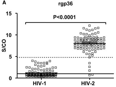
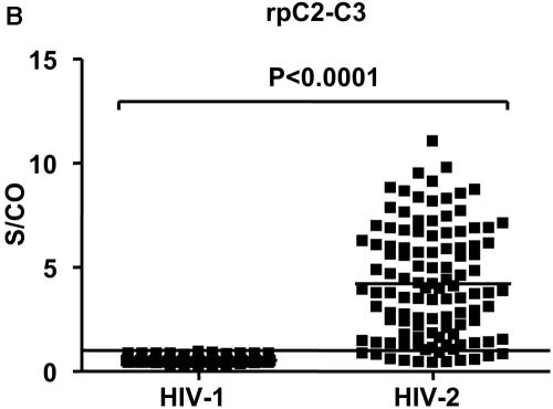
All 106 HIV-2 samples reacted with rgp36, and 99 (93.4%) samples reacted also with rpC2-C3 (Fig. 1A and B). The 100% clinical sensitivity and specificity obtained with rgp36 indicate that the ELISA-HIV2 assay can be used in the serodiagnosis of HIV-2 infection. The mean S/CO ratio was significantly higher for the rgp36 antigen than that for rpC2-C3 (8.27 [SD, 1.49] versus 4.89 [SD, 2.51]; P < 0.0001). These results suggest that the gp36 ectodomain is the immunodominant antigenic region in the HIV-2 envelope and are consistent with previous studies showing that recombinant gp36 proteins derived from several laboratory strains of HIV-2 are highly immunogenic (18, 40, 50). Most commercial and homemade ELISAs report similar sensitivities using substantially more sera per reaction compared to our assay (50 to 200 μl versus 1 μl) (38, 43, 48). However, several fourth-generation mixed HIV-1/2 assays performed poorly with diluted (up to 1:1,000) HIV-2 samples, suggesting that they may not detect the low levels of antibodies present at seroconversion and early infection (29). The higher sensitivity of the ELISA-HIV2 assay suggests that it may permit improved detection of HIV-2 seroconversions and recent infections. Testing of longitudinal specimens from recently infected individuals would be needed to support this claim. Such studies are, however, difficult to perform due to the low incidence of HIV-2 infection (20).
FIG.1.
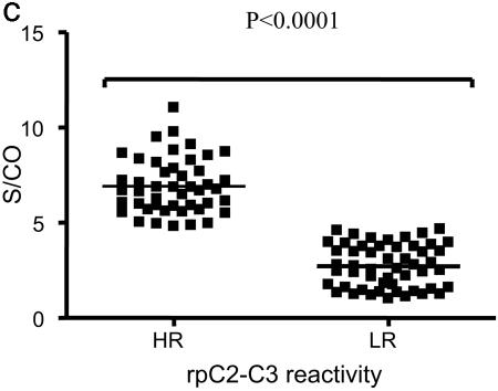
Patterns of reactivity of HIV plasma samples with rgp36 and rpC2-C3. Reactivity of HIV-1 and HIV-2 samples with rgp36 (A) and rpC2-C3 (B). (C) Low (LR) and high (HR) rpC2-C3 responders. S/CO, ODsample/ODcutoff ratio. Reactivity to rgp36 and rpC2-C3 is indicated by open (□) and black (▪) squares, respectively. The horizontal solid line represents the cutoff value; samples with S/CO values of ≥1 are considered reactive. The dotted line represents the lower S/CO value obtained with HIV-2 samples for rgp36. Student's t test was used to compare mean S/CO OD values obtained for both antigens.
The finding that 93.4% of the HIV-2 samples reacted also with the rpC2-C3 polypeptide contrasts with the low immunoreactivity (below 81%) reported for recombinant proteins encoded by corresponding sequences in HIV-2 strains SBL6669 (6), ROD (40), NIHZ (50), and ST (18). One explanation for this discrepancy is that rpC2-C3 may comprise epitopes which are more antigenic than the corresponding regions in HIV-2 strains SBL6669, ROD, NIHZ, and ST, all of which are laboratory-adapted isolates. Therefore, antibodies present in the infected immune sera may recognize the HIV-2ALI antigen better.
HIV-2 patients could be clustered into high immune responders and low immune responders according to the level of antibodies to rpC2-C3 (Fig. 1C). Conflicting reports exist on the prognostic value of gp120 antibody responses. Nevertheless, high gp120 binding antibody titers were negatively correlated to immune functions and viremia control in chronically HIV-1-infected patients (46). It will be important to investigate the correlations between the titer of C2-C3 binding antibodies, viremia, and immune functions, including neutralizing antibody response, in HIV-2 infection.
Antibodies to the envelope gp41 develop early in HIV-1 infection, while antibodies to the V3 region of gp120 develop later in infection. Therefore, the different antibody responses to rpC2-C3 may also be due to the timing of infection (32, 36). Further testing of longitudinal specimens from seroconverters will be needed to study the kinetics of antibody responses to this envelope protein and to assess the usefulness of this information to date the timing of HIV-2 infection.
All 95 HIV-1 samples analyzed with ELISA-HIV2 gave negative results with rpC2-C3. Thirty-one (32.6%) samples cross-reacted with rgp36, but the reactivity was significantly weaker than that of HIV-2 samples (mean S/CO ratio, 2.42 [SD, 0.85] versus 8.27 [SD, 1.49]; P < 0.0001) (Fig. 1A). These results suggested that the ELISA-HIV2 assay could be useful to discriminate between HIV-1 and HIV-2 infection in individuals with dual-positive serology. Seven HIV-1 and HIV-2 dually reactive serum samples were analyzed by ELISA-HIV2 and PCR amplification of HIV gag and/or env genes. For PCR, proviral DNA was extracted from uncultured peripheral blood mononuclear cells with the Wizard genomic DNA purification kit (Promega). For HIV-1, nested PCR was used to amplify a 409-bp fragment from the C2-C3 env region, using outer primer pair JA167 and JA170 and inner primers JA168 and JA169, and a 582-bp fragment from the p17 gag region, using outer primer pair JA152 and JA155 and inner primers JA153 and JA154. Thermal cycling conditions for PCR and primer numbers and positions have been described previously (25). For HIV-2, nested PCR was used to amplify a 378-bp fragment from the HIV-2 C2-C3 env gene region (positions 6949 to 7327 in HIV-2ALI) as described elsewhere (3). The amplified PCR products were visualized by electrophoresis in 2% agarose gel. For each patient, at least two independent PCRs were performed under identical conditions. HIV-1 plasma viral load was determined using the Quantiplex HIV RNA 3.0 (bDNA) kit (Bayer Diagnostics). The PCR and ELISA-HIV2 results indicated that none of the patients was dually infected, four patients being infected with HIV-2 and three with HIV-1 (Table 1). Therefore, the ELISA-HIV2 assay can be used to discriminate between HIV-1 and HIV-2 infections in dually seroreactive patients. In Portugal, the reported rate of dual HIV-1/HIV-2 seropositivity is 1.4% but the true rate of dual infections is unknown (9). The finding that none of the dually seroreactive patients was dually infected suggests that dual HIV-1/HIV-2 infections are rare in Portugal. Earlier reports suggested that most dually seropositive individuals from Guinea-Bissau (78 to 86%) (47), Ivory Coast, and The Gambia (72%) (19) were indeed dually infected. In more recent studies performed in India (22) and Senegal (15), a 40% rate of dual infections was reported among dually seroreactive patients. Although the number of patients was small in all studies, the declining prevalence of dual infections that they document is consistent with the worldwide decreasing incidence and prevalence rates of HIV-2 infection (9, 20).
TABLE 1.
Type of infection in dually HIV-1- and HIV-2-seroreactive individuals determined with ELISA-HIV2 and PCR amplification
| Sample | Result by ELISA-HIV2 (S/CO)
|
HIV-1 viral loada | Result by PCR amplificationb
|
Type of infection | |||
|---|---|---|---|---|---|---|---|
| rgp36 | rpC2-C3 | HIV-1
|
HIV-2 (env) | ||||
| gag | env | ||||||
| 107 | 9.35 | 11.87 | <50 | − | − | + | HIV-2 |
| 108 | 8.53 | 1.89 | <50 | − | − | + | HIV-2 |
| 109 | 8.68 | 4.11 | <50 | − | − | + | HIV-2 |
| 110 | 2.58 | 0.55 | 241 | − | + | − | HIV-1 |
| 111 | 8.25 | 3.32 | <50 | − | − | + | HIV-2 |
| 112 | 0.51 | 0.49 | 47,273 | + | + | − | HIV-1 |
| 113 | 0.80 | 0.50 | 35,123 | + | + | − | HIV-1 |
Number of RNA copies/ml of plasma.
Amplification of p17 (gag) and C2-C3 (env) regions. −, negative; +, positive.
Reactivity against two envelope glycoproteins is the World Health Organization criterion used for the WB confirmation of HIV infection (16). To further investigate the reliability of ELISA-HIV2 as a confirmatory test, we tested a panel of samples (n = 56) that were reactive in the screening assay VIDAS HIV DUO and in the confirmatory assay New LAV Blot II (51 positive and 5 indeterminate). All 51 WB-positive samples reacted as HIV-2 samples in ELISA-HIV2, whereas the indeterminate samples were HIV-2 negative in ELISA-HIV2 (Table 2). Four indeterminate samples reacted as HIV-1 in the HIV-1 Western blot and Peptilav 1-2. One indeterminate sample, which reacted also as HIV-1 in WB, was dually HIV-1/HIV-2 seroreactive in Peptilav 1-2. These results demonstrate that ELISA-HIV2 can be used as a confirmatory assay for the serodiagnosis of HIV-2 infection.
TABLE 2.
Diagnostic performance of ELISA-HIV2 with HIV-1 samples classified as indeterminate in HIV-2 Western blot (New LAV Blot II)
| Sample | Result by:
|
||||
|---|---|---|---|---|---|
| ELISA-HIV2 (S/CO)a
|
New LAV Blot II (band pattern on HIV-2 blot) | Peptilav 1-2b
|
|||
| rgp36 | rpC2-C3 | HIV-1 | HIV-2 | ||
| 53 | 0.77 | 0.45 | p26 | + | − |
| 62 | 0.63 | 0.55 | p68, gp36, p26 | + | − |
| 68 | 1.70 | 0.42 | gp36, p26 | + | − |
| 76 | 2.57 | 0.58 | (gp140, gp105, p68, p26)c | + | + |
| 92 | 3.43 | 0.50 | p68, p26 | + | − |
ODsample/ODcutoff ratio.
+, positive; −, negative.
Faint bands.
In conclusion, the highly sensitive and specific ELISA-HIV2 is an excellent alternative to the available tests for the serologic diagnosis and confirmation of HIV-2 infection. The dual-antigen format adopted in ELISA-HIV2 will permit the qualitative and quantitative characterization of the antibody response to the envelope gp125 and gp36 glycoproteins in HIV-2-infected patients.
Acknowledgments
This work was supported by grant POCTI/ESP/48045/2002 from Fundação para a Ciência e Tecnologia (FCT), Portugal.
REFERENCES
- 1.Andersson, S., Z. da Silva, H. Norrgren, F. Dias, and G. Biberfeld. 1997. Field evaluation of alternative testing strategies for diagnosis and differentiation of HIV-1 and HIV-2 infections in an HIV-1 and HIV-2-prevalent area. AIDS 11:1815-1822. [DOI] [PubMed] [Google Scholar]
- 2.Azevedo-Pereira, J. M., M. H. Lourenço, F. Barin, R. Cisterna, F. Denis, P. Moncharmont, R. Grillo, and M. O. Santos-Ferreira. 1994. Multicenter evaluation of a fully automated screening test, VIDAS HIV 1+ 2, for antibodies to human immunodeficiency virus types 1 and 2. J. Clin. Microbiol. 32:2559-2563. [DOI] [PMC free article] [PubMed] [Google Scholar]
- 3.Barroso, H., F. Araújo, M. H. Gomes, A. Mota-Miranda, and N. Taveira. 2004. Phylogenetic demonstration of two cases of perinatal human immunodeficiency virus type 2 infection diagnosed in adulthood. AIDS Res. Hum. Retrovir. 20:1373-1376. [DOI] [PubMed] [Google Scholar]
- 4.Beelaert, G., G. Vercauteren, K. Fransen, M. Mangelschots, M. De Rooy, S. Garcia-Ribas, and G. van der Groen. 2002. Comparative evaluation of eight commercial enzyme linked immunosorbent assays and 14 simple assays for detection of antibodies to HIV. J. Virol. Methods 105:197-206. [DOI] [PubMed] [Google Scholar]
- 5.Benitez, J., D. Palenzuela, J. Rivero, and J. V. Gavilondo. 1998. A recombinant protein based immunoassay for the combined detection of antibodies to HIV-1, HIV-2 and HTLV-I. J. Virol. Methods 70:85-91. [DOI] [PubMed] [Google Scholar]
- 6.Böttiger, B., A. Karlsson, P. Å. Andreasson, A. Nauclér, C. M. Costa, E. Norrby, and G. Biberfeld. 1990. Envelope cross-reactivity between human immunodeficiency virus types 1 and 2 detected by different serological methods: correlation between cross-neutralization and reactivity against the main neutralizing site. J. Virol. 64:3492-3499. [DOI] [PMC free article] [PubMed] [Google Scholar]
- 7.Centers for Disease Control and Prevention. 1990. Current trends in surveillance for HIV-2 infection in blood donors: United States, 1987-1989. Morb. Mortal. Wkly. Rep. 39:829-831. [PubMed] [Google Scholar]
- 8.Clavel, F., D. Guetard, F. Brun-Vezinet, S. Chamaret, M. A. Rey, M. O. Santos-Ferreira, A. G. Laurent, C. Dauguet, C. Katlama, C. Rouzioux, D. Klatzman, J. L. Champalimaud, and L. Montagnier. 1986. Isolation of a new human retrovirus from West African patients with AIDS. Science 233:343-346. [DOI] [PubMed] [Google Scholar]
- 9.Comissão Nacional de Luta Contra a Sida. 2004. A situação em Portugal a 30 de Dezembro de 2004. Documento SIDA 133/CVEDT. Comissão Nacional de Luta Contra a Sida, Lisbon, Portugal.
- 10.Decker, J. M., F. Bibollet-Ruche, X. Wei, S. Wang, D. N. Levy, W. Wang, E. Delaporte, M. Peeters, C. A. Derdeyn, S. Allen, E. Hunter, M. S. Saag, J. A. Hoxie, B. H. Hahn, P. D. Kwong, J. E. Robinson, and G. M. Shaw. 2005. Antigenic conservation and immunogenicity of the HIV coreceptor binding site. J. Exp. Med. 201:1407-1419. [DOI] [PMC free article] [PubMed] [Google Scholar]
- 11.de Wolf, F., R. H. Meloen, M. Bakker, F. Barin, and J. Goudsmit. 1991. Characterization of human antibody-binding sites on the external envelope glycoprotein of human immunodeficiency virus type 2. J. Gen. Virol. 72:1261-1267. [DOI] [PubMed] [Google Scholar]
- 12.Galli, R. A., S. Castriciano, M. Fearon, C. Major, K. W. Choi, J. Mahony, and M. Chernesky. 1997. Performance characteristics of recombinant enzyme immunoassay to detect antibodies to human immunodeficiency virus type 1 (HIV-1) and HIV-2 and to measure early antibody responses in seroconverting patients. J. Clin. Microbiol. 34:999-1002. [DOI] [PMC free article] [PubMed] [Google Scholar]
- 13.Gnann, J. W., J. B. McCormick, S. Mitchell, J. A. Nelson, and M. B. A. Oldstone. 1987. Synthetic peptide immunoassay distinguishes HIV type 1 and HIV type 2 infections. Science 237:1346-1349. [DOI] [PubMed] [Google Scholar]
- 14.Gonzalez, L., R. W. Boyle, M. Zhang, J. Castillo, S. Whittier, P. Della-Latta, L. M. Clarke, J. R. George, X. Fang, J. G. Wang, B. Hosein, and C. Y. Wang. 1997. Synthetic-peptide-based enzyme-linked immunosorbent assay for screening human serum or plasma for antibodies to human immunodeficiency virus type 1 and type 2. Clin. Diagn. Lab. Immunol. 4:598-603. [DOI] [PMC free article] [PubMed] [Google Scholar]
- 15.Gottlieb, G. S., P. S. Sow, S. E. Hawes, I. Ndoye, A. M. Coll-Seck, M. E. Curlin, C. W. Critchlow, N. B. Kiviat, and J. I. Mullins. 2003. Molecular epidemiology of dual HIV-1/HIV-2 seropositive adults from Senegal, West Africa. AIDS Res. Hum. Retrovir. 19:575-584. [DOI] [PubMed] [Google Scholar]
- 16.Guèye-Ndiaye, A. 2002. Serodiagnosis of HIV infection, p. 121-138. In M. Essex, S. Mboup, P. J. Kanki, R. G. Marlink, and S. D. Tlou (ed.), AIDS in Africa, 2nd ed. Kluwer Academic/Plenum Publishers, New York, N.Y.
- 17.Holmgren, B., Z. da Silva, O. Larsen, P. Vastrup, S. Andersson, and P. Aaby. 2003. Dual infections with HIV-1, HIV-2 and HTLV-I are more common in older women than in men in Guinea-Bissau. AIDS 17:241-253. [DOI] [PubMed] [Google Scholar]
- 18.Huang, M. L., M. Essex, and T.-H. Lee. 1991. Localization of immunogenic domains in the human immunodeficiency virus type 2 envelope. J. Virol. 65:5073-5079. [DOI] [PMC free article] [PubMed] [Google Scholar]
- 19.Ishikawa, K., K. Fransen, K. Ariyoshi, J. N. Nkengasong, W. Janssens, L. Heyndrickx, H. Whittle, M. O. Diallo, P. D. Ghys, I. M. Coulibaly, A. E. Greenberg, J. Piedade, W. Canas-Ferreira, and G. van der Groen. 1998. Improved detection of HIV-2 proviral DNA in dually seroreactive individuals by PCR. AIDS 12:1419-1425. [DOI] [PubMed] [Google Scholar]
- 20.Jaffar, S., A. D. Grant, J. Whitworth, P. G. Smith, and H. Whittle. 2004. The natural history of HIV-1 and HIV-2 infections in adults in Africa: a literature review. Bull. W. H. O. 82:462-469. [PMC free article] [PubMed] [Google Scholar]
- 21.Kanki, P. J., J-L. Sankalé, and S. Mboup. 2002. Biology of human immunodeficiency virus type 2 (HIV-2), p. 74-103. In M. Essex, S. Mboup, P. J. Kanki, R. G. Marlink, and S. D. Tlou (ed.), AIDS in Africa, 2nd ed. Kluwer Academic/Plenum Publishers, New York, N.Y.
- 22.Kannangai, R., S. Ramalingam, K. J. Prakash, O. C. Abraham, R. George, R. C. Castillo, D. H. Schwartz, M. V. Jesudason, and G. Sridharan. 2001. A peptide enzyme linked immunosorbent assay (ELISA) for the detection of human immunodeficiency virus type-2 (HIV-2) antibodies: an evaluation on polymerase chain reaction (PCR) confirmed samples. J. Clin. Virol. 22:41-46. [DOI] [PubMed] [Google Scholar]
- 23.Kulkarni, S., S. Tripathy, K. Agnihotri, N. Jatkar, S. Jadhav, W. Umakanth, K. Dhande, P. Tondare, R. Gangakhedkar, and R. Paranjape. 2005. Indian primary HIV-2 isolates and relationship between V3 genotype, biological phenotype and coreceptor usage. Virology 337:68-75. [DOI] [PubMed] [Google Scholar]
- 24.Laurent, C., K. Seck, N. Coumba, T. Kane, N. Samb, A. Wade, F. Liegeois, S. Mboup, I. Ndoye, and E. Delaporte. 2003. Prevalence of HIV and other sexually transmitted infections, and risk behaviours in unregistered sex workers in Dakar, Senegal. AIDS 17:1811-1816. [DOI] [PubMed] [Google Scholar]
- 25.Leitner, T., D. Escanilla, S. Marquina, J. Wahlberg, C. Brostrom, H. B. Hansson, M. Uhlen, and J. Albert. 1995. Biological and molecular characterization of subtype D, G, and A/D recombinant HIV-1 transmissions in Sweden. Virology 10:136-146. [DOI] [PubMed] [Google Scholar]
- 26.Lizeng, Q., C. Nilsson, S. Sourial, S. Andersson, O. Larsen, P. Aaby, M. Ehnlund, and E. Björling. 2004. Potent neutralizing serum immunoglobulin A (IgA) in human immunodeficiency virus type 2-exposed IgG-seronegative individuals. J. Virol. 78:7016-7022. [DOI] [PMC free article] [PubMed] [Google Scholar]
- 27.Lizeng, Q., P. Skott, S. Sourial, C. Nilsson, S. Andersson, M. Ehnlund, N. Taveira, and E. Bjorling. 2003. Serum immunoglobulin A (IgA)-mediated immunity in human immunodeficiency virus type 2 (HIV-2) infection. Virology 308:225-232. [DOI] [PubMed] [Google Scholar]
- 28.Ly, T. D., S. Laperche, C. Brennan, A. Vallari, A. Ebel, J. Hunt, L. Martin, D. Daghfal, G. Schochetman, and S. Devare. 2004. Evaluation of the sensitivity and specificity of six HIV combined p24 antigen and antibody assays. J. Virol. Methods 122:185-194. [DOI] [PubMed] [Google Scholar]
- 29.Ly, T. D., L. Martin, D. Daghfal, A. Sandridge, D. West, R. Bristow, L. Chalouas, X. Qiu, S. C. Lou, J. C. Hunt, G. Schochetman, and S. G. Devare. 2001. Seven human immunodeficiency virus (HIV) antigen-antibody combination assays: evaluation of HIV seroconversion sensitivity and subtype detection. J. Clin. Microbiol. 39:3122-3128. [DOI] [PMC free article] [PubMed] [Google Scholar]
- 30.Mannervik, M., P. Putkonen, V. Ruden, K. A. Kent, E. Norrby, B. Wahren, and P. A. Broliden. 1992. Identification of B-cell antigenic sites on HIV-2 gp125. AIDS 5:177-187. [PubMed] [Google Scholar]
- 31.Manocha, M., K. T. Chitralekha, M. Thakar, D. Shashikiran, R. S. Paranjape, and D. N. Rao. 2003. Comparing modified and plain peptide linked enzyme immunosorbent assay (ELISA) for detection of human immunodeficiency virus type-1 (HIV-1) and type-2 (HIV-2) antibodies. Immunol. Lett. 85:275-278. [DOI] [PubMed] [Google Scholar]
- 32.McDougal, J. S., C. D. Pilcher, B. S. Parekh, G. Gershy-Damet, B. M. Branson, K. Marsh, and S. Z. Wiktor. 2005. Surveillance for HIV-1 incidence using tests for recent infection in resource-constrained countries. AIDS 19(Suppl. 2):S25-S30. [DOI] [PubMed] [Google Scholar]
- 33.Norrby, E., G. Biberfeld, F. Chiodi, A. von Gegerfeldt, A. Naucler, E. Parks, and R. Lerner. 1987. Discrimination between antibodies to HIV and to related retroviruses using site-directed serology. Nature 329:248-250. [DOI] [PubMed] [Google Scholar]
- 34.Norrby, E., P. Putkonen, B. Bottiger, G. Utter, and G. Biberfeld. 1991. Comparison of linear antigenic sites in the envelope proteins of human immunodeficiency virus (HIV) type 2 and type 1. AIDS Res. Hum. Retrovir. 7:279-285. [DOI] [PubMed] [Google Scholar]
- 35.Paranjape, R. S., S. P. Tripathy, P. A. Menon, S. M. Mehendale, P. Khatavkar, D. R. Joshi, U. Patil, D. A. Gadkari, and J. J. Rodrigues. 1997. Increasing trend of HIV seroprevalence among pulmonary tuberculosis patients in Pune, India. Indian J. Med. Res. 106:207-211. [PubMed] [Google Scholar]
- 36.Pilcher, C. D., S. A. Fiscus, T. Q. Nguyen, E. Foust, L. Wolf, D. Williams, R. Ashby, J. O. O'Dowd, J. T. McPherson, B. Stalzer, L. Hightow, W. C. Miller, J. J. Eron, Jr., M. S. Cohen, and P. A. Leone. 2005. Detection of acute infections during HIV testing in North Carolina. N. Engl. J. Med. 352:1873-1883. [DOI] [PubMed] [Google Scholar]
- 37.Robert-Guroff, M., K. Aldrich, R. Muldoon, T. L. Stern, G. P. Bansal, T. J. Matthews, P. D. Markham, R. C. Gallo, and G. Franchini. 1992. Cross-neutralization of human immunodeficiency virus type 1 and 2 and simian immunodeficiency virus isolates. J. Virol. 66:3602-3608. [DOI] [PMC free article] [PubMed] [Google Scholar]
- 38.Saville, R. D., N. T. Constantine, F. R. Cleghorn, N. Jack, C. Bartholomew, J. Edwards, P. Gomez, and W. A. Blattner. 2001. Fourth-generation enzyme-linked immunosorbent assay for the simultaneous detection of human immunodeficiency virus antigen and antibody. J. Clin. Microbiol. 39:2518-2524. [DOI] [PMC free article] [PubMed] [Google Scholar]
- 39.Schim van der Loeff, M. F., and P. Aaby. 1999. Towards a better understanding of the epidemiology of HIV-2. AIDS 13:S69-S84. [PubMed] [Google Scholar]
- 40.Schulz, T. F., W. Oberhuber, J. M. Hofbauer, P. Hengster, C. Larcher, L. C. Gurtler, R. Tedder, H. Wachter, and M. P. Dierich. 1989. Recombinant peptides derived from the env-gene of HIV-2 in the serodiagnosis of HIV-2 infections. AIDS 3:165-172. [DOI] [PubMed] [Google Scholar]
- 41.Schupbach, J. 1999. Human immunodeficiency viruses, p. 847-870. In P. R. Murray, E. J. Baron, M. A. Pfaller, F. C. Tenover, and R. H. Yolken (ed.), Manual of clinical microbiology, 7th ed. American Society for Microbiology, Washington, D.C.
- 42.Sickinger, E., M. Stieler, B. Kaufman, H.-P. Kapprell, D. West, A. Sandridge, S. Devare, G. Schochetman, J. C. Hunt, D. Daghfal, and AxSYM Clinical Study Group. 2004. Multicenter evaluation of a new, automated enzyme-linked immunoassay for detection of human immunodeficiency virus-specific antibodies and antigen. J. Clin. Microbiol. 42:21-29. [DOI] [PMC free article] [PubMed] [Google Scholar]
- 43.Simon, F., S. Souquiere, F. Damond, A. Kfutwah, M. Makuwa, E. Leroy, P. Rouquet, J. L. Berthier, J. Rigoulet, A. Lecu, P. T. Telfer, I. Pandrea, J. C. Plantier, F. Barre-Sinoussi, P. Roques, M. C. Muller-Trutwin, and C. Apetrei. 2001. Synthetic peptide strategy for the detection of and discrimination among highly divergent primate lentiviruses. AIDS Res. Hum. Retrovir. 17:937-952. [DOI] [PubMed] [Google Scholar]
- 44.Taveira, N. C., F. Bex, A. Burny, D. Robertson, M. O. Ferreira, and J. Moniz-Pereira. 1994. Molecular characterization of the env gene from a non-syncytium-inducing HIV-2 isolate (HIV-2ALI). AIDS Res. Hum. Retrovir. 10:223-224. [DOI] [PubMed] [Google Scholar]
- 45.Thorstensson, R., S. Andersson, S. Lindback, F. Dias, F. Mhalu, H. Gaines, and G. Biberfeld. 1998. Evaluation of 14 commercial HIV-1/HIV-2 antibody assays using serum panels of different geographical origin and clinical stage including a unique seroconversion panel. J. Virol. Methods 70:139-151. [DOI] [PubMed] [Google Scholar]
- 46.Trkola, A., H. Kuster, C. Leemann, A. Oxenius, C. Fagard, H. Furrer, M. Battegay, P. Vernazza, E. Bernasconi, R. Weber, B. Hirschel, S. Bonhoeffer, and H. F. Gunthard. 2004. Humoral immunity to HIV-1: kinetics of antibody responses in chronic infection reflects capacity of immune system to improve viral set point. Blood 104:1784-1792. [DOI] [PubMed] [Google Scholar]
- 47.Walther-Jallow, L., S. Andersson, Z. da Silva, and G. Biberfeld. 1999. High concordance between polymerase chain reaction and antibody testing of specimens from individuals dually infected with HIV types 1 and 2 in Guinea-Bissau, West Africa. AIDS Res. Hum. Retrovir. 15:957-962. [DOI] [PubMed] [Google Scholar]
- 48.Weber, B., A. Berger, H. Rabenau, and H. W. Doerr. 2002. Evaluation of a new combined antigen and antibody human immunodeficiency virus screening assay, VIDAS HIV DUO Ultra. J. Clin. Microbiol. 40:1420-1426. [DOI] [PMC free article] [PubMed] [Google Scholar]
- 49.Weiss, R. A., P. R. Clapham, J. N. Weber, D. Whitby, R. S. Tedder, T. O'Connor, S. Chamaret, and L. Montagnier. 1998. HIV-2 antisera cross-neutralize HIV-1. AIDS 2:95-100. [DOI] [PubMed] [Google Scholar]
- 50.Zuber, M., K. P. Samuel, J. A. Lautenberger, P. J. Kanki, and T. S. Papas. 1990. Bacterially produced HIV-2 env polypeptides specific for distinguishing HIV-2 from HIV-1 infections. AIDS Res. Hum. Retrovir. 6:525-534. [DOI] [PubMed] [Google Scholar]


