Abstract
A sudden increase in neutropenic hematology patients with Candida krusei colonization and bacteremia prompted a longitudinal epidemiological investigation. We identified 39 patients; 13 developed candidemia, and three died; 25 patients carried the same genotype. We intervened by changing antifungal prophylaxis and implementing strict infection control measures. The incidence dropped immediately.
Fungal infections among neutropenic patients are associated with substantial morbidity and mortality (6). The most frequently used antifungal prophylaxis protocols primarily aim at decontamination of the gastrointestinal tract (7, 10). This often includes fluconazole. However, some Candida yeast species are intrinsically resistant or have acquired resistance to fluconazole (3). Candida krusei, for instance, is not susceptible to fluconazole, which facilitates escape from prophylaxis (4, 8, 11). We describe here the control of an outbreak of C. krusei infections among hemato-oncology patients.
All patients were at the Hematology Department of the Erasmus Medical Center, Rotterdam, The Netherlands, consisting of three clinical wards in two separate locations (10, 21, and 16 beds). The study period lasted from July 2001 to January 2004. After notification of the outbreak (18 January 2002), two kinds of interventions were implemented simultaneously: antifungal prophylaxis was changed and strict infection control measures were implemented. Surveillance culturing of the rectum, throat, and vagina was increased from once to twice weekly. When blood cultures were indicated, 10 ml of blood was injected into Mycobottles (Becton Dickinson, Lelystad, The Netherlands).
Before the outbreak, fluconazole prophylaxis (1 daily dose of 200 mg by mouth) was given to patients expected to be neutropenic for more than 10 days. After identifying the outbreak, this was altered to amphotericin B suspension (4 daily doses of 6 ml by mouth). Routine preventive measures before the outbreak included general hand hygiene and, in case of manipulation of the central venous Hickman line, gowns, gloves, and caps were worn.
Upon outbreak notification all C. krusei-positive patients were nursed in single (isolation) rooms. Personnel were cohorted to negative or positive patients. They wore gowns and gloves upon entering the rooms of positive patients. Patients were only allowed to leave the room for diagnostic or medical procedures or for use of sanitary facilities if not present in the isolation room. In that case, the sanitary facilities were cohorted to positive and negative patients. Hand hygiene was improved by supplying colonized patients with an alcohol-based hand rub.
All individual medical histories were checked. Invasive and (radio)diagnostic examinations, dental treatment, and all visits to medical doctors were assessed for transmission risk. Patients not on fluconazole prophylaxis were cultured once for the presence of C. krusei in rectal swabs (January 2002). If health care workers developed skin lesions, these were specifically cultured for yeasts. To investigate food or the environment as a potential source, different foodstuffs, appliances, and medical and dental paraphernalia were cultured for Candida spp.
The differential intake of foods by colonized and noncolonized patients was recorded. A patient was considered colonized if C. krusei was isolated from rectal, throat, or vaginal swabs. Candidemia was defined when one or more blood cultures grew C. krusei. Index cases were defined as each first patient identified with a single fungal genotype, cultured in more than one patient, or those patients identified in time after the first patient with the same genotype, but prior transmission was not plausible.
Materials from swabs or positive Mycobottles were inoculated directly on Sabouroud agar and Brucella blood agar (Becton Dickinson). Plates were incubated for a minimum of 3 days and yeast identification involved Candida Chromagar (Chromagar, Paris, France) and VITEK (bioMérieux, Marcy-l'Etoile, France). MICs were determined with the Sensititre YeastOne method (Trek Diagnostic Systems, East Grinstead, England) at 24 and 48 h postinoculation. CLSI (formerly NCCLS) breakpoints were applied.
For DNA isolation, colonies from a subculture of a single colony were treated with lyticase (Sigma, Zwijndrecht, The Netherlands). DNA from spheroplasts was isolated by the Qiaex DNeasy tissue kit protocol (QIAGEN, Mannheim, Germany). Two different genetic tests involving variable C. krusei repeated sequences were employed for genetic typing (8, 9). This involved PCR and agarose gel electrophoresis. Based on the lengths of the PCR products separate genotypes were assigned. Per strain, two such genotypes were defined, which were combined into a single numerical code. Statistical analyses were performed using two-sided Fisher Exact tests. A P value of less than 0.05 indicated significance.
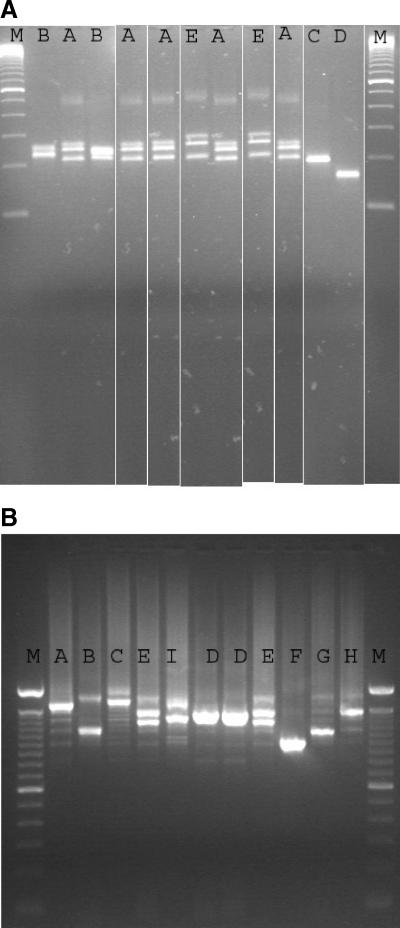
One hundred and thirty-five strains from 39 patients were subjected to genotyping, including all except two of the invasive isolates as those strains were lost or could not be revived after storage. The number of isolates available per patient varied from 1 to 24 and these were all subjected to genotyping. The CKTNR assay generated six genotypes (A through F) and the CKRS1 test identified nine genotypes (A through I) (2). The results from the CKTNR assay compared to those by the CKRS1 test were not always concordant. Therefore, we combined the results of both typing assays and 12 genotypes could be identified, numbered 1 through 12 (Fig. 1).
FIG. 1.
(A) Profiles of CKTNR tests of all but one identified genotypes. The CKTNR assay generated six genotypes (A through F). Lane 1 (M) shows the molecular size markers. Lanes 2 to 11 give genotypes 1 to 10, respectively (genotype 11 is missing); lane 12 shows genotype 12. Genotypes are identified by combining the two tests (see panel B). Mark that there is no distinction between genotypes 1 and 3, genotypes 2, 4, 7, and 9, and genotypes 6 and 8 by using the CKTNR test. (B) Profiles of CKRS1 tests of all but one of the identified genotypes. The CKRS1 test identified nine genotypes (A through I). Lane 1 (M) shows molecular size markers. Lanes 2 to 11, genotypes 1 to 10, respectively (genotype 11 is missing); lane 12 shows genotype 12. Genotypes are identified by combining the two tests. Mark that there is no distinction between genotypes 4 and 8 and genotypes 6 and 7 by using the CKRS1 test.
Genotypes were identified as outbreak isolates when encountered in more than one patient. Genotype 1 was identified as the main outbreak genotype (25 out of the 39 patients). The other genotypes were less prevalent with only genotypes 6 and 8 occurring four and three times, respectively (Table 1). Only two patients were colonized with two different types of C. krusei. The second genotype was in both patients cultured only once. All other patients consistently showed the same genotype, varying from being cultured once to up to 24 times per patient.
TABLE 1.
Outcomes of patients who were colonized or had candidemiaa
| Parameter | Outbreak strain genotype 1 | Outbreak strain genotype 6 | Outbreak strain genotype 8 | Nonoutbreak strains | Total |
|---|---|---|---|---|---|
| No. of patients | 25 | 4 | 3 | 9 | 39b |
| No. colonized (%) | 19 (68) | 1 (3.6) | 0 | 8b (29) | 28 (72) |
| No. with candidemiab (%) | 6c (46) | 3 (23) | 3 (23) | 1 (7.7) | 13 (33) |
| No. with complications (%) | 1 (25) | 1 (25) | 1 (25) | 1 (25) | 4/13 (31) |
| No. (%) of deaths due to infectiond | 0 | 0 | 2 (66) | 1 (33) | 3/13 (23) |
| No. of index cases (%) | 4 (22) | 3 (17) | 2 (11) | 9 (50) | 18/41 (44) |
| No. of secondary cases (%) | 21e (91) | 1 (4.3) | 1 (4.3) | 0 | 23/41 (56) |
Distribution and outcome of 39 hematologic patients colonized or presenting with candidemia (n = 13) by outbreak or nonoutbreak strains of Candida krusei (n= 41). Patients were defined as being colonized if they did not develop candidemia. Index cases were defined as having acquired their strain through selection. Secondary cases were defined as having acquired their strain through transmission. As two patients showed two different genotypes, 41 index and secondary cases were defined.
Two patients were colonized with genotype 1 as well as a nonoutbreak strain. Both had an invasive strain, genotype 1; they were categorized as candidemia by “outbreak strain genotype 1” and as colonized by “nonoutbreak” as well. Of two patients for whom the invasive strains were lost, before candidemia, one was colonized with genotype 1 and the other with genotype 6: we assume candidemia was caused by the colonizing genotype.
P = 0.026 compared to genotype 8.
As defined by an infectious diseases specialist.
P = 0.034 compared to transmission of genotype 6.
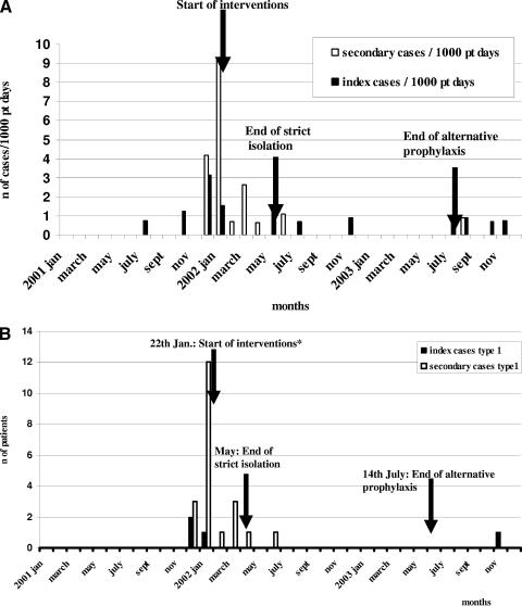
During the study period, 39 patients were diagnosed with carriage and/or candidemia due to C. krusei. Figure 2A shows the incidence of newly colonized and/or infected patients per 1,000 patient days in time; the distribution of C. krusei genotype 1 is shown in Fig. 2B. Patients colonized and/or infected by genotype 1 acquired their strain significantly more frequently (21 of 25) through transmission compared to those colonized by genotype 6 (one of four, P = 0.034). Of the four index cases with genotype 1, three were responsible for transmission to 21 secondary cases (Fig. 1B). Rectal carriage of C. krusei was not detected in nonneutropenic patients.
FIG. 2.
(A) Incidence per 1,000 patient days of index cases and secondary cases in relation to interventions. Interventions were: culture frequency change from one to two times/week, prophylactic regimen changed from fluconazole to amphotericin B oral solution, and strict isolation procedures (see text). Index cases are those in whom C. krusei is most likely selected from their own flora by the use of fluconazole and defined as the first patient in time with a new genotype, a unique genotype, or those with a genotype encountered more than once, but acquisition by transmission was not plausible. Secondary cases are those admitted simultaneously on the same ward with an index case colonized with the same genotype. (B) Index cases (n = 4) and secondary cases (n = 21) colonized by genotype 1 over time in relation to interventions.
Candidemia occurred in 13 patients of which three died. Five patients, of whom three had complications, developed renal insufficiency. A mean of 7 days elapsed between colonization and infection. In 4 out of 12 patients, colonization before infection went undetected. Candidemia after colonization by genotype 1 was significantly less frequent than candidemia after colonization by genotype 8 (P = 0,026), although genotype 1 was most frequently cultured from blood (6 of 13) (Table 1).
Cultures of the environment, food, and dental and medical paraphernalia were negative and no differences in food intake or medical treatment were identified. None of the health care workers reported skin lesions. All strains were susceptible to amphotericin B (MIC, 0.5 to 2 mg/liter), voriconazole (MIC, 0.5 to 1 mg/liter) and ketoconazole (MIC, 1 to 4 mg/liter), but resistant to 5-flucytosine (MIC, 16 to 32 mg/liter). All but one strain was resistant to fluconazole (MIC, 8 to 258 mg/liter). Strains belonging to the same genotype were similar in antibiogram (data not shown).
We present here a large outbreak of C. krusei involving 39 patients, of which 13 developed candidemia. The intervention strategies led to a sharp decrease in the incidence. Before December 2001, several distinct genotypes were selected, but transmission was not detected. However, immediately after selection of genotype 1, transmission occurred. Twenty-one out of 23 secondary cases were due to genotype 1. In 4 out of 13 fungemic patients, no colonization was previously detected. This can be due to either a short colonization period before candidemia occurred or direct inoculation of the strain into the vascular system during catheter manipulation.
Nosocomial C. krusei infections were noted decades ago and were, at that time, considered extremely rare (1). In 1991, Wingard et al. (11) found a sevenfold higher frequency of C. krusei infection and a significantly higher proportion of colonized patients in those who received fluconazole. They concluded that the prophylactic use of fluconazole may permit intrinsically resistant Candida spp. to emerge (11).
Genetic typing of C. krusei was used to clarify a partly polyclonal outbreak (5). We used here a combination of two recent molecular typing tools. Shemer et al. (9) targeted a polymorphic microsatellite target with a discriminatory index of 0.96. The discriminatory power of CKRS-1 typing was even higher: the value of 1.0 suggests optimal discrimination (2). We here show that a combination of the results of two separate tests is certainly worthwhile. Since 12 combination types were documented, merging data clearly enhances the typing resolution. Therefore, we assume that identical genotypes accurately reflect clonal spread. Further studies on transmission factors and determinants are needed, as strains were disseminated through as yet unidentified mechanisms. In order to be able to install early prevention measures and prevent transmission, we suggest that real-time surveillance and molecular typing of C. krusei is needed when using fluconazole on a large scale.
Acknowledgments
We thank the Department of Hematology for adequately performing strict infection control measures and Marian Humphrey for reviewing the manuscript for English syntax and grammatical errors.
This study was not financially supported.
REFERENCES
- 1.Berger, C., R. Frei, A. Gratwohl, A. Tichelli, B. Hafliger, and B. Speck. 1988. A Candida krusei epidemic in a hematology department. Schweiz. Med. Wochenschr. 118:37-41. [PubMed] [Google Scholar]
- 2.Carlotti, A., F. Chaib, A. Couble, N. Bourgeois, V. Blanchard, and J. Villard. 1997. Rapid identification and fingerprinting of Candida krusei by PCR based amplification of the species-specific repetitive polymorphic sequence CKRS-1. J. Clin. Microbiol. 35:1337-1343. [DOI] [PMC free article] [PubMed] [Google Scholar]
- 3.Eggimann, P., J. Garbino, and D. Pittet. 2003. Management of Candida species infections in critically ill patients. Lancet Infect. Dis. 3:772-785. [DOI] [PubMed] [Google Scholar]
- 4.Marr, K. A., K. Seidel, T. C. White, and R. Bowden. 2000. Candidemia in allogenic blood and marrow transplant patients: evolution of risk factors after the adoption of prophylactic fluconazole. J. Infect. Dis. 181:309-316. [DOI] [PubMed] [Google Scholar]
- 5.Noskin, G. A., J. Lee, D. M. Hacek, M. Postelnick, B. E. Reisberg, V. Stosor, S. A. Weisman, and L. R. Peterson. 1996. Molecular typing for investigating an outbreak of Candida krusei. Diagn. Microbiol. Infect. Dis. 26:117-123.12. [DOI] [PubMed] [Google Scholar]
- 6.Pfaller, M. A. 1996. Nosocomial candidiasis: emerging species, reservoirs, and modes of transmission. Clin. Infect. Dis. 22:S89-S94. [DOI] [PubMed]
- 7.Puzniak, L., S. Teutsch, W. Powderly, and L. Polish. 2004. Has the epidemiology of nosocomial candidemia changed? Infect. Control Hosp. Epidemiol. 25:628-633. [DOI] [PubMed] [Google Scholar]
- 8.Rex, J. H., T. J. Walsh, J. D. Sobel, et al. 2000. Practice guidelines for the treatment of candidiasis. Infectious Diseases Society of America. Clin. Infect. Dis. 30:662-678. [DOI] [PubMed] [Google Scholar]
- 9.Shemer, R., Z. Weissman, N. Hashman, and D. Kornitzer. 2001. A highly polymorphic degenerate microsatellite for molecular strains typing of Candida krusei. Microbiology 147:2021-2028. [DOI] [PubMed] [Google Scholar]
- 10.Wingard, J. R. 1995. Importance of Candida species other than C. albicans as pathogens in oncology patients. Clin. Infect. Dis. 20:115-125. [DOI] [PubMed] [Google Scholar]
- 11.Wingard, J. R., W. G. Merz, M. G. Rinaldi, T. R. Johnson, J. E. Karp, and R. Saral. 1991. Increase in Candida krusei infection among patients with bone marrow transplantation and neutropenia treated prophylactically with fluconazole. N. Engl. J. Med. 325:1274-1277. [DOI] [PubMed] [Google Scholar]


