

Abstract
1. The problem of colour reception is that we do not know the action spectra of the visual pigments involved, the nature of the signals generated nor the interaction between these signals. We only know the incident light and the electric results of interaction.
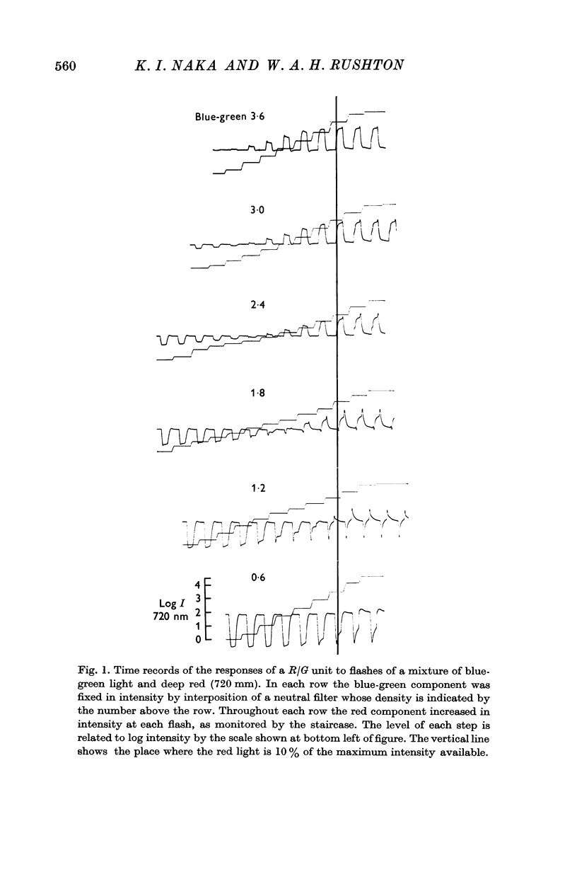
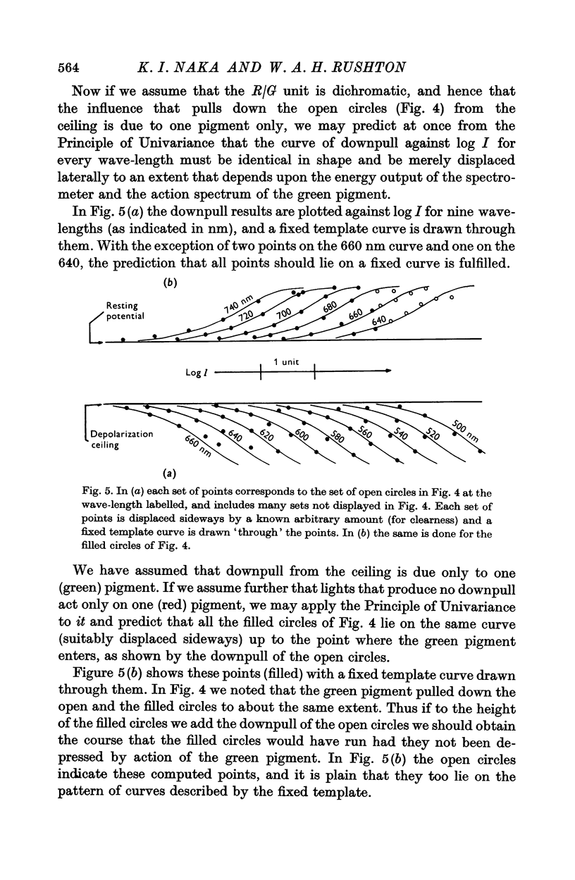
2. In Part 1 we show that S-potentials from red/green (R/G) units saturated with deep red light show this property: added green light pulls down the ceiling of depolarization, but more added red had no power to raise it again. Thus lights that depress the deep red ceiling equally stimulate the green pigment equally. From this the action spectrum of the green pigment can be obtained.
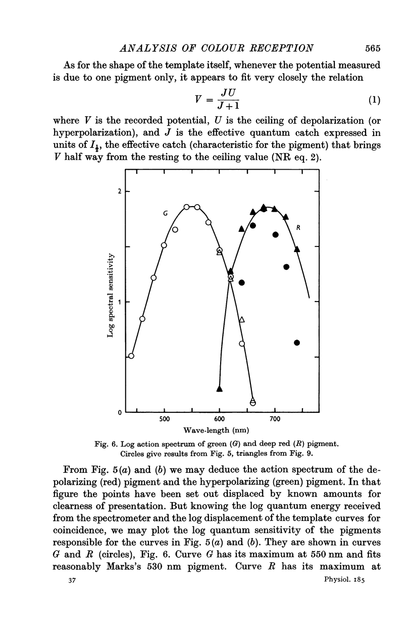
3. If we assume that only two visual pigments are involved in the R/G unit, and that lights which do not pull down the deep red ceiling are below the threshold for green cones, then in this range only the red pigment is excited and we may obtain its action spectrum. Its maximum is at 680 nm where no visual pigment so far has been found.
4. In Part 2 we consider the following mathematical problem: `Is it possible that two pigments of given action spectra could combine their outputs in such a way that the resultant would be identical with the output of a third pigment of given action spectrum, for every intensity of every monochromatic light?' The solution shows that this is always mathematically possible, and the necessary interaction function is deduced.
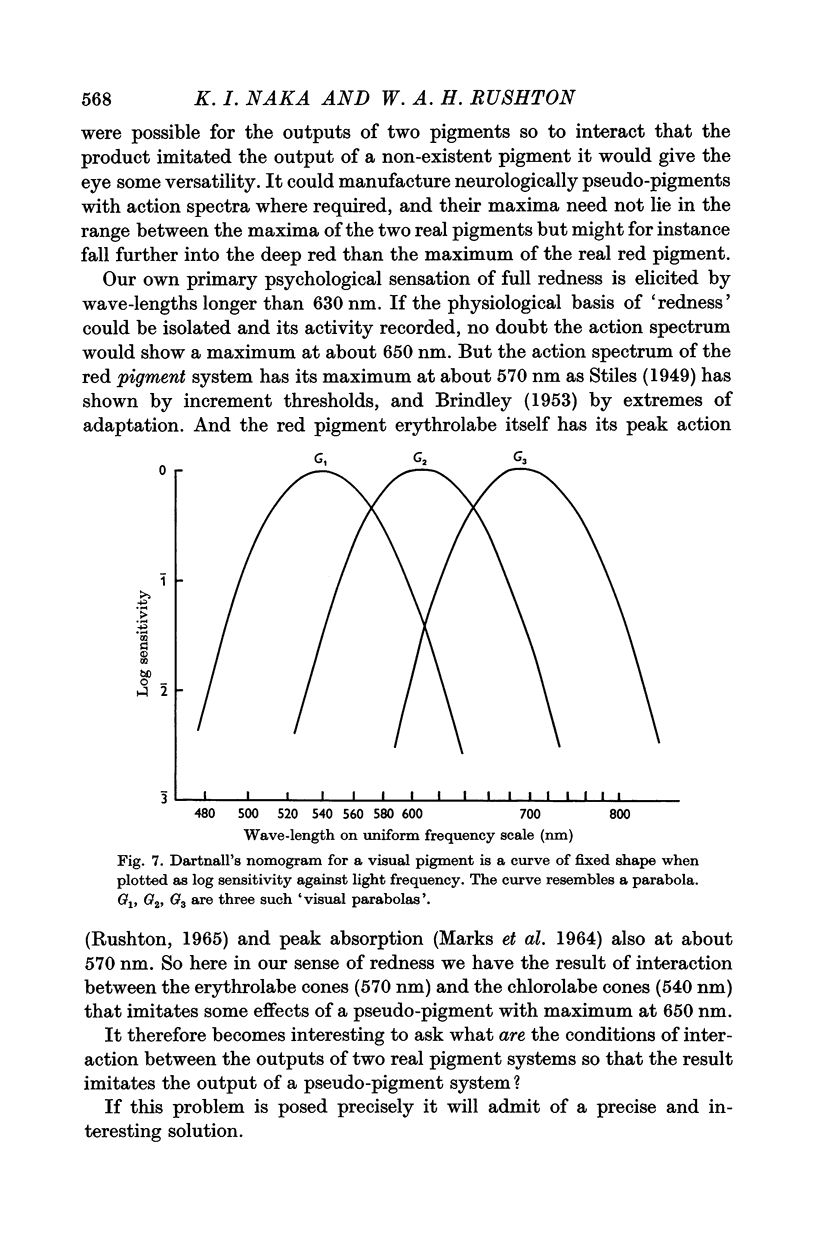
5. It is shown further that if the log action spectra are the `visual parabolas' that resemble Dartnall's nomogram, then the interaction function is simply a linear transform such as Hartline & Ratliff (1957) have found with lateral inhibition in Limulus and Donner & Rushton (1959) with silent substitution in the frog.
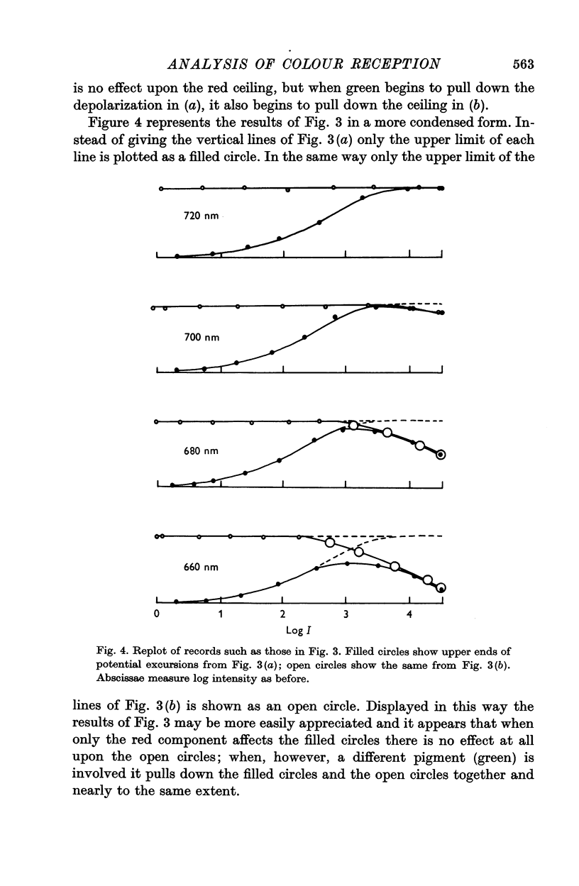
6. An interaction that matches a single pigment to perfection for all monochromatic lights will not match it for certain mixtures. By this criterion the 680 nm excitability is a pigment and not the resultant of two other pigments, i.e. pigments more excitable in other spectral regions.
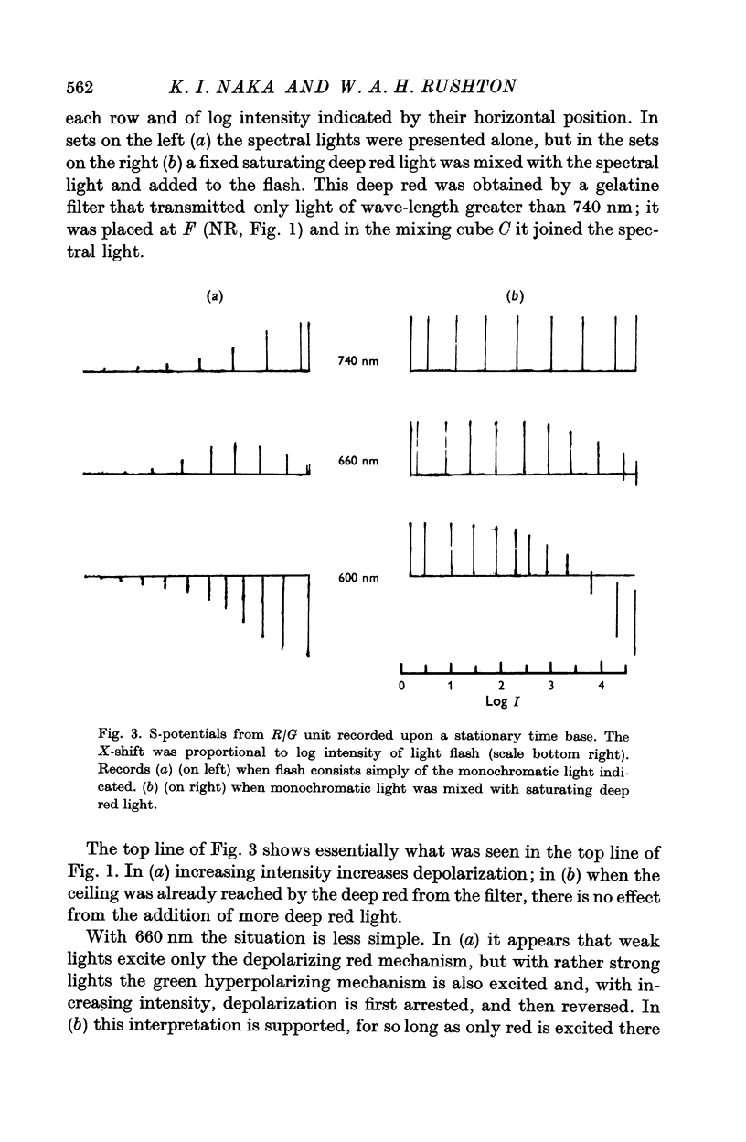
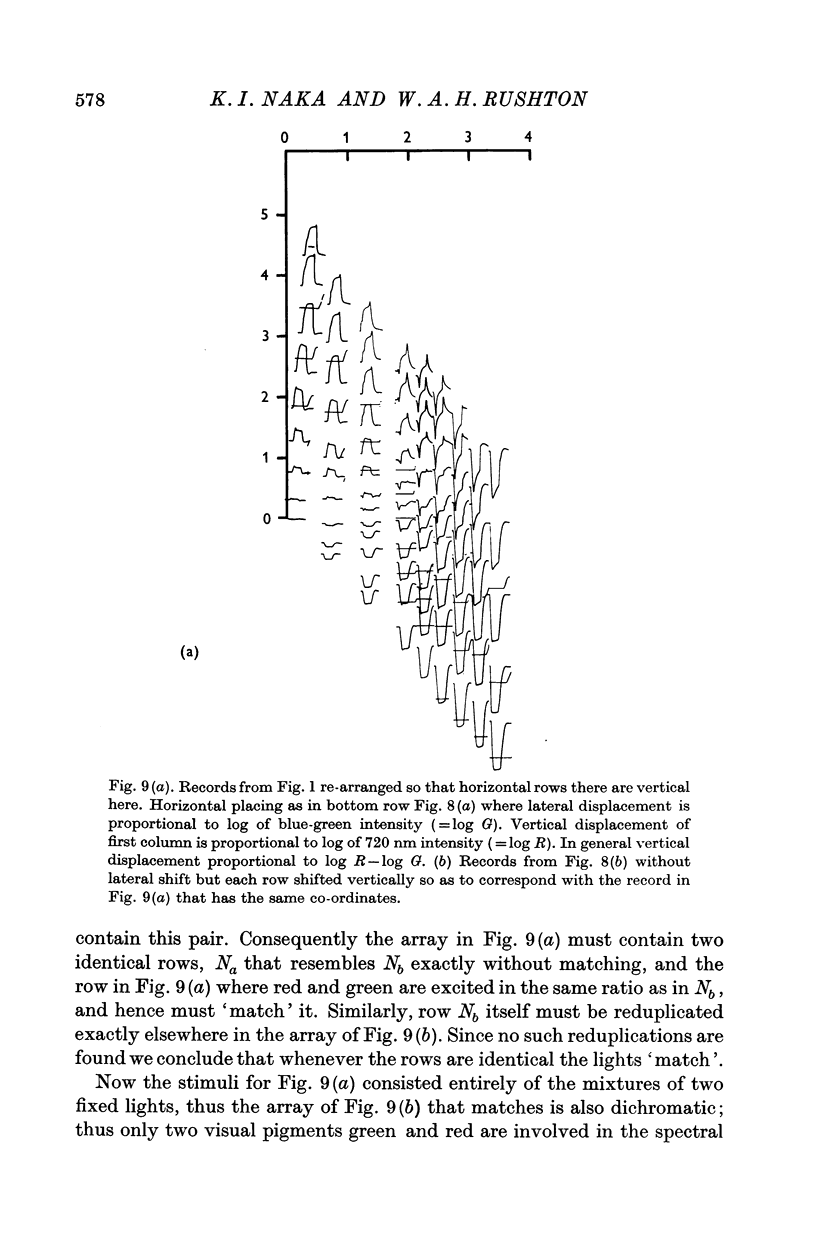
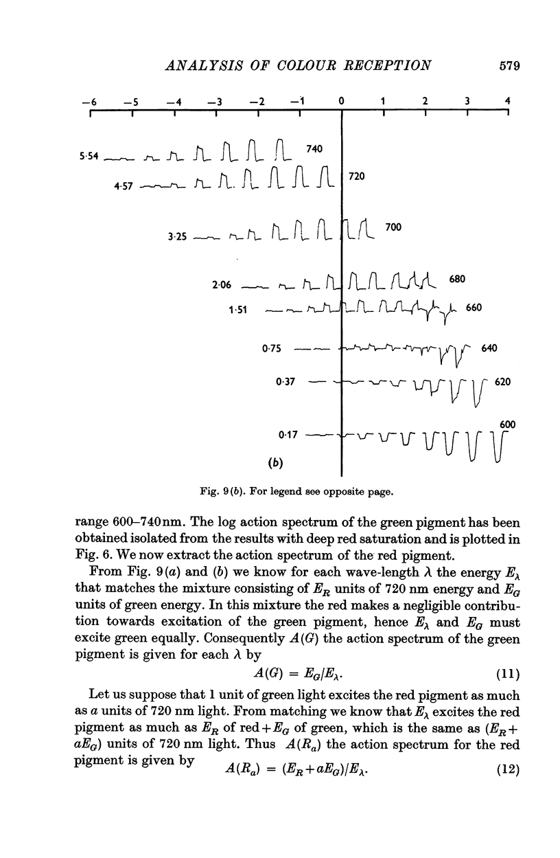
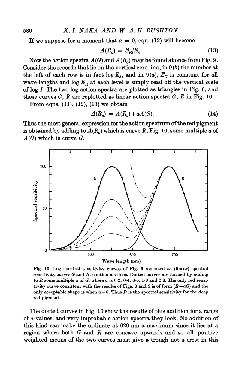
7. In Part 3 monochromatic lights are matched by red+green mixtures that give identical responses. From this the action spectrum of the red pigment may be obtained without involving nerve organization (except as a null detector). The result, which has one arbitrary constant, is given by the curves of Fig. 10, the continuous curve R or one of the dotted curves. Of these only curve R is acceptable.
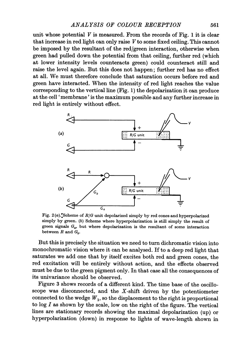
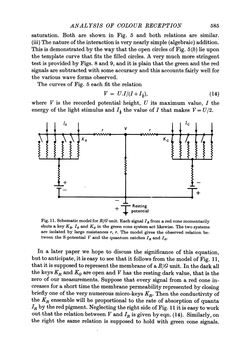
8. Knowing the action spectra for red and green cones we may consider what signals are generated and how they interact to give the records. Figure 11 suggests a model that will account for the size and sign of S-potentials as function of the quantum catch by the two pigments. It does not embrace the time or space parameters which can be very complex.
Full text
PDF

Selected References
These references are in PubMed. This may not be the complete list of references from this article.
- BRINDLEY G. S. The effects on colour vision of adaptation to very bright lights. J Physiol. 1953 Nov 28;122(2):332–350. doi: 10.1113/jphysiol.1953.sp005003. [DOI] [PMC free article] [PubMed] [Google Scholar]
- DONNER K. O., RUSHTON W. A. Retinal stimulation by light substitution. J Physiol. 1959 Dec;149:288–302. doi: 10.1113/jphysiol.1959.sp006340. [DOI] [PMC free article] [PubMed] [Google Scholar]
- FUORTES M. G. Electric activity of cells in the eye of Limulus. Am J Ophthalmol. 1958 Nov;46(5 Pt 2):210–223. doi: 10.1016/0002-9394(58)90800-6. [DOI] [PubMed] [Google Scholar]
- HARTLINE H. K., RATLIFF F. Inhibitory interaction of receptor units in the eye of Limulus. J Gen Physiol. 1957 Jan 20;40(3):357–376. doi: 10.1085/jgp.40.3.357. [DOI] [PMC free article] [PubMed] [Google Scholar]
- Liebman P. A., Entine G. Sensitive low-light-level microspectrophotometer: detection of photosensitive pigments of retinal cones. J Opt Soc Am. 1964 Dec;54(12):1451–1459. doi: 10.1364/josa.54.001451. [DOI] [PubMed] [Google Scholar]
- MACNICHOL E. F., Jr Subthreshold excitatory processes in the eye of Limulus. Exp Cell Res. 1958;14(Suppl 5):411–425. [PubMed] [Google Scholar]
- MARKS W. B., DOBELLE W. H., MACNICHOL E. F., Jr VISUAL PIGMENTS OF SINGLE PRIMATE CONES. Science. 1964 Mar 13;143(3611):1181–1183. doi: 10.1126/science.143.3611.1181. [DOI] [PubMed] [Google Scholar]
- MARKS W. B. VISUAL PIGMENTS OF SINGLE GOLDFISH CONES. J Physiol. 1965 May;178:14–32. doi: 10.1113/jphysiol.1965.sp007611. [DOI] [PMC free article] [PubMed] [Google Scholar]
- Naka K. I., Rushton W. A. S-potentials from colour units in the retina of fish (Cyprinidae). J Physiol. 1966 Aug;185(3):536–555. doi: 10.1113/jphysiol.1966.sp008001. [DOI] [PMC free article] [PubMed] [Google Scholar]
- Naka K. I., Rushton W. A. S-potentials from luminosity units in the retina of fish (Cyprinidae). J Physiol. 1966 Aug;185(3):587–599. doi: 10.1113/jphysiol.1966.sp008003. [DOI] [PMC free article] [PubMed] [Google Scholar]
- RUSHTON W. A. A FOVEAL PIGMENT IN THE DEUTERANOPE. J Physiol. 1965 Jan;176:24–37. doi: 10.1113/jphysiol.1965.sp007532. [DOI] [PMC free article] [PubMed] [Google Scholar]
- RUSHTON W. A. Excitation pools in the frog's retina. J Physiol. 1959 Dec;149:327–345. doi: 10.1113/jphysiol.1959.sp006343. [DOI] [PMC free article] [PubMed] [Google Scholar]