Abstract
Despite representing the majority of the world's foot-and-mouth disease (FMD)-susceptible livestock, sheep and goats have generally been neglected with regard to their epidemiological role in the spread of FMD. In the present investigations, FMD virus quadrivalent double emulsion (Montanide ISA 206) vaccines were tested in sheep. The oil adjuvant elicited a better immune response at any time than did aluminum hydroxide gel vaccine, and the response developed quicker. The animals maintained their neutralizing antibody titers at >3 log10 for the duration of the trial (90 days). Sheep were found to be late responders to serotypes A, C, and Asia-1; a clear upward shift in titer was observed at 60 days postvaccination. However, development of the immune response to serotype O in sheep was superior to that in cattle and goats.
Foot-and-mouth disease (FMD) is an acute, febrile, and highly contagious vesicular disease affecting cloven-hoofed mammals, including cattle, sheep, goats, deer, and pigs. However, the typical severity of the disease and the level and duration of infectiousness vary widely among hosts, with sheep showing less clinical evidence of infection than cattle or pigs (25). Despite representing the majority of the world's FMD-susceptible livestock, sheep and goats have generally been neglected with regard to their epidemiological role in the spread of FMD (30). All of the most recent outbreaks of FMD within and around the European Union member states have involved sheep (10, 11, 15, 31), and in North Africa, a definite predilection for sheep has been reported (16). In Turkey, 18.5% of the total FMD cases reported in 1996 were associated with small ruminants (31), and in Greece, during the 1996 epidemic, 5,000 sheep and goats were destroyed (15). In the 2001 epidemic in Great Britain, the first species infected on the affected farms was almost always sheep (53%) or cattle (45%) rather than pigs (1%) (11). The health hazards to the small-ruminant population of the Middle East posed by the trade in live sheep and goats has also been pointed out by some researchers (26).
Pigs are considered important hosts in the dissemination of FMD virus (FMDV), as they excrete large quantities of airborne virus, but sheep pose problems of a different kind. Unlike FMD in cattle and pigs, FMD in sheep is frequently mild or inapparent so that infection and subsequent transmission can often go unobserved (25). The most common clinical sign observed in sheep is lameness, but even this is not frequent. Airborne excretion of virus from subclinically infected sheep and recovered animals further contributes to the problem of control (3, 25). An outbreak of FMD in sheep, which remains undiagnosed until after the disease has spread, particularly where mixed animal husbandry is practiced, could have devastating consequences. In addition, a disease outbreak due to mixed viral serotypes is also possible (12). It is therefore of primary importance to attain protective immunity in susceptible sheep flocks, thus reducing the likelihood of disease transmission.
Emulsified vaccines based on mineral oils have been reported to provide a high level of immunity for a prolonged period (1, 2, 4, 14). In this study, we have attempted to evaluate the efficiency of double emulsion quadrivalent vaccines formulated with virus concentrates using polyethylene glycol (PEG) and those with conventional aluminum hydroxide gel-saponin (AGS) vaccines.
FMDV type O (Ind R2/75), A22 (Ind 17/77), C (Ind 1/64), and Asia-1 (Ind 63/72) vaccine strains maintained at the Indian Veterinary Research Institute, Bangalore, were used for vaccine production. The virus strains were grown in baby hamster kidney 21 (BHK-21) cell line cl 13 cells, and culture supernatants from infected monolayer were collected 16 to 18 h postinfection. The viruses were treated with 1% (vol/vol) chloroform at 4°C for 1 h, clarified at 6,000 × g for 30 min at 4°C, and stored for further use. Each vaccine strain (O, A, C, and Asia-1) was passaged once in cattle tongue epithelium and then adapted to a BHK-21 monolayer. The virus at the sixth monolayer passage level was used for further propagation in BHK-21 Razi suspension cells grown in a monolayer. This virus was used as the seed virus to infect BHK-21 Razi suspension cells. Clarified cell culture supernatant containing FMDV was collected from a virus-infected Razi suspension culture. The virus was concentrated by 8% PEG 6000 and inactivated by binary ethyleneimine at a final concentration of 0.001 M. The efficiency of viral concentration was analyzed by complement fixation test and infectivity assay. Infectivity titration (50% tissue culture infective dose [TCID50] determination) was performed with BHK-21 monolayer cells before and after virus concentration (Table 1).
TABLE 1.
Efficiency of FMDV concentration as measured by complement fixation test and infectivity assay
| Virus serotype | Titer before concentration
|
Titer after concentration
|
||
|---|---|---|---|---|
| CFTa | TCID50 | CFT | TCID50 | |
| O | 160 | 4.2 | 2,560 | 5.2 |
| A | 160 | 5.9 | 2,560 | 6.4 |
| C | 80 | 5.25 | 1,280 | 6.25 |
| Asia-1 | 80 | 4.2 | 2,560 | 4.9 |
CFT, complement fixation test.
The inactivated antigen was diluted in sodium phosphate buffer (pH 7.6) to get the required final concentration of 3.5 μg (146 S) per dose per serotype. The oil adjuvant used for vaccine preparation was ready-to-formulate Montanide ISA 206 (Seppic, Paris, France). The ratio of the aqueous antigen to the oil adjuvant was 50:50. The mixture was stirred to form a water-in-oil-in-water blend. To obtain an extremely stable emulsion, slow-shear mixing at 300 rpm for 5 min was performed and was followed by a brief cycle of mixing at the same speed for 24 h at 4°C. Vaccine for all four of the FMDV serotypes was prepared, and equal quantities of all of them were mixed to make a polyvalent vaccine. The stability of the emulsions was tested by the drop test. AGS vaccine, prepared in the vaccine production unit of the Indian Veterinary Research Institute, that contained 3.5 μg of 146 S antigen per serotype per dose was used for comparison. It was prepared by mixing inactivated PEG-concentrated virus (60 parts), aluminum hydroxide gel (30 parts), and Glycocoll buffer as required to adjust the pH to 8.7 (20). The vaccines were stored at 4°C until use.
The potency test was done with a group of 10 healthy FMDV antibody-free calves (aged 12 to 18 months) for each serotype of the virus. They were injected with 1 ml of monovalent vaccine subcutaneously, and two unvaccinated calves were kept as controls. The geometric mean virus-neutralizing titers (28) of vaccinated calves for FMD vaccine types O, A, C, and Asia-1 were 2.35 ± 0.4, 2.65 ± 0.23, 2.75 ± 0.18, and 1.8 ± 0.3 log10, respectively, at 21 days postvaccination (dpv). Challenge was at 21 dpv, when 104 50% cattle infective doses of the homologous virus were injected by the intradermolingual route. Protection was assessed over a period of 10 days, and the protection criterion was failure of the virus to spread beyond the challenge site. Virus spreading was assessed by the appearance of secondary lesions in the fore and hind limbs, and the degree of protection was expressed as a percentage of the total vaccinated group. The oil vaccine conferred 90% protection against serotypes A and C and 80% protection against serotypes O and Asia-1.
Sixty-five crossbreed sheep of either sex aged 1 to 2 years that had not been vaccinated against FMD were selected. Three weeks before the vaccination, all of the animals under study were dewormed (Fenbendazole at 10 mg/kg). The animals were grouped into twos, and one received 2 ml of oil-adjuvanted vaccine and the other received 5 ml of AGS vaccine subcutaneously on the side of the neck. Serum was collected on 0, 30, 60, and 90 dpv. We also vaccinated 62 goats aged 1 to 2 years with 2 ml and 60 crossbreed cattle with 4 ml of double emulsion quadrivalent vaccine. The immune responses elicited by the quadrivalent vaccine were studied by using a virus microneutralization test (28) and a liquid-phase sandwich (blocking) enzyme-linked immunosorbent assay (ELISA) (17).
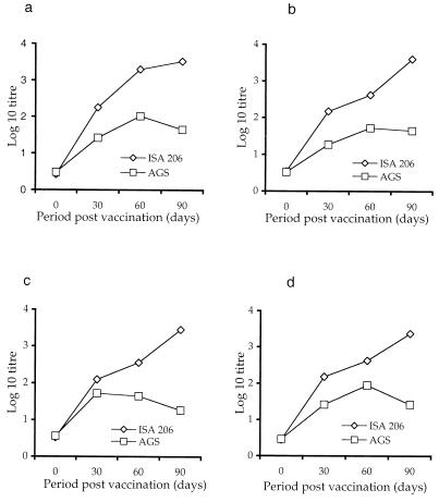
The neutralizing antibody responses elicited in sheep by the oil emulsion and AGS vaccines are shown in Fig. 1. Although we collected serum samples from a limited number of animals, the data obtained are of value for the planning of FMD prophylactic measures. Sheep were early responders to serotype O, and the serum neutralizing (SN) antibody titers were well above 3 at 60 dpv in all of the animals screened. During the subsequent period, the titers increased moderately. For serotypes A and C, the initial magnitude of the response was lower than that to serotype O and the titer was ≥2.5 after 60 dpv (Table 2). However, a clear upward shift was observed only at 60 dpv, reaching ≥3.5 by 90 dpv. The response to serotype Asia-1 showed a constant rise throughout the observation period. The overall antibody responses were considerably lower in AGS-vaccinated animals (Fig. 1). Although the titers increased by 60 dpv, they tended to decline during the later period. The difference between the mean neutralizing antibody titers of the two adjuvant groups was statistically significant for all four of the serotypes (P < 0.05; unpaired Student t test). Interestingly, in sheep that received the AGS vaccine, the response to serotype C was weaker than that to serotype O although the stability of the viral capsid structures is greater in serotype C than in serotype O (27). This establishes the poor adjuvanticity of AGS vaccine in sheep (19-21).
FIG. 1.
Serum antibody responses of sheep to individual viral components measured by microneutralization assay following vaccination with FMDV quadrivalent ISA206 or AGS adjuvanted vaccine. Following vaccination, serum samples were collected from six animals at 0, 30, 60, and 90 dpv. The virus-neutralizing antibody titers toward FMDV serotype O (Ind R2/75) (a), FMDV serotype A22 (Ind 17/77) (b), FMDV serotype C (Ind 1/64) (c), and FMDV serotype Asia-1 (Ind 63/72) (d) were measured by microneutralization test.
TABLE 2.
Comparison of geometric mean neutralizing antibody responses of sheep, goats, and cattle to quadrivalent FMD vaccines formulated with ISA 206 adjuvanta
| FMDV serotype | Period (days) | ISA 206
|
P value (sheep vs goats) | ISA206 (Cattle) | P value (sheep vs cattle) | |
|---|---|---|---|---|---|---|
| Sheep | Goats | |||||
| O | 30 | 2.21 | 1.44 | 0.002b | 2.62 | 0.02b |
| 60 | 3.33 | 1.8 | <0.0001b | 3.15 | 0.44 | |
| 90 | 3.52 | 2.76 | 0.001b | 3.45 | 0.06 | |
| A | 30 | 2.15 | 2.34 | 0.25 | 2.62 | 0.003b |
| 60 | 2.64 | 2.76 | 0.39 | 3 | 0.006b | |
| 90 | 3.62 | 2.94 | <0.0001b | 3.37 | 0.01b | |
| C | 30 | 2.12 | 2.02 | 0.35 | 2.52 | 0.0001b |
| 60 | 2.54 | 2.55 | 0.56 | 3.15 | 0.006b | |
| 90 | 3.45 | 3 | 0.008b | 3.37 | 0.3 | |
| Asia-1 | 30 | 2.22 | 2.1 | 0.79 | 2.55 | 0.01b |
| 60 | 2.60 | 2.63 | 0.76 | 3.15 | 0.01b | |
| 90 | 3.40 | 3.22 | 0.08 | 3.3 | 0.84 | |
Six serum samples were collected at 30, 60, and 90 dpv. Virus-neutralizing antibody titers were measured by microneutralization test. The neutralizing antibody titer of the serum was expressed as the reciprocal of the dilution that neutralized 50% of the virus. The values shown are geometric mean titers expressed in log10, and statistical significance was determined by unpaired Student t test.
P < 0.05.
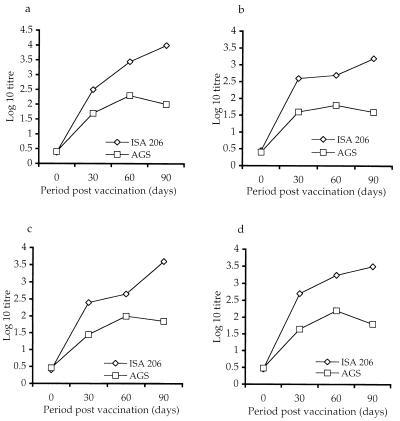
The mean ELISA titers observed with each of the viral components are presented in Fig. 2. A striking similarity between the SN and ELISA titers was a sharp decline in the titer after 60 dpv for serotypes O and Asia-1 in AGS vaccine (Fig. 2a). With the oil adjuvant, the ELISA titers for all four serotypes rose steadily for the duration of the trial.
FIG. 2.
ELISA antibody responses of sheep for various FMDV serotypes following vaccination with binary ethyleneimine-inactivated quadrivalent FMDV vaccines against FMDV serotype O (Ind R2/75) (a), FMDV serotype A22 (Ind 17/77) (b), FMDV serotype C (Ind 1/64) (c), and FMDV serotype Asia-1 (Ind 63/72) (d). The animals were vaccinated with either oil-adjuvanted (ISA 206) or aluminum hydroxide gel-adjuvanted FMDV vaccine, and serum samples were collected at 0, 30, 60, and 90 dpv. Antibody levels were determined by liquid-phase sandwich ELISA.
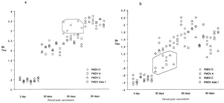
The neutralizing antibody titers of six serum samples for various viral serotypes were plotted by univariate scattergram (Fig. 3a and b). The early neutralizing antibody titers of oil adjuvant from six serum samples were in the range of 1.75 to 2.5 (30 dpv), and the profiles obtained with all four of the viral serotypes were similar. However, the scattergram plotted by utilizing SN antibody titers measured at 60 dpv distinguished two different groups among the four viral serotypes. The A, C, and Asia-1 serotypes clustered apart from serotype O, confirming a strong immune response to FMDV serotype O in all six of the animals tested (Fig. 3a; marked area). Further, a 90-dpv response analysis confirmed a homogeneous response to all four of the serotypes, the titers being in the range of 3 to 3.5 for all of the samples tested. The animals vaccinated with AGS vaccine showed different patterns of clustering (Fig. 3b). During the 30-dpv period, serotypes O and Asia-1 grouped away from serotypes A and C, confirming a poor early immune response to the latter serotypes. Of six serum samples, five had titers of 0.7 to 1.2 for FMDV serotype C and three had titers in the range of 0.8 to 1.2 for FMDV serotype A (Fig. 3b; marked area). Although a moderate upward shift was observed during the 60-dpv period (1.4 to 2.2), the neutralizing response showed a tendency to decline by the end of observation period (1.2 to 1.8; 90 dpv).
FIG. 3.
Univariate scattergram of neutralizing antibody responses of sheep vaccinated with FMDV quadrivalent vaccine formulated with ISA 206 (a) or aluminum hydroxide gel-saponin (b) adjuvant. The neutralizing antibody titers of six animals for each of the viral components (O, A, C, and Asia-1) were plotted against time points of 0, 30, 60, and 90 days, and a univariate scattergram was plotted by using the StatView software package.
We compared neutralizing antibody responses elicited by double emulsion quadrivalent FMD vaccine among ruminants. The quantum and rate of development of immune responses were greater in cattle (22) than those observed in small ruminants for all four serotypes (Table 2). Cattle showed an early neutralizing immune response to the vaccine, followed by a modest increase during the later phase. The geometric mean SN titers for all four serotypes were >2.5 log10 at 30 dpv (P < 0.05). During the subsequent period, a steady titer increase was observed that was maintained at ≥3.3 at 90 dpv. However, small ruminants exhibited variations in the immune response elicited. Goats were found to be late responders to serotype O, and the neutralizing antibody titers were significantly lower than those of sheep during the study period (23). The initial magnitude of the neutralizing antibody response of goats to serotype O was lower than those of sheep and cattle (1.44 at 30 dpv) (Table 2), and the titer was 1.8 at 60 dpv. A peak in the immune response was attained only after 60 dpv, and the titer was maintained at 2.76 by 90 dpv. In contrast, sheep were early responders to serotype O and the SN titer was superior to those of cattle and goats by 60 dpv (3.3) (Table 2). Further, goats vaccinated with the oil adjuvant showed a constant rise in titers in response to serotypes A, C, and Asia-1 and the titers were maintained at ≥3 at 90 dpv. However, sheep experienced a clear upward shift in their titers in response to serotypes A, C, and Asia-1 after 60 dpv and the neutralizing antibody titers were almost similar to (FMDV C and Asia-1; P > 0.05) or better than (FMDV A; P < 0.05) those of cattle by 90 dpv.
It is well established that immunity to FMD correlates with a neutralizing antibody titer directed against the structural proteins of the viral capsid (13, 24). The experiments done by Rweyemamu and coworkers (29) with cattle showed that 1.12 μg of 146 S FMDV type O1 antigen in aqueous FMD vaccines is equivalent to one 50% protective dose. However, in a later study (24) with the same vaccine virus, 2.2 μg of the antigen was required to obtain one 50% protective dose although protection was observed even with a smaller antigen dose (29). Similar correlations have also been established for other FMDV serotypes (5, 9, 18, 24). Studies by Doel (9) revealed that the useful operational limits of the antigen payload were between 1.5 and 9.2 μg of 146 S. Our data reveal that even vaccines having a payload of 3.5 μg were able to elicit a good SN titer, but this was dependent on the adjuvant used for vaccine preparation and the animal species. Thus, the protective immune response seems to be dictated by the use of superior adjuvants in the vaccine formulations.
The mode of action of oil adjuvant was attributed to depot formation at the site of injection, a vehicle for transport of the antigen throughout the lymphatic system, and slow antigen release with the stimulation of antibody-producing cells. Moreover, being a water-in-oil-in-water emulsion, Montanide ISA 206 had various advantages, like lower viscosity, easy administration, greater stability, and production of smaller nodules at the site of injection (2), compared to other oil adjuvants, making it an ideal adjuvant candidate for FMD vaccines. Recently, Cox and coworkers (6) demonstrated that ISA 206 could induce an SN antibody response within 4 dpv and protect sheep against viremia following an airborne challenge with homologous FMDV as early as 3 dpv. The work by Barnett and Cox (2) has indicated the superiority of the Montanide ISA 206 preparation for longer-term protection of cattle. Taken together, our results support the utility of polyvalent double emulsion vaccines in the FMD control program.
Univariate scattergrams confirmed the high-quality immune response of sheep to serotype O virus, although serotype O is more labile than the other three. This shows that Montanide ISA 206 could prevent the degradation of viral capsid structures, as the poor adjuvanticity of the AGS vaccine was attributed to this factor also. It was surmised that the loss of potency was due to the proteolysis of VP1 or possibly the physical breakdown of the virus following adsorption to the aluminum hydroxide gel (7, 8). Whether this strong immune response to serotype O at 60 dpv is specific to sheep or is common to other animal species is not clear, as cattle and goats were found to be late responders to serotype O. More trials may be necessary to confirm this observation.
Acknowledgments
P.K.P is the recipient of a fellowship from the Council of Scientific and Industrial Research, Government of India. We are grateful to the staff of IVRI, Bangalore, for technical support. Cooperation from veterinary officers and farmers is greatly appreciated. Thanks to Srini V. Kaveri and Sébastien Lacroix-Desmazes (INSERM U430, Hôpital Broussais, Paris, France) for critical reading of the manuscript.
REFERENCES
- 1.Bahnemann, H. G., and J. A. Mesquita. 1987. Oil-adjuvant vaccine against foot-and-mouth disease. Boletin del Centro Panamericano Fiebre Aftosa 53:25-30. [Google Scholar]
- 2.Barnett, P. V., L. Pullen, L. Williams, and T. R. Doel. 1996. International bank for foot-and-mouth disease vaccine: assessment of Montanide ISA 25 and 206, two commercially available oil adjuvants. Vaccine 14:1187-1198. [DOI] [PubMed] [Google Scholar]
- 3.Barnett, P. V., and S. J. Cox. 1999. The role of small ruminants in the epidemiology and transmission of foot-and-mouth disease. Vet. J. 158:6-13. [DOI] [PubMed] [Google Scholar]
- 4.Barteling, S. J., and J. Vreeswijk. 1991. Developments in foot-and-mouth disease vaccines. Vaccine 9:75-88. [DOI] [PubMed] [Google Scholar]
- 5.Black, L., M. J. Francis, M. M. Rweyemamu, O. Umehara, and A. Boge. 1984. The relationship between serum antibody titres and protection from foot-and-mouth disease in pigs after oil emulsion vaccination. J. Biol. Stand. 12:379-389. [DOI] [PubMed] [Google Scholar]
- 6.Cox, S. J., P. V. Barnett, P. Dani, and J. S. Salt. 1999. Emergency vaccination of sheep against foot-and-mouth disease: protection against disease and reduction in contact transmission. Vaccine 17:1858-1868. [DOI] [PubMed] [Google Scholar]
- 7.Doel, T. R., and R. F. Staple. 1982. The elution of foot-and-mouth disease virus from vaccines adjuvanted with aluminium hydroxide and with saponin. J. Biol. Stand. 10:185-195. [DOI] [PubMed] [Google Scholar]
- 8.Doel, T. R., and L. Pullen. 1990. International bank for foot-and-mouth disease vaccine: stability studies with virus concentrates and vaccines prepared from them. Vaccine 8:473-478. [DOI] [PubMed] [Google Scholar]
- 9.Doel, T. R. 1999. Optimisation of the immune response to foot-and-mouth disease vaccines. Vaccine 17:1767-1771. [DOI] [PubMed] [Google Scholar]
- 10.Donaldson, A. I., and T. R. Doel. 1992. Foot-and-mouth disease: the risk for Great Britain after 1992. Vet. Rec. 131:114-120. [DOI] [PubMed] [Google Scholar]
- 11.Ferguson, N. M., C. A. Donnelly, and R. M. Anderson. 2001. The foot-and-mouth epidemic in Great Britain: pattern of spread and impact of interventions. Science 292:1155-1160. [DOI] [PubMed] [Google Scholar]
- 12.Gajendragad, M. R., K. Prabhudas, S. Gopalakrishna, V. V. S. Suryanarayana, and C. Natarajan. 1999. A note on outbreaks caused by mixed foot-and-mouth disease. Acta Virol. 43:49-52. [PubMed] [Google Scholar]
- 13.Ganesh, R. M., G. Butchaiah, and A. K. Sen. 1994. Antibody response to 146 S particle, 12 S protein subunit and isolated VP 1 polypeptide of foot-and-mouth disease virus type Asia 1. Vet. Microbiol. 39:135-143. [DOI] [PubMed] [Google Scholar]
- 14.Iyer, A. V., S. Ghosh, S. N. Singh, and R. A. Deshmukh. 2000. Evaluation of three "ready to formulate' oil adjuvants for foot-and-mouth disease vaccine production. Vaccine 19:1097-1105. [DOI] [PubMed] [Google Scholar]
- 15.Kitching, R. P. 1996. A recent history of foot-and-mouth disease. J. Comp. Pathol. 118:89-108. [DOI] [PubMed] [Google Scholar]
- 16.Mackay, D. 1994. Foot-and-mouth disease in North Africa. Foot Mouth Dis. Newsl. 1:24-28. [Google Scholar]
- 17.McCullough, K. C., J. R. Crowther, and W. C. Butcher. 1985. A liquid phase blocking ELISA and its use in the identification of epitopes on foot-and-mouth disease virus antigens. J. Virol. Methods 11:329-338. [DOI] [PubMed] [Google Scholar]
- 18.McCullough, K. C., L. Bruckner, R. Schaffner, W. Fraefel, H. K. Muller, and U. Khim. 1992. Relationship between the anti-FMD virus antibody reaction as measured by different assays and protection in vivo against challenge infection. Vet. Microbiol. 30:99-112. [DOI] [PubMed] [Google Scholar]
- 19.Nair, S. P., and A. K. Sen. 1992. A comparative study on the immune response of sheep to foot and mouth disease virus vaccine type Asia-1 prepared with different inactivants and adjuvants. Comp. Immunol. Microbiol. Infect. Dis. 15:117-124. [DOI] [PubMed] [Google Scholar]
- 20.Nair, S. P., and A. K. Sen. 1993. A comparative study of the immune responses of sheep against foot-and-mouth disease virus types Asia-1 and O PEG-concentrated aluminium hydroxide gel and oil-adjuvanted vaccines. Vaccine 11:782-786. [DOI] [PubMed] [Google Scholar]
- 21.Nair, S. P., and A. K. Sen. 1994. Studies on the immune response of foot-and-mouth disease virus vaccine type Asia-1 in pregnant ewes, lambs and evaluation of type O vaccine by challenge. Acta Virol. 38:257-261. [PubMed] [Google Scholar]
- 22.Patil, P. K., J. Bayry, S. P. Nair, S. Gopalakrishna, C. M. Sajjanar, L. D. Misra, and C. Natarajan. 2002. Early antibody responses of cattle for foot-and-mouth disease quadrivalent double oil emulsion vaccine. Vet. Microbiol. 87:103-109. [DOI] [PubMed] [Google Scholar]
- 23.Patil, P. K., J. Bayry, C. Ramakrishna, B. Hugar, L. D. Misra, and C. Natarajan. 2002. Immune responses of goats against foot-and-mouth disease quadrivalent vaccine: comparison of double oil emulsion and aluminium hydroxide gel vaccines in eliciting immunity. Vaccine 20:2781-2789. [DOI] [PubMed] [Google Scholar]
- 24.Pay, T. W. F., and P. J. Hingley. 1987. Correlation of 140 S antigen dose, the serum neutralising antibody response and the level of protection induced in cattle by foot-and-mouth disease vaccines. Vaccine 5:60-64. [DOI] [PubMed] [Google Scholar]
- 25.Pay, T. W. F. 1988. Foot-and-mouth disease in sheep and goats: a review. FMD Bull. 26:2-13. [Google Scholar]
- 26.Rapoport, E., and A. Shimshony. 1997. Health hazards to the small ruminant population of the Middle East posed by the trade of sheep and goat meat. Rev. Sci. Tech. Off. Int. Epiz. 16:57-64. [DOI] [PubMed] [Google Scholar]
- 27.Razdan, R., A. K. Sen, B. V. Rao, and V. V. Suryanarayana. 1996. Stability of foot-and-mouth disease virus, its genome and protein at 37°C. Acta Virol. 40:9-14. [PubMed] [Google Scholar]
- 28.Rweyemamu, M., J. C. Booth, M. Head, and T. W. F. Pay. 1978. Micro neutralisation test for the serological typing and subtyping of the foot and mouth disease virus strains. J. Hyg. 81:107-123. [DOI] [PMC free article] [PubMed] [Google Scholar]
- 29.Rweyemamu, M. M., L. Black, A. Boge, A. C. Thorne, and G. M. Terry. 1984. The relationship between the 146 S antigen dose in aqueous foot-and-mouth disease vaccines and the serum antibody response in cattle. J. Biol. Stand. 12:111-120. [DOI] [PubMed] [Google Scholar]
- 30.Sharma, S. K. 1981. Foot-and-mouth disease in sheep and goats. Vet. Res. J. 4:1-21. [Google Scholar]
- 31.Taylor, M. N., and M. Tufan. 1996. Detailed investigations using farmer interviews to assess the losses caused by FMD outbreaks in Turkey. Report of Turkish-German Animal Health Information Project (GTZ), Ministry of Agriculture and Rural Affairs, Republic of Turkey. Deutsche Gesellschaft für Technische Zusammenarbeit (GTZ) GmbH—German Technical Cooperation, Eschborn, Germany.


