Abstract
The molecular chaperone HSP90 regulates stability and function of multiple protein kinases. The HSP90-binding drug geldanamycin interferes with this activity and promotes proteasome-dependent degradation of most HSP90 client proteins. Geldanamycin also binds to GRP94, the HSP90 paralog located in the endoplasmic reticulum (ER). Because two of three ER stress sensors are transmembrane kinases, namely IRE1α and PERK, we investigated whether HSP90 is necessary for the stability and function of these proteins. We found that HSP90 associates with the cytoplasmic domains of both kinases. Both geldanamycin and the HSP90-specific inhibitor, 514, led to the dissociation of HSP90 from the kinases and a concomitant turnover of newly synthesized and existing pools of these proteins, demonstrating that the continued association of HSP90 with the kinases was required to maintain their stability. Further, the previously reported ability of geldanamycin to stimulate ER stress-dependent transcription apparently depends on its interaction with GRP94, not HSP90, since geldanamycin but not 514 led to up-regulation of BiP. However, this effect is eventually superseded by HSP90-dependent destabilization of unfolded protein response signaling. These data establish a role for HSP90 in the cellular transcriptional response to ER stress and demonstrate that chaperone systems on both sides of the ER membrane serve to integrate this signal transduction cascade.
The endoplasmic reticulum (ER) is the primary organelle within the cell in which secreted and transmembrane proteins are synthesized and modified to attain their proper tertiary structure. Various environmental stresses (e.g., glucose deprivation, disturbance in intracellular Ca2+ stores, and inhibition of protein glycosylation) lead to the accumulation of incorrectly folded proteins in the ER lumen (18). Further, the pathobiology of some diseases has been linked to the deleterious effects of accumulated mutant or misfolded proteins in the ER (4, 14). In eukaryotic cells, the response to unfolded protein accumulation in the ER (termed the unfolded protein response or UPR) involves three distinct aspects (reviewed in references 15 and 21): (i) translational attenuation, which reduces the burden of newly synthesized proteins to be folded by the ER; (ii) transcriptional induction of ER resident molecular chaperones and related stress response proteins, including BiP/GRP78 and GRP94; and (iii) removal of misfolded proteins from the ER by retrograde transport coupled to their degradation by 26S proteasomes at or near the cytoplasmic face of the ER membrane.
The UPR signal transduction pathway was first elucidated for Saccharomyces cerevisiae. In yeast, an ER-resident transmembrane protein kinase, Ire1p, is the sole proximal sensor of ER stress leading to activation of the UPR (8, 23, 29). The N terminus of Ire1p senses the perturbed environment within the ER lumen, resulting in oligomerization and trans phosphorylation of its cytoplasmically located kinase domain. This in turn is thought to activate an adjacent endoribonuclease activity in the C terminus of Ire1p that excises a translation-inhibitory intron from the mRNA encoding the transcription factor Hac1. When Hac1 is efficiently translated, it binds to the unfolded protein response element in the promoters of ER chaperones and other UPR targets and up-regulates their transcription (5, 16, 22, 24).
The mammalian UPR is more complex. In mammals, the ER contains two Ire1p homologues, the ubiquitously expressed IRE1α (38-40) and IRE1β, which is expressed primarily in gut epithelium (40). In addition, a third resident transmembrane ER kinase, PERK, which is related to the yeast cytosolic Gcn2 kinase and to the mammalian PKR kinase, phosphorylates eIF-2α, thereby mediating translational repression in response to UPR activation (11, 34). Surprisingly, BiP induction by ER stress is not impaired either in IRE1α−/− IRE1β−/− double knockout cells obtained from mouse embryos (39) or in murine embryonic stem cells in which the IRE1α gene was inactivated by gene disruption (19), suggesting the presence of IRE-independent or -compensating pathways in mammalian cells that regulate ER stress-induced transcriptional responses.
The ER resident transmembrane transcription factor ATF6 is an additional transactivator of the UPR. The cytosolic portion of this protein encodes a transcriptional activation domain that, when cleaved from the membrane in response to ER stress, migrates to the nucleus, where it is able to up-regulate the BiP gene and other stress-responsive genes (12, 20, 41, 45). Whether ATF6 and IRE1 function as serial components of a single pathway or instead represent parallel but overlapping pathways remains under investigation.
A number of key regulatory kinases, including the soluble serine/threonine kinases Raf1 (32, 36, 42), Akt (31, 35), Gcn2 (10), PKR (9), and the type I transmembrane tyrosine kinase p185/ErbB2 (6, 43), depend on interaction with the molecular chaperone HSP90 for stability and function. Where investigated, HSP90 has been found to associate with the kinase domains of these proteins, and interference with this association by the HSP90 binding drug geldanamycin (GA) results in their rapid destabilization and proteasome-mediated degradation (for a review, see reference 25). HSP90 has not previously been implicated in the ER stress response. However, because both PERK and IRE1 are type I transmembrane kinases, and given the HSP90 dependence of both Gcn2 and PKR, we decided to investigate the possibility that HSP90 may participate in the mammalian UPR by regulating the stability and function of IRE1 and/or PERK. In addition, we sought to determine whether the recently reported ability of GA to induce only a transient UPR (17) was due to disruption of HSP90-mediated effects on the UPR signaling pathway.
Here we show that HSP90 does indeed associate with the cytoplasmic domains of both ER transmembrane kinases and that its continued association is required for the stability of these proteins. Finally, our data show that the ability of GA to transiently induce the UPR depends not on its binding to HSP90 but on its interaction with GRP94, the ER-resident paralog of HSP90. This effect is time limited because of the GA-induced loss of HSP90-dependent IRE1 activity.
MATERIALS AND METHODS
Cell culture and transient transfections.
NIH 3T3 fibroblasts and HEK 293 and COS7 cells were grown in Dulbecco's modified Eagle's medium containing 10% fetal bovine serum, 2 mM l-glutamine, and 1% Fungizone (BioWhittaker, Walkersville, Md.). AR42J rat pancreatic tumor cells were maintained in F12K medium (American Type Culture Collection, Manassas, Va.) supplemented with 15% fetal bovine serum, 2 mM l-glutamine, and 1% Fungizone.
For transient-transfection studies, HEK 293 and COS7 cells were grown in 6-cm-diameter dishes. Cells were transfected with 0.5 to 1 μg each of the Flag-tagged cDNAs encoding either full-length human IRE1α (amino acids 1 to 1115) or ΔIRE1 (lacking the cytoplasmic domain; amino acids 725 to 1115), using Lipofectamine-Plus (Gibco BRL, Rockville, Md.) according to the manufacturer's instructions. When necessary, 24 h after transfection, cells were treated with GA (according to the experiment) and then lysed with cold TNESV buffer (50 mM TRIS-HCl [pH 7.4], 1% Nonidet P-40, 2 mM EDTA, 100 mM NaCl, 1 mM sodium orthovanadate) containing protease inhibitors. The full-length IRE1α and ΔIRE1 cDNAs, cloned in pcDNA3.1 plasmid, were a kind gift from K. Imaizumi (Osaka University, Osaka, Japan) and have been described previously (13).
The ATF6 expression plasmid, pCGN-ATF6 (containing a hemagglutinin [HA] tag at the N terminus), was obtained from R. Prywes (Columbia University, New York, N.Y.), and has been previously described (41). COS7 cells were transiently transfected as described above. At 20 to 24 h after transfection, cells were treated with GA, the GA derivative 514, or tunicamycin, according to experimental conditions. Both cleaved and uncleaved forms of transiently expressed HA-tagged ATF6 protein were detected in cell lysates by sodium dodecyl sulfate-polyacrylamide gel electrophoresis (SDS-PAGE) and Western blotting as described previously (43).
Antibodies and immunoprecipitations.
HSP90 antibodies, SPA-830 and SPA-835, were purchased from StressGen Biotechnologies Corp. (Victoria, British Columbia, Canada). HA, BiP, and XBP1 antibodies were purchased from Santa Cruz Biotechnologies (Santa Cruz, Calif.). IRE1 and PERK polyclonal antisera were obtained from D. Ron (New York University, New York, N.Y.). Anti-FLAG-M2 antibody was purchased from Upstate Biotechnology (Lake Placid, N.Y.). Immunoprecipitation assays were performed as described previously (43). Briefly, soluble proteins (400 μg for each condition) were immunoprecipitated with 1 to 3 μg of anti-Flag, anti-PERK, or anti-IRE1 antibodies, and immune complexes were bound on protein A-Sepharose beads for 6 h at 4°C (with rotation). The beads were washed four times with TNESV buffer, and bound proteins were resolved by SDS-7.5 or 10% PAGE and transferred to nitrocellulose membranes for Western blot analysis.
Stress inducers.
GA and its HSP90-preferring derivative 514 (NSC#658514) were obtained from the Developmental Therapeutics Program (National Cancer Institute), dissolved in dimethyl sulfoxide at stock concentrations of 2 and 5 mM, respectively, and diluted to their final concentrations using aqueous media. Tunicamycin, dithiothreitol (DTT), and thapsigargin were purchased from Sigma (St. Louis, Mo.) and dissolved in dimethyl sulfoxide or water at stock concentrations of 2.5 mg/ml, 1 M, and 1 mM, respectively.
GST-HSP90 binding of IRE1.
Equal amounts of protein lysate from transfected cells or from cells containing high levels of endogenous IRE1 (AR42J) were incubated with glutathione Sepharose 4B beads (Amersham Pharmacia, Uppsala, Sweden) that had been previously loaded with purified glutathione S-transferase (GST)-HSP90 (a gift of D. Toft, Mayo Clinic, Rochester, Minn.). The samples were rotated at 4°C for 2 h and then washed thoroughly with TNESV buffer. Washed beads were boiled in protein loading buffer and analyzed by SDS-PAGE. Western blotting was performed with either anti-Flag or anti-IRE 1 antibodies.
Determination of IRE1 half-life in the presence or absence of GA.
COS7 cells were transfected with IRE1 plasmids as described above. These cells or AR42J cells (expressing abundant endogenous IRE1) were treated with GA for 8 h or as indicated. The cells were then starved for 30 min in methionine/cysteine-free media and pulse-labeled with 100 μCi of [35S]methionine/cysteine (Tran 35S-label; ICN, Costa Mesa, Calif.)/ml for 60 min. The cells were washed twice and further incubated in complete Dulbecco's modified Eagle's medium supplemented with 10% fetal calf serum in the presence or absence of GA (1 μM). At various times after being returned to complete medium, cells were lysed and IRE1 proteins were immunoprecipitated with either anti-Flag antibody or anti-IRE1 antisera. Samples were analyzed by SDS-PAGE and autoradiography. Band densities were quantified by NIH Image software using a Macintosh 9500 computer.
RESULTS
Association of HSP90 with IRE1.
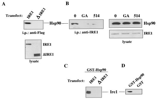
Plasmids expressing Flag-tagged full-length IRE1α or IRE1α lacking the cytoplasmic kinase and RNase domains (ΔIRE1) were transfected into COS7 cells. Cells were lysed 24 h later, and protein lysates were subjected to immunoprecipitation using an anti-Flag antibody. After SDS-PAGE and electrotransfer to nitrocellulose, blots were probed with an antibody to HSP90 (Fig. 1A). HSP90 was clearly coprecipitated with full-length IRE1 but not with ΔIRE1, even though ΔIRE1 was reproducibly expressed to a greater degree than was wild-type IRE1. By transfecting 1/10 the amount of ΔIRE1 as wild-type IRE1, approximately equivalent steady-state levels (Fig. 1A, bottom panel) were obtained. In order to rule out the possibility that association of HSP90 with IRE1 was a transfection artifact, we immunoprecipitated endogenous IRE1α from AR42J, a pancreatic cell line with a well-developed ER. HSP90 readily coprecipitated with IRE1 from untreated cells (Fig. 1B), while a brief exposure (3 h) of AR42J cells to GA disrupted IRE1-HSP90 association (Fig. 1B) without altering the steady-state levels of either IRE1 or HSP90 (Fig. 1B, right panels). We recently identified a chemically similar benzoquinone ansamycin, here designated 514, that has a 90-fold better binding affinity for HSP90 than for the chaperone's ER paralog, GRP94 (43). Using this compound, we also observed disruption of association of IRE1 with HSP90 (Fig. 1B). Last, we used purified GST-HSP90 protein to pull down both tagged IRE1 exogenously expressed in HEK293 cells and endogenous IRE1 in AR42J cells (Fig. 1C and D). Consistent with the in vivo binding data, GST-HSP90 was unable to pull down equivalent amounts of ΔIRE1 transiently expressed in HEK293 cells (Fig. 1C). Taken together, these data support the hypothesis that full-length IRE1 associates with HSP90 via IRE1's cytoplasmic domain and that GA binding to HSP90 disrupts this association.
FIG. 1.
HSP90 associates with IRE1 but not with cytoplasmic domain-deleted IRE1. Cos7 cells were transfected with IRE1-FLAG or ΔIRE1-FLAG plasmids and lysed in TNESV after 24 h. Untransfected AR42J cells were treated or with GA or its derivative 514 (1 μM) of left untreated (lane 0) for 3 h, and then the cells were lysed similarly. Soluble proteins (400 μg) were immunoprecipitated with either 3 μg of anti-Flag antibody (A) or IRE1 antibody (B), and complexes were bound on protein A-Sepharose beads for 2 h at 4°C. The beads were washed four times with TNESV, bound proteins were resolved by SDS-10% PAGE and transferred to nitrocellulose membranes. Western analysis was performed with either anti-HSP90 or anti-IRE antibodies. Expression levels of transiently transfected wild-type IRE1 and ΔIRE1 are shown for comparison in panel A, and HSP90 and IRE1 steady-state levels in cell lysates are shown for comparison in panel B. HEK293 cells were transfected with IRE1-FLAG or ΔIRE1-FLAG plasmids. After 24 h, the cells were lysed and 1 mg of total proteins from each condition was subjected to GST-HSP90 binding in the presence of 400 μl of glutathione-Sepharose 4B, which had been previously saturated with GST-HSP90 (C). The binding of native IRE1 (in AR42J lysates) to either GST-HSP90 or GST alone is shown in panel D.
Disruption of HSP90 association coincides with loss of IRE1 protein.
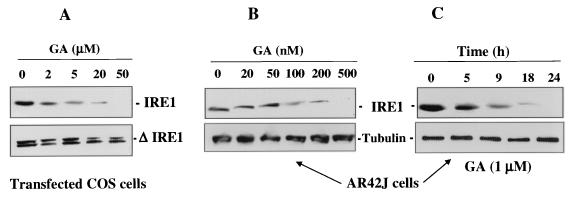
Both COS7 cells transiently transfected with either full-length IRE1 or ΔIRE1 and AR42J cells expressing endogenous IRE1 were treated with increasing concentrations of GA for 24 h. Steady-state levels of Flag-tagged IRE1 (COS7 cells) or endogenous IRE1 (AR42J) proteins were monitored by Western blotting. The data in Fig. 2 show dose- and time-dependent depletion of both transfected and endogenous IRE1, while ΔIRE1 remained completely resistant to GA. Thus, GA sensitivity of IRE1 paralleled HSP90 binding, since ΔIRE1 did not bind HSP90 and was not sensitive to GA. These results are identical to those obtained with another type I transmembrane kinase, ErbB2 (43).
FIG. 2.
Geldanamycin destabilizes IRE1 but not cytoplasmic domain-deleted IRE1. Cos7 cells were transfected with full-length IRE1-FLAG or ΔIRE1-FLAG. Cos7 cells (A) or AR42J cells (expressing endogenous IRE1) (B) were incubated for 24 h with increasing concentrations of GA. (C) AR42J cells were exposed to 1 μM GA for the time periods shown. Cells were lysed, and equivalent amounts of protein were analyzed by SDS-PAGE and Western blotting with Flag antibody (A) or IRE1 antibody (B and C). In panels B and C, tubulin was blotted to demonstrate equal loading of protein.
GA reduces the half-life of IRE1 protein.
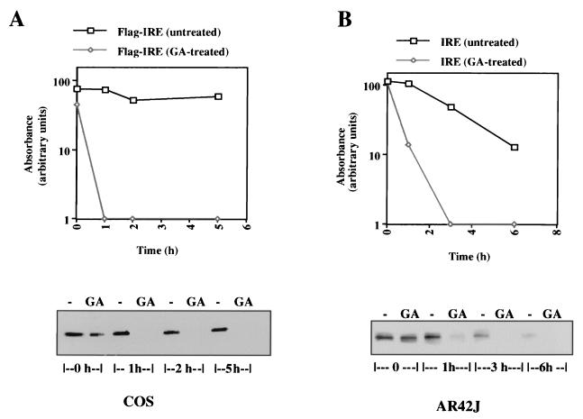
Because GA did not affect IRE1 mRNA levels (data not shown), we expected that its effect on IRE1 protein was posttranslational. Thus, we examined drug effects on the IRE1 protein half-life. COS7 cells transiently transfected with full-length IRE1 and AR42J cells expressing the endogenous protein were pulse-labeled with [35S]methionine/cysteine as described in Materials and Methods and then chased for increasing times in nonradioactive complete medium. In each case, one set of cells was continuously exposed to GA while another set remained untreated. The data show that synthesis of either exogenously expressed or endogenous IRE1 in the presence of GA resulted in a protein with a markedly reduced half-life (Fig. 3). In COS7 cells the half-life of Flag-IRE1 was reduced from >5 h to <1 h, while in AR42J cells the half-life of IRE1α was reduced from approximately 3 h to <1 h. These results are consistent with the effect of GA on other HSP90 client proteins (26).
FIG. 3.
GA decreases the half-life of newly synthesized IRE1. Transiently transfected Cos7 (A) and untransfected AR42J (B) cells were treated with GA (1 μM) 12 h after transfection or left untreated. After an additional 8 h, the medium was replaced with methionine/cysteine-free medium for 30 min, and cells were pulse-labeled with 100 μCi of [35S]methionine/cysteine/ml for 60 min. At 1 h, cells were placed in complete, nonradioactive medium (time zero) and chased for the times shown. GA remained in all drug-treated samples for the duration of the experiment. Proteins were immunoprecipitated with either anti-Flag (A) or anti-IRE1 (B) antibodies. Samples were analyzed by SDS-PAGE and autoradiography. Band densities of scanned films were quantified using NIH Image software to create the graphs shown.
Binding of benzoquinone ansamycin to GRP94 induces an ER stress response, while binding to HSP90 eventually abrogates such a response.
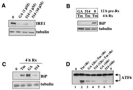
Since GA depletes IRE1 protein within several hours, we suspected that the drug would interfere with the ER's transcriptional response to stress-inducing stimuli. On the other hand, Lawson et al. had previously shown that GA itself induces an ER stress response, although unlike agents such as thapsigargin or tunicamycin, the GA-induced stress response was relatively short-lived (17). In the present experiments, we first confirmed that the level of IRE1 protein was significantly reduced in cells exposed to either GA or 514 for 12 h (Fig. 4A). Concomitantly, exposure to either HSP90 inhibitor for 12 h abrogated the induction of BiP protein synthesis following a 4-h challenge with tunicamycin (TM) (Fig. 4B). Inhibition of the response to TM was not due to inhibition of general protein synthesis in cells exposed to either GA or 514 for 12 h. Thus, untreated cells pulsed briefly with radiolabeled methionine incorporated 1.77 × 10⁶ cpm/mg of protein (trichloroacetic acid-precipitable counts), while cells exposed to GA or 514 for 12 h incorporated 1.78 × 10⁶ and 1.87 × 10⁶ cpm/mg of protein, respectively. In contrast to the results obtained following prolonged exposure to HSP90 inhibitors, a 4-h exposure to GA itself produced a robust BiP response, equivalent to that caused by TM (Fig. 4C). However, 4-h challenge with 514 caused only a negligible BiP response (Fig. 4C). Thus, although both GA and 514 blocked the stress response after prolonged exposure, after a brief exposure, only GA was able to promote a stress response.
FIG. 4.
Both GA and 514, a GA derivative that has a 2-log-greater affinity for HSP90 than for GRP94, inhibit the UPR after prolonged exposure, but only GA induces the UPR after short exposure. (A) AR42J cells (which express high levels of IRE1) were exposed to either GA (1 μM) or 514 (1 or 10 μM) for 12 h. IRE1 and tubulin proteins were detected in whole-cell lysates by Western blotting. (B) AR42J cells were pretreated with either the GA derivative 514 or GA for 12 h before the stress-inducer TM was added (for an additional 4 h). BiP protein was detected by [35S]methionine pulse-labeling for 30 min, and tubulin protein was detected by Western blotting. (C) AR42J cells were exposed to TM, GA, or 514 for 4 h and processed for BiP and tubulin protein detection as was done for panel A. (D) Cos7 cells were transfected with pCGN-ATF6, an ATF6 expression plasmid containing a HA epitope tag. Twenty hours after transfection, cells were either left untreated (lanes 1 and 7), treated with TM for 4 h (lane 2), pretreated with GA for 12 h, followed by TM for an additional 4 h (lane 3), pretreated with 514 for 12 h, followed by TM for 4 h (lane 4), treated with only GA for 12 h (lane 5), or treated with only 514 for 12 h (lane 6). In order to better detect the ATF6 cleavage product induced by ER stress, we treated all cells with the proteasome inhibitor PS-341 (0.5 μM) during exposure to the ER stress agent. In lanes 1, 5, 6, and 7, PS-341 was added for the final 4 h of incubation prior to lysis. Following cell lysis, equivalent amounts of soluble proteins were separated by SDS-PAGE and analyzed for stress-induced ATF6 cleavage by Western blotting using an anti-HA antibody.
GA inhibits ER stress-induced cleavage and activation of ATF6.
IRE1 is not the sole mediator of the transcriptional component of the UPR. Indeed, the BiP response to ER stress remains essentially intact in IRE1−/− cells (19). Thus, the dramatic inhibition of ER stress-mediated BiP induction by prolonged inhibition of HSP90 requires additional explanation. ATF6, an ER transmembrane localized transcription factor, has also been implicated in the transcriptional response to ER stress, since its cytoplasmic domain can activate BiP transcription upon cleavage from the membrane (12, 20, 41, 45). Since ATF6 cleavage is necessary for its transcriptional activity, we transfected cells with an ATF6 expression plasmid, and 20 h later we determined the effects of GA and 514 on cleavage of ATF6 in response to TM. A proteasome inhibitor was included in these incubations, since the cleaved fragment of ATF6 is very unstable but can be protected by proteasome inhibition (44). Although exogenous overexpression of ATF6 can lead to spontaneous cleavage, we successfully titered the amount of plasmid transfected to avoid this problem (Fig. 4D, lanes 1 and 7). While TM at 4 h caused easily detectable cleavage of ATF6 (Fig. 4D, compare lanes 1 and 2), prior exposure to both HSP90 inhibitors for 12 h resulted in much less ATF6 cleavage following TM challenge (Fig. 4D, compare lane 2 with lanes 3 and 4). The HSP90 inhibitors alone caused minimal ATF6 cleavage (Fig. 4D, lanes 5 and 6). Thus, prevention of stress-induced ATF6 cleavage is an additional means by which HSP90 inhibition affects the transcriptional arm of the UPR.
GA promotes BiP dissociation from IRE1, but other stressors do not disrupt the HSP90-IRE1 interaction.
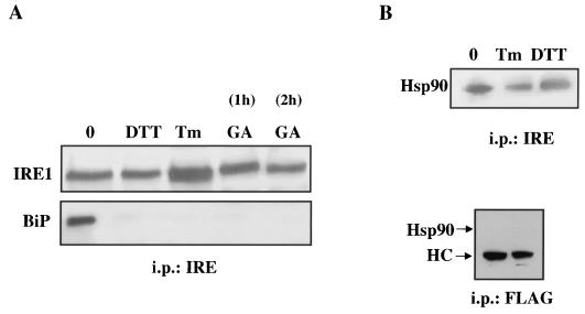
Agents that induce ER stress have been shown to disrupt association of the ER chaperone BiP with the lumenal domain of IRE1 (2, 28). We wished to determine whether GA had a similar activity and at the same time determine if ER stress generally results in dissociation of HSP90 from the cytoplasmic domain of IRE1. Therefore, we exposed AR42J cells to DTT for 30 min, to TM for 2 h, or to GA for 1 or 2 h. After immunoprecipitation of endogenous IRE1, coprecipitation of BiP was examined by Western blotting. We found that each of the three agents efficiently disrupted BiP-IRE1 association when present for the times shown (Fig. 5A). In contrast, and unlike GA, neither TM nor DTT dissociated HSP90 from IRE1 (Fig. 5B). Specificity of the HSP90 coimmunoprecipitation with anti-IRE antibody was confirmed by the inability of anti-FLAG antibody to coimmunoprecipitate HSP90 (Fig. 5B, bottom panel, and Fig. 1A). Thus, although GA mimics the ability of other ER stress agents in disrupting BiP/IRE1 complexes, disruption of HSP90-IRE1 association is neither a general feature nor a prerequisite of the ER stress response.
FIG. 5.
ER stressors, including GA, dissociate IRE1 from BiP but do not dissociate HSP90 from IRE1. (A) AR42J cells were treated with GA (2 μM) for 1 or 2 h, TM (2.5 μg/ml) for 2 h, or DTT (10 mM) for 30 min. After lysis, 400 μg of soluble proteins were immunoprecipitated with 2 μg of IRE1 antibody, and the complexes were bound on protein A-Sepharose beads. Bound proteins were resolved by SDS-7.5% PAGE and transferred to nitrocellulose membranes. Western analysis was performed with anti-IRE1 and anti-BiP antibodies. (B) In a similar experiment, HSP90 coprecipitation following anti-IRE1 immunoprecipitation was examined (by an HSP90 Western blot) in cells treated with TM or DTT. The specificity of HSP90 coprecipitation with IRE1 is shown by lack of an HSP90 signal in anti-FLAG immunoprecipitates.
The ER transmembrane kinase PERK also associates with HSP90 and is destabilized by GA.
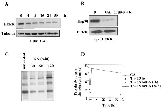
We next examined whether the other ER stress-activated kinase PERK was also an HSP90 client protein. As shown in Fig. 6A and B, HSP90 does coprecipitate with PERK in AR42J cells. Brief exposure to GA disrupts this association, and prolonged exposure to GA results in a decline in PERK protein level (due to a decreased protein half-life; data not shown).
FIG. 6.
The ER transmembrane kinase PERK associates with HSP90 but is functionally less sensitive to GA than is IRE1. (A) AR42J cells were treated with GA (1 μM) for the times indicated, cells were lysed, and proteins were resolved by SDS- 10% PAGE and transferred to nitrocellulose membranes. Western analysis of PERK steady-state levels was performed with an anti-PERK antibody. Tubulin was blotted to demonstrate equal loading of protein. (B) AR42J cells were left untreated or treated with GA (1 μM for 4 h) and then lysed, and 400 μg of soluble proteins were immunoprecipitated with 2 μg of anti-PERK antibody. Antibody complexes were bound on protein A-Sepharose beads, washed, eluted, and resolved by SDS-10% PAGE. After transfer to a nitrocellulose membrane, Western analysis of coprecipitated HSP90 was performed. The immunoprecipitates were also blotted for PERK to demonstrate its equivalent pulldown in the presence and absence of GA. (C) AR42J cells were treated with GA (1 μM) for the indicated times. The cells were then pulse-labeled with 100 μCi of [35S]methionine/cysteine/ml for 30 min and lysed. Equal amounts of total soluble proteins were analyzed by SDS-PAGE and autoradiography. (D) AR42J cells were treated with thapsigargin (Th; 0.5 μM) for 0.5 h (filled diamond), GA (1 μM) for 0.5, 1, 2, or 24 h (filled circles) or with a combination of GA and Th (filled square and filled triangle; GA was added for the indicated time, followed by Th for 30 min). Cells were then pulse-labeled and processed as for panel C. Equal amounts of total cell proteins were analyzed by SDS-PAGE and autoradiography.
GA fails to block thapsigargin-induced translational inhibition.
PERK activation leads to translational attenuation (11, 34). In order to determine whether PERK function requires PERK-HSP90 association, we tested whether GA can block stress-induced translational inhibition. Using a radioactive methionine pulse to measure translation, we observed that GA itself caused a noticeable but transient attenuation of translation (Fig. 6C). However, preexposure of cells to GA for 1 or 24 h did not prevent the translational attenuation observed following a 30-min exposure to thapsigargin (Fig. 6D). These data show that although PERK stability is reduced when it is disassociated from HSP90, resulting in a slow decline in its steady-state level, the activity of the remaining PERK protein is sufficient to inhibit protein synthesis during activation of the ER stress response.
DISCUSSION
Our data demonstrate the involvement of HSP90 in the transcriptional arm of the UPR. HSP90 associates with both exogenously introduced and endogenously expressed IRE1α, and the HSP90 inhibitor GA promotes dissociation of the chaperone from IRE1 concomitant with a marked reduction in the half-life of the kinase. Since IRE1 lacking its cytosolic domain neither associates with HSP90 nor is sensitive to GA, it is likely that drug sensitivity is dependent on chaperone interaction with the kinase domain of IRE1, as is the case with other HSP90 client transmembrane kinases.
In accord with the dependence of IRE1α stability on HSP90 binding, prolonged exposure of cells to GA inhibits BiP induction caused by ER stress. However, in agreement with and extension of the study of Lawson et al. (17), short-term exposure to GA mimics the activity of other ER stressors in that it stimulates BiP protein synthesis and promotes the dissociation of IRE1α from BiP. However, the GA-induced disassociation of HSP90 from IRE1 is not a prerequisite for IRE1 activation, since other stress agents, including DTT and Tm, do not share this property with GA.
How can GA both induce and inhibit ER stress-dependent transcription, depending on the length of exposure? Our present data offer an explanation for the transience of the GA-induced UPR (17). GA interacts with similar affinities with both HSP90 and its ER paralog, GRP94 (6, 43). If the binding of GA to GRP94 interfered with this chaperone's role in protein maturation, thereby increasing the requirement for BiP to bind to “stalled” incompletely folded proteins transiting the ER, the resultant release of BiP from the luminal domain of IRE1 would induce the ER stress response (2). Indeed, GA is reported to interfere with the maturation and secretion of at least two proteins, bile-salt-dependent lipase (27) and immunoglobulin lambda light chain (1), that both interact with GRP94 during their transit through the ER. A third protein, the toll receptor, also depends on GRP94 for maturation as it transits the ER (30); however, possible effects of GA on the toll receptor have not yet been examined. At the same time, the deleterious effect of GA on IRE1 should eventually interfere with the cell's transcriptional response to ER stress. The combined but opposing effects of GA on both HSP90 and GRP94 could explain the transitory stress response induced by this drug. This proposed mechanism is supported by the data obtained with the GA derivative 514. Unlike GA, 514 binds to GRP94 with a Kd(GRP94) of 90 μM, compared to a Kd(HSP90) of 1 μM for HSP90 (43). Since 514 proved to be a poor inducer of BiP synthesis upon short exposure but an efficient inhibitor of BiP response to TM upon prolonged treatment, the data are consistent with the hypothesis that the former event depends on binding of drug to GRP94 while the later process requires binding of drug to HSP90.
Given that BiP responds to ER stress in cells obtained from IRE1−/− mice, it is unlikely that the ability of GA to significantly impair BiP induction is due solely to its effect on IRE1. The transcription factor ATF6 may represent a bifurcation of the transcriptional arm of the UPR in mammalian cells. Recent data from several laboratories suggest that IRE- and ATF6-mediated signaling pathways converge to maximally induce the UPR transcriptional response (3, 7, 19, 46). In response to ER stress, ATF6 migrates from ER to Golgi, where it is cleaved from a 90-kDa membrane-associated form to an approximately 50-kDa soluble active fragment (7), which migrates to the nucleus and activates transcription. Although we were unable to detect significant GA- or 514-induced proteolytic cleavage of transfected ATF6, pretreatment with both HSP90 inhibitors markedly reduced the ability of TM to cause ATF6 cleavage. Thus, by demonstrating that GA can interfere with both transcriptional arms (IRE1 and ATF6) of the UPR, these data offer an explanation as to how GA can so dramatically impair the BiP response to ER stress. However, complicating interpretation of these findings, GA did not affect the steady-state level of unprocessed ATF6, nor did we observe HSP90 in association with either form of ATF6 (data not shown). Also, BiP has been reported to bind to the luminal domain of ATF6, much as it does to IRE1 and PERK, and removal of BiP binding sites results in constitutive translocation of ATF6 to the Golgi, where it is cleaved (33). Although based on this paradigm we would have expected GA to activate ATF6, it is possible that the GA effect on ATF6 is indirect and that the drug interferes elsewhere in the ATF6 cleavage pathway to inhibit processing of the transcription factor. One possibility currently being explored is that the inhibition by GA of stress-induced ATF6 activation is dependent on drug activity toward one or more upstream HSP90 client kinases whose phosphorylation of ATF6 is a prerequisite for its cleavage (or transport from ER to Golgi). Indeed, phosphorylation of ATF6 by p38 mitogen-activated protein kinase has been shown to modulate its transcriptional activity in the cellular context of cardiac myocytes (37). Whether p38 kinase is activated by ER stress and is sensitive to GA, and whether ATF6 phosphorylation is a necessary signal for either its transit to the Golgi or its proteolytic cleavage, are currently under investigation.
PERK, a mediator of ER stress-induced translational inhibition, is a second ER transmembrane kinase found in mammalian cells. We show here that PERK also associates with HSP90 and that GA disrupts this association, slowly reducing the steady-state level of the protein over many hours. However, unlike IRE1α, PERK activity is much less dependent on HSP90. Thus, even long-term exposure to GA, although it significantly reduces the steady-state level of the PERK protein, does not prevent an ER stressor from stimulating PERK autophosphorylation (data not shown) or PERK-mediated translational inhibition. These data suggest that PERK levels are in excess of what is necessary for its activity to be manifest. The lack of sensitivity of PERK to both HSP90 and GRP94 inhibition by GA is somewhat surprising given the apparent interchangeability of the luminal domains of PERK and IRE (2). In addition, two cytosolic kinases related to PERK, Gcn2 in yeast and PKR in mammals, both depend on HSP90 for their activity and stability (9, 10). Further examination of the nature of PERK-HSP90 and PERK-BiP interaction in AR42J and in other cell types may ultimately explain these discrepancies.
An interesting possibility suggested by our data is that all proximal sensors of ER stress are not equally sensitive to an incoming signal. Disruption of GRP94 function by GA may cause only a limited increase in protein load in the ER and thus may be recognized as a minor stress, not a strong enough stimulus to warrant shutting down general protein translation or activating ATF6 but significant enough to moderately stimulate transcription of ER chaperones to compensate. The IRE1α signaling pathway may thus represent the cellular response to a low level of ER stress. In this model, activation of PERK and/or ATF6 would occur only when the stress is prolonged or severe enough to require more than a moderate increase in the level of ER chaperones. Such a graded response model, recently proposed by several groups (19, 46), would help to explain why, when compared to yeast, the regulation of the UPR has become so complex in mammalian cells.
In summary, our data demonstrate that HSP90 is an important component of the transcriptional arm of the UPR, much as the chaperone plays an important role in the cellular response to cytoplasmic stress. However, while nonspecific binding to misfolded or unfolded proteins is thought to be the primary function of HSP90 during cytoplasmic stress, its role in the promulgation of the ER stress response depends instead on HSP90's specific association with at least one ER resident transmembrane kinase, IRE1α.
REFERENCES
- 1.Argon, Y., and B. B. Simen. 1999. GRP94, an ER chaperone with protein and peptide binding properties. Semin. Cell Dev. Biol. 10:495-505. [DOI] [PubMed] [Google Scholar]
- 2.Bertolotti, A., Y. Zhang, L. M. Hendershot, H. P. Harding, and D. Ron. 2000. Dynamic interaction of BiP and ER stress transducers in the unfolded-protein response. Nat. Cell Biol. 2:326-332. [DOI] [PubMed] [Google Scholar]
- 3.Calfon, M., H. Zeng, F. Urano, J. H. Till, S. R. Hubbard, H. P. Harding, S. G. Clark, and D. Ron. 2002. IRE1 couples endoplasmic reticulum load to secretory capacity by processing the XBP-1 mRNA. Nature 415:92-96. [DOI] [PubMed] [Google Scholar]
- 4.Carrell, R. W., and D. A. Lomas. 1997. Conformational disease. Lancet 350:134-138. [DOI] [PubMed] [Google Scholar]
- 5.Chapman, R., C. Sidrauski, and P. Walter. 1998. Intracellular signaling from the endoplasmic reticulum to the nucleus. Annu. Rev. Cell Dev. Biol. 14:459-485. [DOI] [PubMed] [Google Scholar]
- 6.Chavany, C., E. Mimnaugh, P. Miller, R. Bitton, P. Nguyen, J. Trepel, L. Whitesell, R. Schnur, J. Moyer, and L. Neckers. 1996. p185erbB2 binds to GRP94 in vivo. Dissociation of the p185erbB2/GRP94 heterocomplex by benzoquinone ansamycins precedes depletion of p185erbB2. J. Biol. Chem. 271:4974-4977. [DOI] [PubMed] [Google Scholar]
- 7.Chen, X., J. Shen, and R. Prywes. 2002. The lumenal domain of ATF6 senses ER stress and causes translocation of ATF6 from the ER to the Golgi. J. Biol. Chem. 30:13045-13052. [DOI] [PubMed] [Google Scholar]
- 8.Cox, J. S., C. E. Shamu, and P. Walter. 1993. Transcriptional induction of genes encoding endoplasmic reticulum resident proteins requires a transmembrane protein kinase. Cell 73:1197-1206. [DOI] [PubMed] [Google Scholar]
- 9.Donze, O., T. Abbas-Terki, and D. Picard. 2001. The Hsp90 chaperone complex is both a facilitator and a repressor of the dsRNA-dependent kinase PKR. EMBO J. 20:3771-3780. [DOI] [PMC free article] [PubMed] [Google Scholar]
- 10.Donze, O., and D. Picard. 1999. Hsp90 binds and regulates Gcn2, the ligand-inducible kinase of the alpha subunit of eukaryotic translation initiation factor 2 [corrected]. Mol. Cell. Biol. 19:8422-8432. [DOI] [PMC free article] [PubMed] [Google Scholar]
- 11.Harding, H. P., Y. Zhang, and D. Ron. 1999. Protein translation and folding are coupled by an endoplasmic-reticulum-resident kinase. Nature 397:271-274. [DOI] [PubMed] [Google Scholar]
- 12.Haze, K., H. Yoshida, H. Yanagi, T. Yura, and K. Mori. 1999. Mammalian transcription factor ATF6 is synthesized as a transmembrane protein and activated by proteolysis in response to endoplasmic reticulum stress. Mol. Biol. Cell 10:3787-3799. [DOI] [PMC free article] [PubMed] [Google Scholar]
- 13.Imaizumi, K., K. Miyoshi, T. Katayama, T. Yoneda, M. Taniguchi, T. Kudo, and M. Tohyama. 2001. The unfolded protein response and Alzheimer's disease. Biochim. Biophys. Acta 1536:85-96. [DOI] [PubMed] [Google Scholar]
- 14.Katayama, T., K. Imaizumi, N. Sato, K. Miyoshi, T. Kudo, J. Hitomi, T. Morihara, T. Yoneda, F. Gomi, Y. Mori, Y. Nakano, J. Takeda, T. Tsuda, Y. Itoyama, O. Murayama, A. Takashima, P. St George-Hyslop, M. Takeda, and M. Tohyama. 1999. Presenilin-1 mutations downregulate the signalling pathway of the unfolded-protein response. Nat. Cell Biol. 1:479-485. [DOI] [PubMed] [Google Scholar]
- 15.Kaufman, R. J. 1999. Stress signaling from the lumen of the endoplasmic reticulum: coordination of gene transcriptional and translational controls. Genes Dev. 13:1211-1233. [DOI] [PubMed] [Google Scholar]
- 16.Kohno, K., K. Normington, J. Sambrook, M. J. Gething, and K. Mori. 1993. The promoter region of the yeast KAR2 (BiP) gene contains a regulatory domain that responds to the presence of unfolded proteins in the endoplasmic reticulum. Mol. Cell. Biol. 13:877-890. [DOI] [PMC free article] [PubMed] [Google Scholar]
- 17.Lawson, B., J. W. Brewer, and L. M. Hendershot. 1998. Geldanamycin, an hsp90/GRP94-binding drug, induces increased transcription of endoplasmic reticulum (ER) chaperones via the ER stress pathway. J. Cell Physiol. 174:170-178. [DOI] [PubMed] [Google Scholar]
- 18.Lee, A. S. 1992. Mammalian stress response: induction of the glucose-regulated protein family. Curr. Opin. Cell Biol. 4:267-273. [DOI] [PubMed] [Google Scholar]
- 19.Lee, K., W. Tirasophon, X. Shen, M. Michalak, R. Prywes, T. Okada, H. Yoshida, K. Mori, and R. J. Kaufman. 2002. IRE1-mediated unconventional mRNA splicing and S2P-mediated ATF6 cleavage merge to regulate XBP1 in signaling the unfolded protein response. Genes Dev. 16:452-466. [DOI] [PMC free article] [PubMed] [Google Scholar]
- 20.Li, M., P. Baumeister, B. Roy, T. Phan, D. Foti, S. Luo, and A. S. Lee. 2000. ATF6 as a transcription activator of the endoplasmic reticulum stress element: thapsigargin stress-induced changes and synergistic interactions with NF-Y and YY1. Mol. Cell. Biol. 20:5096-5106. [DOI] [PMC free article] [PubMed] [Google Scholar]
- 21.Mori, K. 2000. Tripartite management of unfolded proteins in the endoplasmic reticulum. Cell 101:451-454. [DOI] [PubMed] [Google Scholar]
- 22.Mori, K., T. Kawahara, H. Yoshida, H. Yanagi, and T. Yura. 1996. Signalling from endoplasmic reticulum to nucleus: transcription factor with a basic-leucine zipper motif is required for the unfolded protein-response pathway. Genes Cells 1:803-817. [DOI] [PubMed] [Google Scholar]
- 23.Mori, K., W. Ma, M. J. Gething, and J. Sambrook. 1993. A transmembrane protein with a cdc2+/CDC28-related kinase activity is required for signaling from the ER to the nucleus. Cell 74:743-756. [DOI] [PubMed] [Google Scholar]
- 24.Mori, K., A. Sant, K. Kohno, K. Normington, M. J. Gething, and J. F. Sambrook. 1992. A 22 bp cis-acting element is necessary and sufficient for the induction of the yeast KAR2 (BiP) gene by unfolded proteins. EMBO J. 11:2583-2593. [DOI] [PMC free article] [PubMed] [Google Scholar]
- 25.Neckers, L., E. Mimnaugh, and T. W. Schulte. 1999. Hsp90 as an anti-cancer target. Drug Resist. Update 2:165-172. [DOI] [PubMed] [Google Scholar]
- 26.Neckers, L., T. W. Schulte, and E. Mimnaugh. 1999. Geldanamycin as a potential anti-cancer agent: its molecular target and biochemical activity. Investig. New Drugs 17:361-373. [DOI] [PubMed] [Google Scholar]
- 27.Nganga, A., N. Bruneau, V. Sbarra, D. Lombardo, and J. Le Petit-Thevenin. 2000. Control of pancreatic bile-salt-dependent-lipase secretion by the glucose-regulated protein of 94 kDa (Grp94). Biochem. J. 352(Pt. 3):865-874. [PMC free article] [PubMed] [Google Scholar]
- 28.Okamura, K., Y. Kimata, H. Higashio, A. Tsuru, and K. Kohno. 2000. Dissociation of Kar2p/BiP from an ER sensory molecule, Ire1p, triggers the unfolded protein response in yeast. Biochem. Biophys. Res. Commun. 279:445-450. [DOI] [PubMed] [Google Scholar]
- 29.Patil, C., and P. Walter. 2001. Intracellular signaling from the endoplasmic reticulum to the nucleus: the unfolded protein response in yeast and mammals. Curr. Opin. Cell Biol. 13:349-355. [DOI] [PubMed] [Google Scholar]
- 30.Randow, F., and B. Seed. 2001. Endoplasmic reticulum chaperone gp96 is required for innate immunity but not cell viability. Nat. Cell Biol. 3:891-896. [DOI] [PubMed] [Google Scholar]
- 31.Sato, S., N. Fujita, and T. Tsuruo. 2000. Modulation of Akt kinase activity by binding to Hsp90. Proc. Natl. Acad. Sci. USA 97:10832-10837. [DOI] [PMC free article] [PubMed] [Google Scholar]
- 32.Schulte, T. W., M. V. Blagosklonny, C. Ingui, and L. Neckers. 1995. Disruption of the Raf-1-Hsp90 molecular complex results in destabilization of Raf-1 and loss of Raf-1-Ras association. J. Biol. Chem. 270:24585-24588. [DOI] [PubMed] [Google Scholar]
- 33.Shen, J., X. Chen, L. Hendershot, and R. Prywes. 2002. ER stress regulation of ATF6 localization by dissociation of BiP/GRP78 binding and unmasking of Golgi localization signals. Dev. Cell 3:99-111. [DOI] [PubMed] [Google Scholar]
- 34.Shi, Y., K. M. Vattem, R. Sood, J. An, J. Liang, L. Stramm, and R. C. Wek. 1998. Identification and characterization of pancreatic eukaryotic initiation factor 2 alpha-subunit kinase, PEK, involved in translational control. Mol. Cell. Biol. 18:7499-7509. [DOI] [PMC free article] [PubMed] [Google Scholar]
- 35.Soga, S., S. V. Sharma, Y. Shiotsu, M. Shimizu, H. Tahara, K. Yamaguchi, Y. Ikuina, C. Murakata, T. Tamaoki, J. Kurebayashi, T. W. Schulte, L. M. Neckers, and S. Akinaga. 2001. Stereospecific antitumor activity of radicicol oxime derivatives. Cancer Chemother. Pharmacol. 48:435-445. [DOI] [PubMed] [Google Scholar]
- 36.Stancato, L. F., Y. H. Chow, K. A. Hutchison, G. H. Perdew, R. Jove, and W. B. Pratt. 1993. Raf exists in a native heterocomplex with hsp90 and p50 that can be reconstituted in a cell-free system. J. Biol. Chem. 268:21711-21716. [PubMed] [Google Scholar]
- 37.Thuerauf, D. J., N. D. Arnold, D. Zechner, D. S. Hanford, K. M. DeMartin, P. M. McDonough, R. Prywes, and C. C. Glembotski. 1998. p38 mitogen-activated protein kinase mediates the transcriptional induction of the atrial natriuretic factor gene through a serum response element. A potential role for the transcription factor ATF6. J. Biol. Chem. 273:20636-20643. [DOI] [PubMed] [Google Scholar]
- 38.Tirasophon, W., K. Lee, B. Callaghan, A. Welihinda, and R. J. Kaufman. 2000. The endoribonuclease activity of mammalian IRE1 autoregulates its mRNA and is required for the unfolded protein response. Genes Dev. 14:2725-2736. [DOI] [PMC free article] [PubMed] [Google Scholar]
- 39.Urano, F., A. Bertolotti, and D. Ron. 2000. IRE1 and efferent signaling from the endoplasmic reticulum. J. Cell Sci. 113(Pt 21):3697-3702. [DOI] [PubMed] [Google Scholar]
- 40.Wang, X. Z., H. P. Harding, Y. Zhang, E. M. Jolicoeur, M. Kuroda, and D. Ron. 1998. Cloning of mammalian Ire1 reveals diversity in the ER stress responses. EMBO J. 17:5708-5717. [DOI] [PMC free article] [PubMed] [Google Scholar]
- 41.Wang, Y., J. Shen, N. Arenzana, W. Tirasophon, R. J. Kaufman, and R. Prywes. 2000. Activation of ATF6 and an ATF6 DNA binding site by the endoplasmic reticulum stress response. J. Biol. Chem. 275:27013-27020. [DOI] [PubMed] [Google Scholar]
- 42.Wartmann, M., and R. J. Davis. 1994. The native structure of the activated Raf protein kinase is a membrane-bound multi-subunit complex. J. Biol. Chem. 269:6695-6701. [PubMed] [Google Scholar]
- 43.Xu, W., E. Mimnaugh, M. F. Rosser, C. Nicchitta, M. Marcu, Y. Yarden, and L. Neckers. 2001. Sensitivity of mature Erbb2 to geldanamycin is conferred by its kinase domain and is mediated by the chaperone protein Hsp90. J. Biol. Chem. 276:3702-3708. [DOI] [PubMed] [Google Scholar]
- 44.Ye, J., R. B. Rawson, R. Komuro, X. Chen, U. P. Dave, R. Prywes, M. S. Brown, and J. L. Goldstein. 2000. ER stress induces cleavage of membrane-bound ATF6 by the same proteases that process SREBPs. Mol. Cell 6:1355-1364. [DOI] [PubMed] [Google Scholar]
- 45.Yoshida, H., K. Haze, H. Yanagi, T. Yura, and K. Mori. 1998. Identification of the cis-acting endoplasmic reticulum stress response element responsible for transcriptional induction of mammalian glucose-regulated proteins. Involvement of basic leucine zipper transcription factors. J. Biol. Chem. 273:33741-33749. [DOI] [PubMed] [Google Scholar]
- 46.Yoshida, H., T. Matsui, A. Yamamoto, T. Okada, and K. Mori. 2001. XBP1 mRNA is induced by ATF6 and spliced by IRE1 in response to ER stress to produce a highly active transcription factor. Cell 107:881-891. [DOI] [PubMed] [Google Scholar]


