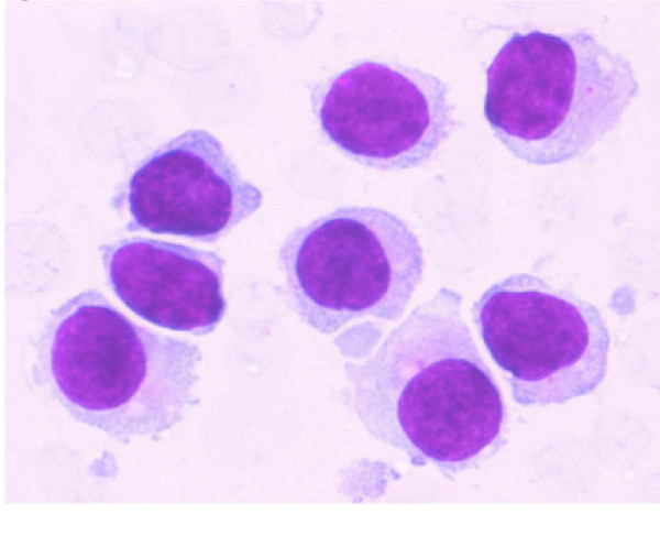
Figure 2.
A higher magnification of the aspirate from same area as shown in Figure 1 shows small to intermediate sized mononuclear cells with moderate cytoplasm, round to oval nuclei with smooth nuclear borders, a stippled chromatin pattern and occasional single nucleoli. The cytoplasm is pale grey and granular with hair-like and short, blunt, cytoplasmic projections. (Stain: Diff Quik, Magnification: × 60).

